The purpose of this study was to observe the morphological pattern by CT angiography
and risk factors for development of peripheral vascular disease in Bangladeshi patient suffering
from peripheral vascular disease using a multidetector scanner in the evaluation of patients with
peripheral vascular disease.
Keunikan anatomi small vessel of the brain dan neurovascular unit, kontroversi peran stganasi vena dalam patofisiologi, klasifikasi small vessel disease, variasi kriteria diagnostik, pitfall dalam neuroimaging, pilihan antiplatelet untuk prevensi sekundar, dampaknya bagi outcome pasien, hubungannya dengan gangguan fungsi kognitif.
Hmm, apa lagi nih yang baru?
Carotid Endarterectomy in Stroke Prevention UpdateDenise Crute
Neurocritical Care lecture on the role of carotid artery surgery in stroke prevention, reviewing the latest studies and evidence-based updates, along with neurosurgical techniques.
A Review of Atherectomy in Peripheral Arterial Diseaseasclepiuspdfs
Atherectomy involves exciting technology and offers expanded treatment options for PAD. Data are scant so far in most lower extremity territories to support its use over other interventions, but newer results are promising. There is still a financial benefit to choosing atherectomy in the outpatient setting that likely drives much of its popularity among interventionalists. Atherectomy is an exciting technology for peripheral vascular intervention. Its use has greatly increased over the last decade. Data on its superiority to angioplasty or angioplasty with stenting are scant. Here, we review atherectomy techniques and principles along with results and controversy surrounding its use.
Vertebral artery pseudo-aneurysms and dissections are known to occur as a result of mechanical
manipulations of the cervical region, traumatic injury, spontaneously and iatrogenic injury because of central
venous catheterization. Central venous lines have become an integral part of patient care, but they are
not without complications. Vertebral artery injury (leading to pseudo-aneurysm and dissection) is one of
the rarer complications of central venous catheter placement. We report a case of inadvertent vertebral
artery catheterization during a dialysis catheter placement which subsequently demonstrated arterial
blood. Duplex ultrasound and computed tomographic (CT) scan confirmed vertebral artery catheterization.
It was successfully treated with open surgical technique by the vascular surgeon because of the size of
catheter and subsequent requirement of artery repair. There were no neurological sequelae. Open surgical
repair remains the gold standard of treatment. Endovascular repair of vertebral artery pseudo-aneurysms
has been described with promising outcomes, but long-term results are lacking. This case report describes
the rare iatrogenic event of vertebral artery injury and reviews its etiology, diagnosis, complications, and management.
An Overview of Filter-Protected Carotid Artery Stentinggailms
These slides give an overview of cerebral protection devices used today in carotid artery stenting, with special emphasis on distal protection filters. Previous work in the field, results from our laboratory, and future directions of device development are covered.
Introduction: There is growing evidence that Obstructive Sleep Apnea (OSA) is a risk factor for Pulmonary Embolism (PE). This
association represents a major public health burden.Aims and Objectives: To investigate Computed Tomography Obstruction Index (CTOI) and the Right Ventricular (RV) to Left Ventricular (LV) diameter ratio with OSA severity. Materials and Methods: 46 Patients with (PE) were evaluated for OSA. Pulmonary Artery Obstruction Index (PAOI) and RV/ LV diameter ratio was measured by pulmonary angiography. Pulmonary Embolism Severity Index (PESI) was determined. Epworth Sleepiness Scale (ESS) and Polysomnography (PSG) was performed for all patients. Based on the PAOI, patients divided into (< 15%, 15-50%, > 50%).
PAD can be diagnosed in asymptomatic individuals by a combination of physical examination and simple, noninvasive Doppler ultrasonography to measure the ankle–brachial index
Our project, our experience and our results at December 31 st 2013
Il nostro progetto, la nostra esperienza ed i nostri risultati aggiornati al 31.12.2013
(Angiologia-Chirurgia Vascolare-ULSS 15 Alta Padovana)
(Angiology- Vascular Surgery -ULSS 15 Alta Padovana)
IMAGES OF A COMPLEX CASE OF MULTIPLE ANEURYSMAL DISEASE IN A 58 YEAR OLD MAN
IMMAGINI DI UN CASO COMPLESSO DI MALATTIA POLINEURISMATICA
(Chirurgia Vascolare-ULSS 15 Alta Padovana)
(Vascular Surgery -ULSS 15 Alta Padovana)
Thermal Imaging for the Diagnosis of Early Vascular Dysfunctions: A Case Reportasclepiuspdfs
Diseases of blood vessels (referred in this article as vascular dysfunction) cause more morbidity and mortality, than combined impact of any other major non-communicable disease including cancer. We strongly feel that the development of a therapy system based on the management of disease of the vessel than management of the risk factors will yield better results and provide greater opportunity for individualized therapy. Detection of early vascular changes before clinical manifestations of endothelial dysfunction, hardening of the arteries, increased intima-media thickness, is of great importance for early identification of individuals with increased risk of accelerated atherosclerosis.
Regional variation in chronic kidney disease and associated factors in hypert...M A Hasnat
We aimed to determine the prevalence of chronic
kidney disease (CKD) and its cross-country variation among
hypertensive individuals in rural Bangladesh, Pakistan and
Sri Lanka.We also explored the factors associated with CKD in
these populations.
HIGH SENSITIVE C-REACTIVE PROTEIN (hs-CRP) AND ITS CORRELATION WITH ANGIOGRAP...M A Hasnat
Association between the plasma hs-CRP levels and the severity of coronary
stenosis in subjects remains controversial. This cross sectional study was performed in the
Department of Cardiology, Dhaka Medical College during July 2008 to December 2009, to determine whether the concentrations of hs-CRP correlate with the coronary atherosclerotic disease assessed by coronary angiography.
Percutaneous Transvenous Mitral Commissurotomy in 71 Years Old Woman with Mit...M A Hasnat
AAS Majumder, MA Hasnat, AKMM Islam, M Ullah, MZ Rahman, S Zaman, MM Rahman, MH Alamgir.
Department of Cardiology, NICVD, Dhaka.
(Cardiovasc. j. 2013; 6(1): 68-70)
Title: Sense of Smell
Presenter: Dr. Faiza, Assistant Professor of Physiology
Qualifications:
MBBS (Best Graduate, AIMC Lahore)
FCPS Physiology
ICMT, CHPE, DHPE (STMU)
MPH (GC University, Faisalabad)
MBA (Virtual University of Pakistan)
Learning Objectives:
Describe the primary categories of smells and the concept of odor blindness.
Explain the structure and location of the olfactory membrane and mucosa, including the types and roles of cells involved in olfaction.
Describe the pathway and mechanisms of olfactory signal transmission from the olfactory receptors to the brain.
Illustrate the biochemical cascade triggered by odorant binding to olfactory receptors, including the role of G-proteins and second messengers in generating an action potential.
Identify different types of olfactory disorders such as anosmia, hyposmia, hyperosmia, and dysosmia, including their potential causes.
Key Topics:
Olfactory Genes:
3% of the human genome accounts for olfactory genes.
400 genes for odorant receptors.
Olfactory Membrane:
Located in the superior part of the nasal cavity.
Medially: Folds downward along the superior septum.
Laterally: Folds over the superior turbinate and upper surface of the middle turbinate.
Total surface area: 5-10 square centimeters.
Olfactory Mucosa:
Olfactory Cells: Bipolar nerve cells derived from the CNS (100 million), with 4-25 olfactory cilia per cell.
Sustentacular Cells: Produce mucus and maintain ionic and molecular environment.
Basal Cells: Replace worn-out olfactory cells with an average lifespan of 1-2 months.
Bowman’s Gland: Secretes mucus.
Stimulation of Olfactory Cells:
Odorant dissolves in mucus and attaches to receptors on olfactory cilia.
Involves a cascade effect through G-proteins and second messengers, leading to depolarization and action potential generation in the olfactory nerve.
Quality of a Good Odorant:
Small (3-20 Carbon atoms), volatile, water-soluble, and lipid-soluble.
Facilitated by odorant-binding proteins in mucus.
Membrane Potential and Action Potential:
Resting membrane potential: -55mV.
Action potential frequency in the olfactory nerve increases with odorant strength.
Adaptation Towards the Sense of Smell:
Rapid adaptation within the first second, with further slow adaptation.
Psychological adaptation greater than receptor adaptation, involving feedback inhibition from the central nervous system.
Primary Sensations of Smell:
Camphoraceous, Musky, Floral, Pepperminty, Ethereal, Pungent, Putrid.
Odor Detection Threshold:
Examples: Hydrogen sulfide (0.0005 ppm), Methyl-mercaptan (0.002 ppm).
Some toxic substances are odorless at lethal concentrations.
Characteristics of Smell:
Odor blindness for single substances due to lack of appropriate receptor protein.
Behavioral and emotional influences of smell.
Transmission of Olfactory Signals:
From olfactory cells to glomeruli in the olfactory bulb, involving lateral inhibition.
Primitive, less old, and new olfactory systems with different path
micro teaching on communication m.sc nursing.pdfAnurag Sharma
Microteaching is a unique model of practice teaching. It is a viable instrument for the. desired change in the teaching behavior or the behavior potential which, in specified types of real. classroom situations, tends to facilitate the achievement of specified types of objectives.
Lung Cancer: Artificial Intelligence, Synergetics, Complex System Analysis, S...Oleg Kshivets
RESULTS: Overall life span (LS) was 2252.1±1742.5 days and cumulative 5-year survival (5YS) reached 73.2%, 10 years – 64.8%, 20 years – 42.5%. 513 LCP lived more than 5 years (LS=3124.6±1525.6 days), 148 LCP – more than 10 years (LS=5054.4±1504.1 days).199 LCP died because of LC (LS=562.7±374.5 days). 5YS of LCP after bi/lobectomies was significantly superior in comparison with LCP after pneumonectomies (78.1% vs.63.7%, P=0.00001 by log-rank test). AT significantly improved 5YS (66.3% vs. 34.8%) (P=0.00000 by log-rank test) only for LCP with N1-2. Cox modeling displayed that 5YS of LCP significantly depended on: phase transition (PT) early-invasive LC in terms of synergetics, PT N0—N12, cell ratio factors (ratio between cancer cells- CC and blood cells subpopulations), G1-3, histology, glucose, AT, blood cell circuit, prothrombin index, heparin tolerance, recalcification time (P=0.000-0.038). Neural networks, genetic algorithm selection and bootstrap simulation revealed relationships between 5YS and PT early-invasive LC (rank=1), PT N0—N12 (rank=2), thrombocytes/CC (3), erythrocytes/CC (4), eosinophils/CC (5), healthy cells/CC (6), lymphocytes/CC (7), segmented neutrophils/CC (8), stick neutrophils/CC (9), monocytes/CC (10); leucocytes/CC (11). Correct prediction of 5YS was 100% by neural networks computing (area under ROC curve=1.0; error=0.0).
CONCLUSIONS: 5YS of LCP after radical procedures significantly depended on: 1) PT early-invasive cancer; 2) PT N0--N12; 3) cell ratio factors; 4) blood cell circuit; 5) biochemical factors; 6) hemostasis system; 7) AT; 8) LC characteristics; 9) LC cell dynamics; 10) surgery type: lobectomy/pneumonectomy; 11) anthropometric data. Optimal diagnosis and treatment strategies for LC are: 1) screening and early detection of LC; 2) availability of experienced thoracic surgeons because of complexity of radical procedures; 3) aggressive en block surgery and adequate lymph node dissection for completeness; 4) precise prediction; 5) adjuvant chemoimmunoradiotherapy for LCP with unfavorable prognosis.
Ozempic: Preoperative Management of Patients on GLP-1 Receptor Agonists Saeid Safari
Preoperative Management of Patients on GLP-1 Receptor Agonists like Ozempic and Semiglutide
ASA GUIDELINE
NYSORA Guideline
2 Case Reports of Gastric Ultrasound
- Video recording of this lecture in English language: https://youtu.be/lK81BzxMqdo
- Video recording of this lecture in Arabic language: https://youtu.be/Ve4P0COk9OI
- Link to download the book free: https://nephrotube.blogspot.com/p/nephrotube-nephrology-books.html
- Link to NephroTube website: www.NephroTube.com
- Link to NephroTube social media accounts: https://nephrotube.blogspot.com/p/join-nephrotube-on-social-media.html
Explore natural remedies for syphilis treatment in Singapore. Discover alternative therapies, herbal remedies, and lifestyle changes that may complement conventional treatments. Learn about holistic approaches to managing syphilis symptoms and supporting overall health.
Tom Selleck Health: A Comprehensive Look at the Iconic Actor’s Wellness Journeygreendigital
Tom Selleck, an enduring figure in Hollywood. has captivated audiences for decades with his rugged charm, iconic moustache. and memorable roles in television and film. From his breakout role as Thomas Magnum in Magnum P.I. to his current portrayal of Frank Reagan in Blue Bloods. Selleck's career has spanned over 50 years. But beyond his professional achievements. fans have often been curious about Tom Selleck Health. especially as he has aged in the public eye.
Follow us on: Pinterest
Introduction
Many have been interested in Tom Selleck health. not only because of his enduring presence on screen but also because of the challenges. and lifestyle choices he has faced and made over the years. This article delves into the various aspects of Tom Selleck health. exploring his fitness regimen, diet, mental health. and the challenges he has encountered as he ages. We'll look at how he maintains his well-being. the health issues he has faced, and his approach to ageing .
Early Life and Career
Childhood and Athletic Beginnings
Tom Selleck was born on January 29, 1945, in Detroit, Michigan, and grew up in Sherman Oaks, California. From an early age, he was involved in sports, particularly basketball. which played a significant role in his physical development. His athletic pursuits continued into college. where he attended the University of Southern California (USC) on a basketball scholarship. This early involvement in sports laid a strong foundation for his physical health and disciplined lifestyle.
Transition to Acting
Selleck's transition from an athlete to an actor came with its physical demands. His first significant role in "Magnum P.I." required him to perform various stunts and maintain a fit appearance. This role, which he played from 1980 to 1988. necessitated a rigorous fitness routine to meet the show's demands. setting the stage for his long-term commitment to health and wellness.
Fitness Regimen
Workout Routine
Tom Selleck health and fitness regimen has evolved. adapting to his changing roles and age. During his "Magnum, P.I." days. Selleck's workouts were intense and focused on building and maintaining muscle mass. His routine included weightlifting, cardiovascular exercises. and specific training for the stunts he performed on the show.
Selleck adjusted his fitness routine as he aged to suit his body's needs. Today, his workouts focus on maintaining flexibility, strength, and cardiovascular health. He incorporates low-impact exercises such as swimming, walking, and light weightlifting. This balanced approach helps him stay fit without putting undue strain on his joints and muscles.
Importance of Flexibility and Mobility
In recent years, Selleck has emphasized the importance of flexibility and mobility in his fitness regimen. Understanding the natural decline in muscle mass and joint flexibility with age. he includes stretching and yoga in his routine. These practices help prevent injuries, improve posture, and maintain mobilit
Knee anatomy and clinical tests 2024.pdfvimalpl1234
This includes all relevant anatomy and clinical tests compiled from standard textbooks, Campbell,netter etc..It is comprehensive and best suited for orthopaedicians and orthopaedic residents.
Pulmonary Thromboembolism - etilogy, types, medical- Surgical and nursing man...VarunMahajani
Disruption of blood supply to lung alveoli due to blockage of one or more pulmonary blood vessels is called as Pulmonary thromboembolism. In this presentation we will discuss its causes, types and its management in depth.
Ethanol (CH3CH2OH), or beverage alcohol, is a two-carbon alcohol
that is rapidly distributed in the body and brain. Ethanol alters many
neurochemical systems and has rewarding and addictive properties. It
is the oldest recreational drug and likely contributes to more morbidity,
mortality, and public health costs than all illicit drugs combined. The
5th edition of the Diagnostic and Statistical Manual of Mental Disorders
(DSM-5) integrates alcohol abuse and alcohol dependence into a single
disorder called alcohol use disorder (AUD), with mild, moderate,
and severe subclassifications (American Psychiatric Association, 2013).
In the DSM-5, all types of substance abuse and dependence have been
combined into a single substance use disorder (SUD) on a continuum
from mild to severe. A diagnosis of AUD requires that at least two of
the 11 DSM-5 behaviors be present within a 12-month period (mild
AUD: 2–3 criteria; moderate AUD: 4–5 criteria; severe AUD: 6–11 criteria).
The four main behavioral effects of AUD are impaired control over
drinking, negative social consequences, risky use, and altered physiological
effects (tolerance, withdrawal). This chapter presents an overview
of the prevalence and harmful consequences of AUD in the U.S.,
the systemic nature of the disease, neurocircuitry and stages of AUD,
comorbidities, fetal alcohol spectrum disorders, genetic risk factors, and
pharmacotherapies for AUD.
Prix Galien International 2024 Forum ProgramLevi Shapiro
June 20, 2024, Prix Galien International and Jerusalem Ethics Forum in ROME. Detailed agenda including panels:
- ADVANCES IN CARDIOLOGY: A NEW PARADIGM IS COMING
- WOMEN’S HEALTH: FERTILITY PRESERVATION
- WHAT’S NEW IN THE TREATMENT OF INFECTIOUS,
ONCOLOGICAL AND INFLAMMATORY SKIN DISEASES?
- ARTIFICIAL INTELLIGENCE AND ETHICS
- GENE THERAPY
- BEYOND BORDERS: GLOBAL INITIATIVES FOR DEMOCRATIZING LIFE SCIENCE TECHNOLOGIES AND PROMOTING ACCESS TO HEALTHCARE
- ETHICAL CHALLENGES IN LIFE SCIENCES
- Prix Galien International Awards Ceremony
Title: Sense of Taste
Presenter: Dr. Faiza, Assistant Professor of Physiology
Qualifications:
MBBS (Best Graduate, AIMC Lahore)
FCPS Physiology
ICMT, CHPE, DHPE (STMU)
MPH (GC University, Faisalabad)
MBA (Virtual University of Pakistan)
Learning Objectives:
Describe the structure and function of taste buds.
Describe the relationship between the taste threshold and taste index of common substances.
Explain the chemical basis and signal transduction of taste perception for each type of primary taste sensation.
Recognize different abnormalities of taste perception and their causes.
Key Topics:
Significance of Taste Sensation:
Differentiation between pleasant and harmful food
Influence on behavior
Selection of food based on metabolic needs
Receptors of Taste:
Taste buds on the tongue
Influence of sense of smell, texture of food, and pain stimulation (e.g., by pepper)
Primary and Secondary Taste Sensations:
Primary taste sensations: Sweet, Sour, Salty, Bitter, Umami
Chemical basis and signal transduction mechanisms for each taste
Taste Threshold and Index:
Taste threshold values for Sweet (sucrose), Salty (NaCl), Sour (HCl), and Bitter (Quinine)
Taste index relationship: Inversely proportional to taste threshold
Taste Blindness:
Inability to taste certain substances, particularly thiourea compounds
Example: Phenylthiocarbamide
Structure and Function of Taste Buds:
Composition: Epithelial cells, Sustentacular/Supporting cells, Taste cells, Basal cells
Features: Taste pores, Taste hairs/microvilli, and Taste nerve fibers
Location of Taste Buds:
Found in papillae of the tongue (Fungiform, Circumvallate, Foliate)
Also present on the palate, tonsillar pillars, epiglottis, and proximal esophagus
Mechanism of Taste Stimulation:
Interaction of taste substances with receptors on microvilli
Signal transduction pathways for Umami, Sweet, Bitter, Sour, and Salty tastes
Taste Sensitivity and Adaptation:
Decrease in sensitivity with age
Rapid adaptation of taste sensation
Role of Saliva in Taste:
Dissolution of tastants to reach receptors
Washing away the stimulus
Taste Preferences and Aversions:
Mechanisms behind taste preference and aversion
Influence of receptors and neural pathways
Impact of Sensory Nerve Damage:
Degeneration of taste buds if the sensory nerve fiber is cut
Abnormalities of Taste Detection:
Conditions: Ageusia, Hypogeusia, Dysgeusia (parageusia)
Causes: Nerve damage, neurological disorders, infections, poor oral hygiene, adverse drug effects, deficiencies, aging, tobacco use, altered neurotransmitter levels
Neurotransmitters and Taste Threshold:
Effects of serotonin (5-HT) and norepinephrine (NE) on taste sensitivity
Supertasters:
25% of the population with heightened sensitivity to taste, especially bitterness
Increased number of fungiform papillae
New Directions in Targeted Therapeutic Approaches for Older Adults With Mantl...i3 Health
i3 Health is pleased to make the speaker slides from this activity available for use as a non-accredited self-study or teaching resource.
This slide deck presented by Dr. Kami Maddocks, Professor-Clinical in the Division of Hematology and
Associate Division Director for Ambulatory Operations
The Ohio State University Comprehensive Cancer Center, will provide insight into new directions in targeted therapeutic approaches for older adults with mantle cell lymphoma.
STATEMENT OF NEED
Mantle cell lymphoma (MCL) is a rare, aggressive B-cell non-Hodgkin lymphoma (NHL) accounting for 5% to 7% of all lymphomas. Its prognosis ranges from indolent disease that does not require treatment for years to very aggressive disease, which is associated with poor survival (Silkenstedt et al, 2021). Typically, MCL is diagnosed at advanced stage and in older patients who cannot tolerate intensive therapy (NCCN, 2022). Although recent advances have slightly increased remission rates, recurrence and relapse remain very common, leading to a median overall survival between 3 and 6 years (LLS, 2021). Though there are several effective options, progress is still needed towards establishing an accepted frontline approach for MCL (Castellino et al, 2022). Treatment selection and management of MCL are complicated by the heterogeneity of prognosis, advanced age and comorbidities of patients, and lack of an established standard approach for treatment, making it vital that clinicians be familiar with the latest research and advances in this area. In this activity chaired by Michael Wang, MD, Professor in the Department of Lymphoma & Myeloma at MD Anderson Cancer Center, expert faculty will discuss prognostic factors informing treatment, the promising results of recent trials in new therapeutic approaches, and the implications of treatment resistance in therapeutic selection for MCL.
Target Audience
Hematology/oncology fellows, attending faculty, and other health care professionals involved in the treatment of patients with mantle cell lymphoma (MCL).
Learning Objectives
1.) Identify clinical and biological prognostic factors that can guide treatment decision making for older adults with MCL
2.) Evaluate emerging data on targeted therapeutic approaches for treatment-naive and relapsed/refractory MCL and their applicability to older adults
3.) Assess mechanisms of resistance to targeted therapies for MCL and their implications for treatment selection
The prostate is an exocrine gland of the male mammalian reproductive system
It is a walnut-sized gland that forms part of the male reproductive system and is located in front of the rectum and just below the urinary bladder
Function is to store and secrete a clear, slightly alkaline fluid that constitutes 10-30% of the volume of the seminal fluid that along with the spermatozoa, constitutes semen
A healthy human prostate measures (4cm-vertical, by 3cm-horizontal, 2cm ant-post ).
It surrounds the urethra just below the urinary bladder. It has anterior, median, posterior and two lateral lobes
It’s work is regulated by androgens which are responsible for male sex characteristics
Generalised disease of the prostate due to hormonal derangement which leads to non malignant enlargement of the gland (increase in the number of epithelial cells and stromal tissue)to cause compression of the urethra leading to symptoms (LUTS
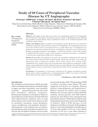
Study of 89 Cases of Peripheral Vascular Disease by CT Angiography
1. 155 Vol.-3, No.-2, January 2011 Cardiovas Journal
Introduction
The peripheral arterial disease (PAD) generally
refers to a disorder that obstructs the blood supply
to upper and lower extremities, most commonly
caused by atherosclerosis but may also result
from thrombosis, embolism, vasculities,
fibromascular dysplasia or entrapment. Where as
the peripheral vascular disease includes PAD and
other atherosclerotic conditions such as renal
artery disease and carotid artery disease as well
as vasculities, vasospasm, venous thrombosis,
venous insufficiency and lymphatic disorder.1
Atherosclerosis is the leading cause of occlusive
arterial disease of the extremities in patients over
40 years old, the highest incidence occurs in sixth
and seventh decades of life.2 The pathology of PAD
is similar to coronary artery disease. The most
important risk factors are smoking, diabetes
mellitus, hyperlipidaemia and hypertension.3
Tobacco increases the relative risk 2-4 times,
Diabetes also increase relative risk for PAD like
smoking. Among patients with PAD diabetic
patients are more likely to have amputation than
non diabetic patients.4 Distal disease affecting the
tibial and peroneal arteries occurs more frequently
in diabetes. Risk of PAD increase if total
cholesterol and LDL are elevated. In Framingham
study it is seen that probability of claudication in
70 years male smoker is 2.5% per 4 year, Which
increases to 24% per 4 year when smoking is
Study of 89 Cases of Peripheral Vascular
Disease by CT Angiography
M Faruque1, AEMM Islam1, S Haque1, MT Islam2, MG Kibria1, M Hossain3, MA Salam1,
F Rahman4, MA Hasnat1, HIL Rahman Khan1
1Department of Cardiology, Dhaka Medical college Hospital, 2 Department Radiology & Imaging, Holy
Family Red Crescent Hospital, Dhaka 3 Department of Vascular surgery, NICVD, Dhaka
4 Department of Cardiology, BSMMU, Dhaka
Abstract:
Objective: The purpose of this study was to observe the morphological pattern by CT angiography
and risk factors for development of peripheral vascular disease in Bangladeshi patient suffering
from peripheral vascular disease using a multidetector scanner in the evaluation of patients with
peripheral vascular disease.
Subject and Method: Eighty nine patients with peripheral vascular disease who were referred for
evaluation of peripheral vascular disease underwent CT angiography. We scanned patients from the
level of the cerebral arteries to the pedal arteries in a single helical scan. CT angiograms were
produced using maximum-intensity-projection, multiplanous reformation and reconstructions.
Findings were graded according to nine categories: 1, normal (0% stenosis); 2, mild (1-49% stenosis);
3, moderate (50-74% stenosis); 4, severe (>75% stenosis); 5, tortuosity; 6, aneurysm, 7, calcification,
8, Arteriovenous malformation (AVM), and 9, haematoma.
Results: We found Most of the patients in our study were male (69 out of 89 patients). The mean age
was 54.49 ±18.36 in male and 49.45 ±17.89 for female. Commonest risk factor in our study was
hypertension 46.1%, followed by diabetes 30.3%, family history 27% smoking 23.6%,
dyslipidaemia13.5%. Stenosis (5.61%) was the predominate lesion followed by haematoma (4.49%),
Arterio –venous malformation (4.49%). Abdominal aorta was mostly affected in the studied population
(58.43%) followed by Lower limb (37.08%), Carotid (22.47%), Renal (7.87%) and Upper limb arteries
(4.49%).
Conclusion: CT angiography is a noninvasive technique for the imaging of peripheral vascular
disease. Since no data is available from a well designed study in PVD in our country, till then the
data obtained from this study can be used in Bangladesh.
(Cardiovasc. j. 2011; 3(2): 155-162)
Key words:
CT angiogram,
Peripheral
vascular disease
(PVD).
Address of Correspondence: Prof. M Faruque, Department of Cardiology, NICVD, Dhaka. Bangladesh.
2. 156 Vol.-3, No.-2, January 2011 Cardiovas Journal
associated with hypertension, hypercholes
trolaemia and diabetes mellitus.5 PAD affects large
number of people world wide but more common in
USA. It is estimated that 10 million people with
symptomatic disease another 20-30 million people
are asymptomatic. It is estimated that 10% of
people over 60 years of age are affected and
prevalence continues to increase with age. The
cardinal symptoms of PAD include intermittent
claudication and rest pain .The location of the
symptom often relates to the site of the most
proximal stenosis. Buttock, hip or thigh
claudication typically occurs in patients with
obstruction of aorta and iliac arteries. Calf
claudication characterizes femoral or popliteal
artery stenosis.Ankle and pedal claudication occurs
in patients with tibial and peroneal artery disease.
Similarly stenosis of the subclavian, axillary or
brachial arteries may cause shoulder, biceps or
forearm claudication respectively. Symptoms
should resolve several minutes after cessation of
effort. There may be impotence in male. Rest pain
in legs worse at night relieved by hanging the leg
over the edge of the bed. Extremities may be cold.
It may be cyanosed; there may be pain, ulceration
and gangrene in 50-100 % patients. On
examination the limb is cold with dry skin and
lack of hair. Pulse is diminished or absent. There
may be ulceration; gangrene or dark discoloration
usually starts at the toes. Death due to PAD is
rare. Death occurs usually secondary to CAD or
CVD. Relative risk of all cause of death is 2-6 fold
higher than general people. Death rate increases
with decrease of ABI. Five years mortality with
ABI < 0.85 is 10% where as mortality approaches
> 50% when ABI is < 0.40. Patients may be
diagnosed by duplex ultrasonography, CT
angiogram, MR angiogram. Conventional
angiogram is gold standard for diagnosis.4
Methods:
Study Population
Peripheral C-T Angiogram was performed in 89
patients from 2006-2007 patients were referred
for vascular insufficiency, severe back pain, neck
swelling, uncontrolled hypertension and vertigo
from outdoor patient department. This non
invasive new Technology of C-T Angiogram
procedure was done on 89 patients who are
suffering from peripheral vascular disease. All the
procedures were done in Modern diagnostic Centre,
Dhanmondi – 8, Dhaka.
We scanned patients fromthe level of the cerebral
arteries to the pedal arteries in a single helical
scan. CT angiograms were produced using
maximum-intensity-projection reconstructions.
Findings weregraded according to nine categories:
1, normal (0% stenosis); 2, mild (1-49% stenosis);
3, moderate (50-74% stenosis); 4, severe (>75%
stenosis); 5, tortuosity; 6, aneurysm, 7, calcification,
8, Arteriovenous malformation (AVM), and 9,
haematoma.6
Inclusion criteria:
(1) Clinically suspected peripheral vascular
disease.
(2) Secondary Hypertension.
(3) Neck swelling of vascular origin.
(4) Vascular injury of Road Traffic Accident.
Exclusion criteria:
(1) Chronic renal failure.
(2) Acute pulmonary oedema (LVF).
(3) Congestive Cardiac Failure.
(4) Valvular Heart disease.
(5) Hypersensitivity to dye.
(6) Non co-operative patient ( Psychosomatic
disease)
(7) Unconscious patient
Procedure
Patient preparation typically involved placing the
patient supine of the CT table without a pillow or
foam wedge under the knees. The rationale for
not putting any support under the knees is that
the field of view should be enlarged to reconstruct
the arteries of interest as they move anteriorly
under the pillow. Any unnecessary increase in the
field-of-view was avoided as this diminishes in-plane
resolution. It may additionally be advantageous to
place tape around the patient‘s knee and ankles.
The rationale for taping is at the necessity to
increase the field-of-view. Taping the feet helped
to remind the patient to keep them immobile.
Somatom Sensation 64 slice (Simens) CT
angiogram machine was used. As soon as the
primary CTA was completed, the reconstruction
Study of 89 Cases of Peripheral Vascular Disease by CT Angiography M Faruque et al.
156
3. 157 Vol.-3, No.-2, January 2011 Cardiovas Journal
of images below the knee should be prioritized and
a quick assessment as the adequacy of these images
made. If insufficient arterial opacification was
present, then the second acquisition could be
triggered at that time. This approach assures that
the pedal vessels will be adequately assessed even
in the setting of substantially prolonged circulation
times. When assessing the peripheral arteries,
contrast medium flow rates of at least 4 ml per
second and preferably 5 ml per second were
desirable. When imaging atherosclerotic occlusive
disease, the scan range was extended from the
supra-renal aorta through the toes in the setting
of limb threatening ischaemia or to the ankles
in the setting of claudication. The presence of
stenosis (considered hemodynamically significant
when greater than fifty percent), occlusions, and
the lengths of these abnormalities are the most
important clinical observations to be made. The
product of scan times and the injection rate
determined total volume of contrast
administered. There were no complication
related to C-T angio and all studies are
technically adequate.
The scan direction was craniocaudal for Lower
Limb CTA, with the range from the level of
infrarenal aorta to the pedal arch; while for Upper
Limb CTA; the scan direction was caudocranial
from the level of aortic arch to the palmer arch.
The patients were instructed to continue quiet
breathing for the duration of scan. The success of
any CTA depends on the calculation of an accurate
delay to start the acquisition of images after the
injection of contrast, to get optimum arterial
enhancement, not contaminated by the venous
phase. The delay was calculated by using the test
bolus technique.
Data Collection & Analysis
The demographic profile of all patients including
age, sex was taken. The major risk factors of
peripheral arterial disease i.e. diabetes,
hypertension, smoking, dyslipidaemia and family
history were taken in each patient. The peripheral
arteries were studied- Carotid arteries, Abdominal
Aorta, Upper limb arteries, Lower limb arteries
and renal arteries. Morphological pattern of lesions
were described in each involved artery. Data
analysis was done in SPSS programme.
Results:
In all of our initial 89 patients, there has been no
technical failure. The procedure has been well
tolerated and in no study was there image
degradation due to motion artifacts. Patients of
peripheral vascular disease, with symptoms of
deceased or absent pulses, claudicaton or rest pain
also underwent CTA.
Table-I
Age distribution of ct angiography patients (n=89)
Age group Sex Total Percentage P value
Male Female 0.61NS
Upto 30 9 4 13 14.6
31-40 7 4 11 12.4
41-50 9 1 10 11.2
51-60 18 4 22 24.7
61-70 14 5 19 21.3
71 & above 12 2 14 15.7
Total 69 20 89 100.0
Table-II
Mean age by sex (Male=69, Female=20)
Sex Mean SD P Value
Male 54.49 18.36 NS
Female 49.45 17.89
Cardiovascular Journal Volume 3, No. 2, 2011
157
4. 158 Vol.-3, No.-2, January 2011 Cardiovas Journal
Table I shows age and sex distribution of the study
population. It shows most of the patients were
above 50yrs of age. Age and sex differences were
not statistically significant.
Table II shows Mean age in male and female were
54.49±18.36 and 49.45±17.89 which was not
significant statistically.
Table III shows distribution of risk factors in both
sexes. Most of the patients were hypertensive
(46.1%) followed by diabetes (30.3%) and smoking
(23.6%). Except smoking other risk factors did not
vary significantly in two sexes.
Table-IVshowsinvolvementoftheperipheralarteries
and their sex distribution. It shows abdominal aorta
was the most affected artery 58.43%, followed by
lower limb arteries 37.08%, carotid arteries 22.47%,
renal arteries 7.87% and upper limb arteries 4.49%.
Involvement of the peripheral arteries did not vary
significantly in both sexes.
Table V shows morphological pattern of involved
carotid arteries and their distribution in both
sexes. Most commonly found lesion was stenosis
(5.61%), followed by haematoma (4.49%), Arterio
–venous malformation (4.49%).
Table-III
Distribution of risk factors of the patients
Risk factors Positive Total Percentage P Value
Male Female
Diabetes 21 6 27 30.3 NS
Hypertension 34 7 41 46.1 NS
Smoking 21 0 21 23.6 S
Dyslipidaemia 9 3 12 13.5 NS
Family history 21 3 24 27 NS
Table-IV
Distribution of the involved peripheral artery diseases by sex
Arteries involved Normal Total Percentage Abnormal Total Percentage
Male Female Male Female
Carotid 56 13 69 77.53 13 7 20 22.47
Abdominal aorta 23 14 37 41.57 46 6 52 58.43
Upper limb 68 17 85 95.51 1 3 4 4.49
Lower limb 37 19 56 62.92 32 1 33 37.08
Renal 64 18 82 92.13 5 2 7 7.87
Table-V
Morphology Pattern of Carotid Artery by sex (n=89)
Carotid Artery Male Female Total Percentage
Mild Stenosis 1 3 4 4.49
Moderate Stenosis 1 0 1 1.12
Tortuous 1 1 2 2.25
Aneurysm 2 1 3 3.37
Calcification 1 1 2 2.25
AVM 3 1 4 4.49
Haematoma 4 0 4 4.49
Normal 56 13 69 77.53
Total 69 20 89 100.00
Study of 89 Cases of Peripheral Vascular Disease by CT Angiography M Faruque et al.
158
5. 159 Vol.-3, No.-2, January 2011 Cardiovas Journal
Fig.-1: Cerebral Angiogram as shown in CT Fig.-2: Cerebral Angiogram as shown in CT
Table-VI
Morphology Pattern of Abdominal Aorta by sex (n=89)
Abdominal aorta Male Female Total Percentage
Mild Stenosis 4 2 6 6.74
Moderate Stenosis 26 2 28 31.46
Severe Stenosis 7 0 7 7.87
Tortuous 0 1 1 1.12
Thrombus 1 0 1 1.12
Aneurysm 3 0 3 3.37
Calcification 5 1 6 6.74
Normal 23 14 37 41.57
Total 69 20 89 100.00
Fig.-3: Abdominal aneurysm affecting supra and
infra renal region
Fig:-4: Extra vascular mass compressing the lower
part of the abdominal aorta near its bifurcation
Cardiovascular Journal Volume 3, No. 2, 2011
159
6. 160 Vol.-3, No.-2, January 2011 Cardiovas Journal
Table-VII
Morphology Pattern of Upper Limb Artery by sex (n=89)
Upper limb artery Male Female Total Percentage
Mild Stenosis 0 1 1 1.12
Moderate Stenosis 0 0 0 0
Severe Stenosis 0 0 0 0
Tortuous 0 2 2 2.25
Thrombus 0 0 0 0
Aneurysm 1 0 1 1.12
Calcification
Normal 68 17 85 95.51
Total 69 20 89 100.00
Table-VIII
Morphology Pattern of Lower Limb Artery by sex (n=89)
Lower limb artery Male Female Total Percentage
Mild Stenosis 2 0 2 2.25
Moderate Stenosis 13 1 14 15.73
Severe Stenosis 3 0 3 3.37
Tortuous 0 0 0 0
Thombus 0 0 0 0
Aneurysm 0 0 0 0
Calcification 14 0 14 15.73
Normal 37 19 56 62.92
Total 69 20 89 100.00
Fig.-5: PVD affecting right and left common iliac
artery.
Fig.-6: Aneurysm affecting infra renal aorta and
right common iliac artery.
Study of 89 Cases of Peripheral Vascular Disease by CT Angiography M Faruque et al.
160
7. 161 Vol.-3, No.-2, January 2011 Cardiovas Journal
Table VI shows morphological pattern of involved
abdominal aorta and their distribution in both
sexes. Most commonly found lesion was stenosis
(46.07), followed by calcification (6.74%), aneurysm
(3.37%).
Table VII shows morphological pattern of involved
upper limb arteries and their distribution in both
sexes. Most commonly found lesion was tortuosity
(2.25%), followed by stenosis (1.12%), aneurysm
(1.12%).
Table VIII shows morphological pattern of involved
lower limb arteries and their distribution in both
sexes. Most commonly found lesion was stenosis
(5.61%), followed by haematoma(4.49%), Arterio –
venous malformation 4.49%.
Table IX shows morphological pattern of involved
carotid arteries and their distribution in both
sexes. Most commonly found lesion was stenosis
(5.61%), followed by haematoma (4.49%), Arterio
–venous malformation 4.49%.
Discussion:
This retrospective observational study was done
to evaluate the pattern of peripheral vascular
diseases and to evaluate CT angiogram as a
diagnostic tool for peripheral vascular diseases. It
is the first study with CT angiogram in Bangladeshi
patient. We want to show not the efficacy of the
procedure but to show the pattern and severity of
the peripheral vascular disease and its correlation
with risk factors.
We evaluated 89 patients with clinical suspicion of
PVD by CT angiogram. Most of the patients in our
study were male (69 out of 89 patients). The mean
age was 54.49 ±18.36 in male and 49.45 ±17.89 for
female. This difference is not significant. Most of
the patients in our series were more than 50yrs of
age (61.7%). This also correlates with the study of
Norgren et al. 7
The commonest risk factor in our study was
hypertension 46.1%, followed by diabetes 30.3%,
family history of atherosclerotic coronary and
peripheral vascular diseases 27% smoking 23.6%,
dyslipidaemia 13.5%. In a prospective study Price
at el have shown that the combined effect of
smoking on the cardiovascular risk factor has
influence on peripheral arterial disease.8
Explanation for the effect of cigarette smoking
on the development of peripheral arterial disease
have been described.9,10 which may reflect the
gender difference in this series as none of the
female subject in this study had history of
smoking. Such mechanisms could include a direct
toxic effect of whole smoke, nicotine and/or
carbon monoxide on endothelial cells, increased
platelet reactivity and agreeability, and/or a
detrimental effect of the elevated white blood cell
count found consistently in smokers.11Diabetes
mellitus was also shown as a major risk factor
for atherosclerotic peripheral arterial disease.
Other risk] factors associated with PVD in this
study also correlates with other studies. 12
Abdominal aorta was mostly affected in the studied
population (58.43%). Lower limb (37.08%), Carotid
(22.47%), Renal (7.87%) and Upper limb arteries
were affected according to decreasing frequency.
Involvements of the peripheral arteries do not vary
Table-IX
Morphology Pattern of Renal Artery by sex (n=89)
Renal artery Male Female Total Percentage
Mild Stenosis 1 0 1 1.12
Moderate Stenosis 1 0 1 1.12
Severe Stenosis 1 0 1 1.12
Tortuous 0 0 0 0
Thrombus 0 0 0 0
Aneurysm 2 2 4 4.49
Calcification 0 0 0 0
Normal 64 18 82 92.13
Total 69 20 89 100.00
Cardiovascular Journal Volume 3, No. 2, 2011
161
8. 162 Vol.-3, No.-2, January 2011 Cardiovas Journal
significantly in both sexes. Stenosis (5.61%) was
the predominate lesion followed by haematoma
(4.49%), Arterio-venous malformation (4.49%) As
atherosclerosis is the main pathogenesis of the
peripheral vascular disease, according to the
observation, morphological prevalence of lesions
can be done from different arterial segments.
Limitation
The major drawback of CTA is large amount of
intravenous contrast and the ionizing radiation
involved. Also, no information is obtained regarding
the flow direction and velocity. In addition, CTA
may fail to demonstrate short segment stenosis,
apart from the fact that horizontally oriented
branches are poorly visualized, thus significant
lesions may be missed in certain instances. Total
number of patients in this study was also small.
Despite these limitations, the vast majority of
examinations we performed were considered
sufficiently diagnostic to avoid more invasive
imaging.
Conclusion:
Our initial experience of CT angiography with
MSCT has shown that it is a promising new, fast
and non-invasive imaging modality that can be
utilized effectively in the evaluation of peripheral
vasculature. Some of the inherent limitations of
the technique and the time consumed in post-
processing can be overcome with future
workstation and technology advances. Thus it
would be appropriate to conclude that CTA is
clearly emerging as a screening tool in patients of
peripheral vascular disease. CT angiography is a
noninvasive techniquefor the imaging of peripheral
vascular disease.Since no data is available from a
well designed study in PVD in our country, till
then the data obtained from this study can be used
in Bangladesh. As the software of CT angiogram
is developing day by day, in near future at least in
some cases it will replace the invasive angiogram.
Acknowledgement :Mr. Mostafizur Rahman, Mr.
Nurul Islam, Mr. Monir Ahmed, Mst. Nasima
Yesmin.
References:
1. Creager M A , Libby P. Peripheral Artery Disease. In:
Libby P, Bonow RO, Mann D L, Zipes D P, Braunwald
E. Eds. Braunwald Heart Disease. A textbook of
Cardiovascular Medicine. 7th edn,Philadelphia: Elsevier
Saunders,2008:1491-1514.
2. Hiatt W R, Medical management of peripheral Arterial
Disease and claudication. N Eng J Med 2001. 344. 1608-
1621.
3. Bloomfield P,Bradbury A,Grubb NR,Newby DE.
Cardiovascular disease. In: Boon NA, Colledge NR,
Walker RB. Eds. Davidson’s Principle and practice of
Medicine. 20th edn, Edinburgh :Churchill Livingstone,
2006: 601-610.
10. Powell JT. Smoking. In: Fowkes FGR, ed. Epidemiology
of Peripheral Vascular Disease. Berlin: Springer-Verlag,
1991:141–53.
11. Friedeman GD, Siegelaub AB, Seltzer CC, Feldman R,
Collen MF. Smoking habits and the leukocyte count.
Arch Environ Health 1973; 26: 137–43.
12. Al-Delaimy WK, Merchant AT, Rimm EB, Will ett WC,
Stampfer MJ, Hu FB. Effect of type 2 diabetes and its
duration on the risk of peripheral arterial disease among
men. Am J Med 2004; 116: 236-40.
Study of 89 Cases of Peripheral Vascular Disease by CT Angiography M Faruque et al.
162