Download to read offline
![2 Case Reports in Critical Care
Figure 1: Figure showing the broken end of knife (yellow arrow).
(a) (b)
(c)
Figure 2: ((a), (b), and (c)) Radiographic images describing the route of the knife and the tip reaching the infratemporal region across the
midline.
injuries and it is a challenging situation both for the initially
evaluating physician as well as for the surgeon as a prompt
evaluation and management of such a condition are essential
to preserve vital visual functions and save life.
Penetrating head and neck injuries have been classified
for long into high velocity and low velocity injuries.
High velocity injuries usually are acquired in war caused
by missile injuries like gun shots and shrapnel wounds and
they cause massive destruction of the craniumand face.
Most of civilian injuries are low velocity and are caused by
otherwise innocent objects.There have beenmultiple reports
of such objects like toys, pencils, stones, wooden sticks,
bicycle brake handle, chopsticks, umbrella ends, thumb tacks,
tooth brushes, crochet hooks, andmetal fence [1–9], while on
the other hand reports on direct transorbital stab injuries by
knife are few [10–14].
The risk of these kinds of injuries is high especially
in the orbital region because the orbital roof and medial](https://image.slidesharecdn.com/transorbitalstab-140924093103-phpapp01/75/Transorbital-stab-injury-with-retained-knife-A-narrow-escape-2-2048.jpg)
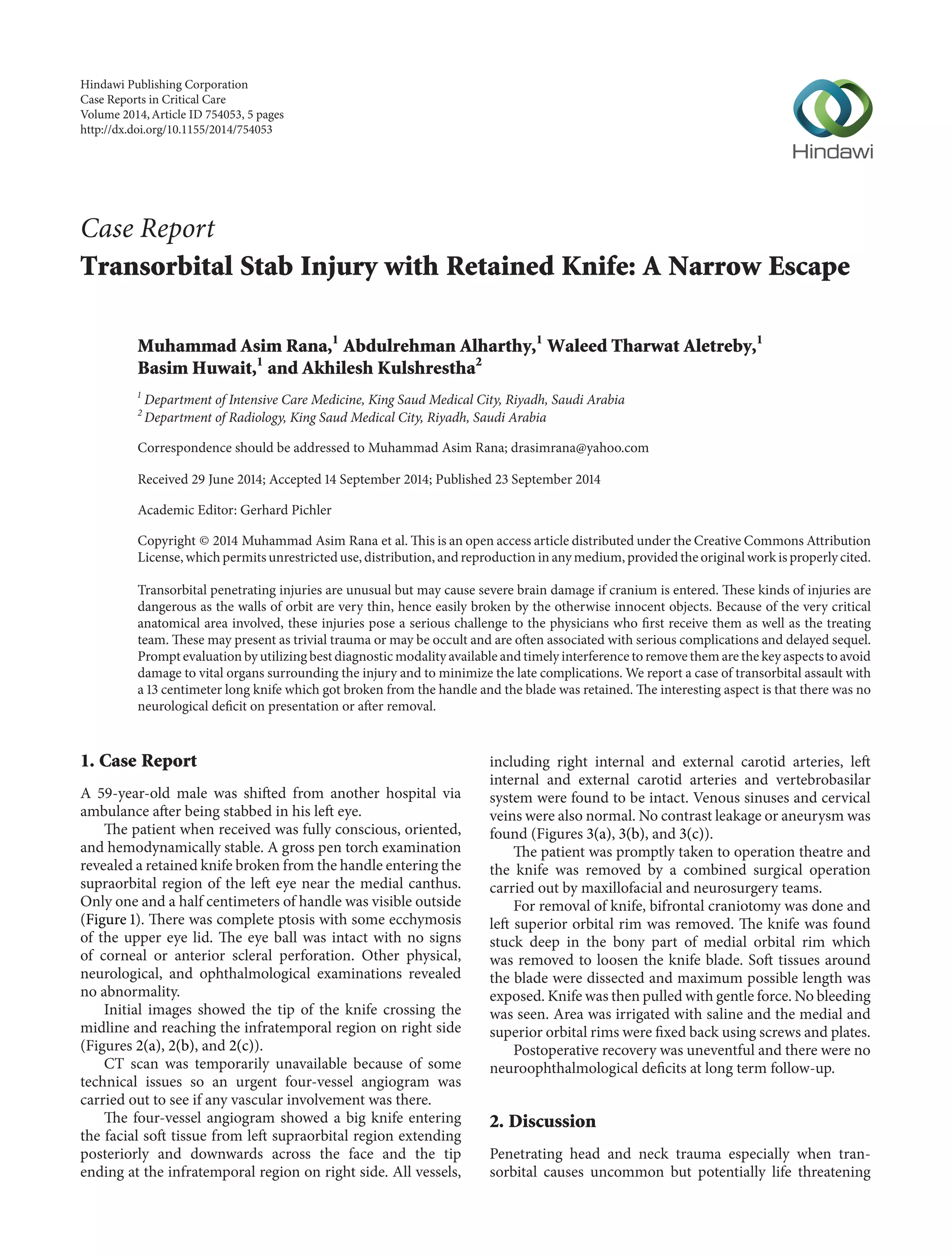
![Case Reports in Critical Care 3
(a) (b)
(c)
Figure 3: ((a), (b), and (c)) Angiogram to evaluate the integrity of vasculature.The tip of knife is visible near left internal carotid artery.The
venous sinus and internal jugular vein appear safe.
wall are relatively thin and the objects even with less force
can easily penetrate deep and cause damage to the globe,
brain, cavernous sinus, paranasal sinuses, and optic nerve.
Hence, the pathophysiological consequences and degree of
permanent neurological deficit in such injuries depend upon
the kinetic energy and the pathway or trajectory of the
offending object, timing to access themedical care, rapidity of
exploration, removal of the object and avoiding the secondary
injury [15, 16]. The consequences of such wounds include
brain contusions, cerebrospinal fluid fistulas, intracerebral,
subdural, and extradural hematomas, and pneumocephalus.
Late sequel can be infectious complications like encephalitis,
meningitis, or cerebral abscess [17–21]. Vascular malforma-tions
although rare can ensue [22, 23]. The outcome of such
injuries is also dependent on primary injury and its associated
complications. Subarachnoid haemorrhage for instance has
been associated with poor outcome [24].
The diagnosis of such injuries is straight forward if the
precise knowledge of the traumatic event and the nature of
the object are available or presence of the foreign body can
be confirmed in the wound. However, diagnosis based on
an incomplete history and in cases of trivial trauma is difficult
and the penetrating injuries may be overlooked [25–27].
In our case, we were able to obtain a detailed history of
the event and the object was visualized in situ. Although the
clinical examination including both ophthalmological and
neurological components is crucial in determining the site
and effect of the injury, imaging techniques are extremely
helpful in making final decision and embarking upon a
method of removal. Computerized tomography is an excel-lent
means of documenting details of orbitocranial trauma
such as extent of soft tissue damage, localization and nature
of the foreign material, and presence of bone fractures and is
indicated in all cases of suspected cranial penetration.
In our case, we could not perform a CT scan because of a
temporary technical issue butwe did a four-vessel angiogram
to rule out any vascular involvement.
As far as the removal of the objects retained in the
orbit with possible cerebral penetration is concerned, the
procedures described in the literature range from simple
extraction to surgical removal depending upon the size,
nature, and location of the object because the considerable](https://image.slidesharecdn.com/transorbitalstab-140924093103-phpapp01/75/Transorbital-stab-injury-with-retained-knife-A-narrow-escape-3-2048.jpg)
![4 Case Reports in Critical Care
differences in the shape and size of the different objects
involved in such accidents make it impossible to establish a
single therapeutic strategy. Some authors have the opinion
that if the object is small and the shape is known, extraction
can be attempted, while surgical removal is advised in other
cases [12, 21, 28–32].
As the wound of entry was high enough through the
supraorbital area, the surgery was attended by the oromax-illofacial
and neurosurgery teams and it proved to be the best
way as they had to do craniotomy and open the orbital rim
for the removal of the knife blade.
In the case, we reported the traumatic cranial damage was
exclusively of primary type and the intracranial extension of
injury which is usually a common occurring in such violent
and dramatic traumas did not take place. It was indeed a
narrow escape.
The case should heighten the awareness of first respon-ders
to such conditions about the possibilities of vascular
injuries or brain stem damage as the inner extent of the
foreign object is not known and they should arrange safe
transfer of such patients to the centers where the treatment
facilities are available. Moreover, diagnostic tools should be
appropriately used and a multidisciplinary team approach
should be sought for the best management.
Conflict of Interests
The authors declare that there is no conflict of interests
regarding the publication of this paper.
Acknowledgment
The authors are indebted to Mrs. Mariam Bandan Latip,
Senior Charge Nurse, ICU, King Saud Medical City, for her
great help in patient follow-up and paper writing.
References
[1] T.-H. Shin, J.-H.Kim, K.-W.Kwak, and S.-H.Kim, “Transorbital
penetrating intracranial injury by a chopstick,” Journal of
Korean Neurosurgical Society, vol. 52, no. 4, pp. 414–416, 2012.
[2] W. B. Huiszoon, P. N. No¨e, and A. Manten, “Fatal transorbital
penetrating intracranial injury caused by a bicycle hand brake,”
International Journal of EmergencyMedicine, vol. 5, no. 1, article
34, 2012.
[3] M. Arslan, M. Eseoglu, B. O. G¨ud¨u, and I. Demir, “Transorbital
orbitocranial penetrating injury caused by a metal bar,” Journal
of Neurosciences in Rural Practice, vol. 3, no. 2, pp. 178–181, 2012.
[4] V. Sams, H. K. Nagarsheth, and T. A. Nickloes, “Transorbital
penetrating intracranial injury caused by sheppard’s hook,”
Journal of Surgical Case Reports, vol. 2010, no. 7, p. 3, 2010.
[5] A. Agrawal, A. Pratap, C. S. Agrawal, A. Kumar, and S.
Rupakheti, “Transorbital orbitocranial penetrating injury due
to bicycle brake handle in a child,” Pediatric Neurosurgery, vol.
43, no. 6, pp. 498–500, 2007.
[6] M. R. Farhadi,M. Becker,C.Stippich,A.W.Unterberg, andK.L.
Kiening, “Transorbital penetrating head injury by a toilet brush
handle,” Acta Neurochirurgica, vol. 151, no. 6, pp. 685–687, 2009.
[7] M.Miscusi, P. Arangio, L. deMartino, F. de-Giorgio,P.Cascone,
and A. Raco, “An unusual case of orbito-frontal rod fence stab
injury with a good outcome,” BMC Surgery, vol. 13, no. 1, article
31, 2013.
[8] G. Satyarthee, S. Borkar, A. Tripathi, and B. Sharma, “Transor-bital
penetrating cerebral injury with a ceramic stone: report of
an interesting case,” Neurology India, vol. 57, no. 3, pp. 331–333,
2009.
[9] J. Skoch, T. L. Ansay, andG.M. Lemole, “Injury to the temporal
lobe via medial transorbital entry of a toothbrush,” Journal of
Neurological Surgery Reports, vol. 74, no. 1, pp. 23–28, 2013.
[10] J. T.Carneiro Jr., A. K. da Silva Tabosa, F. J. de Souza Jr., and E. H.
Shinohara, “Orbitoethmoidal impacted injury by kitchen knife
causing abducens nerve palsy,” Oral and Maxillofacial Surgery,
vol. 15, no. 2, pp. 107–108, 2011.
[11] H. Lichter, M. Snir, K. Segal, and Y. Yassur, “Penetrating
orbitocranial knife injury,” Journal of Pediatric Ophthalmology
and Strabismus, vol. 36, no. 1, pp. 44–46, 1999.
[12] M. Subas¸i, M. P. C¸akar- ¨ Ozdal, P. Nalc¸acioglu-Yuksekkaya, and
A. Alakus¸, “Management of an orbitocranial knife injury: a case
report,” Turkish Journal of Pediatrics, vol. 54, no. 2, pp. 184–186,
2012.
[13] H. Sanaei-Zadeh, K. Aghakhani, and H. Saidi, “Orbito-cerebral
penetrating knife-wound,” Journal of Clinical ForensicMedicine,
vol. 13, no. 3, pp. 146–147, 2006.
[14] O. Okay, E. Da˘glio˘glu, C. Ozdol, O. Uckun, A. Dalgic, and
F. Ergungor, “Orbitocerebral injury by a knife: case report,”
Neurocirugia, vol. 20, no. 5, pp. 467–469, 2009.
[15] L. M. Quinn, R. A. Egan, and W. T. Shults, “Transorbital pene-trating
brainstem injuries,” Archives of Ophthalmology, vol. 124,
no. 6, pp. 915–916, 2006.
[16] D. J. Verret, R. Defatta, and Y. Ducic, “Transorbital penetration
of the skull base with an occult foreign body,” The American
Journal of EmergencyMedicine, vol. 23, no. 7, pp. 901–902, 2005.
[17] J. M. Ecklund, P. K. Mauer, and R. G. Ellenbogen, “Cerebral
abscess after presumed superficial periorbital wound,” Military
Medicine, vol. 164, no. 6, pp. 444–445, 1999.
[18] E. L. Kazarian, N. A. Stokes, and J. T. Flynn, “The orbital punc-ture
wound: intracranial complications of a retained foreign
body,” Journal of Pediatric Ophthalmology and Strabismus, vol.
17, no. 4, pp. 247–250, 1980.
[19] G. P. Duffy and Y. S. Bhandari, “Intracranial complications
following transorbital penetrating injuries.,” British Journal of
Surgery, vol. 56, no. 9, pp. 685–688, 1969.
[20] P. Foy and M. Sharr, “Cerebral abscesses in children after pencil-tip
injuries,” The Lancet, vol. 2, no. 8196, pp. 662–663, 1980.
[21] S. Chibbaro and L. Tacconi, “Orbito-cranial injuries caused
by penetrating non-missile foreign bodies. Experience with
eighteen patients,” Acta Neurochirurgica, vol. 148, no. 9, pp. 937–
942, 2006.
[22] C. F. Kieck and J. C. de Villiers, “Vascular lesions due to tran-scranial
stabwounds,” Journal of Neurosurgery, vol. 60,no. 1, pp.
42–46, 1984.
[23] J. F. Vander and C. C. Nelson, “Penetrating orbital injury with
cavernous sinus involvement.,” Ophthalmic Surgery, vol. 19, no.
5, pp. 328–330, 1988.
[24] C. Peek-Asa, D. McArthur, D. Hovda, and J. Kraus, “Early
predictors of mortality in penetrating compared with closed
brain injury,” Brain Injury, vol. 15, no. 9, pp. 801–810, 2001.
[25] A. D. Shah and C. Decock, “Occult orbito-cranial penetrating
injury by pencil: role of beta tracer protein as a marker for](https://image.slidesharecdn.com/transorbitalstab-140924093103-phpapp01/75/Transorbital-stab-injury-with-retained-knife-A-narrow-escape-4-2048.jpg)
![Case Reports in Critical Care 5
cerebrospinal fluid leakage,” Indian Journal of Ophthalmology,
vol. 59, no. 6, pp. 505–507, 2011.
[26] F. Bilotta, G. Rosa, R. Delfini, R. Pinto, and B. Fiorani, “Unrec-ognized
periorbital penetrating nail in the brain: case report,”
The American Journal of Emergency Medicine, vol. 25, no. 2, pp.
198–199, 2007.
[27] F. Al-Otaibi and S. Baeesa, “Occult orbitocranial penetrating
pencil injury in a child,” Case Reports in Surgery, vol. 2012,
Article ID 716791, 4 pages, 2012.
[28] K. M. Dodson, M. A. Bridges, and E. R. Reiter, “Endoscopic
transnasal management of intracranial foreign bodies,” Archives
of Otolaryngology—Head and Neck Surgery, vol. 130, no. 8, pp.
985–988, 2004.
[29] S. Martin, G. H. Raup, G. Cravens, and C. Arena-Marshall,
“Management of embedded foreign body: penetrating stab
wound to the head,” Journal of Trauma Nursing, vol. 16, no. 2,
pp. 82–86, 2009.
[30] D.Mitilian, B. Charon, F. Brunelle, and F. Di Rocco, “Removal
of a chopstick out of the cavernous sinus, pons, and cerebellar
vermis through the superior orbital fissure,” Acta Neurochirur-gica,
vol. 151, no. 10, pp. 1295–1297, 2009.
[31] F. Ildan, H. Bagdatoglu, B. Boyar,M. Doganay, E. Cetinalp, and
A. Karadayi, “The nonsurgical management of a penetrating
orbitocranial injury reaching the brain stem: case report,”
Journal of Trauma, vol. 36, no. 1, pp. 116–118, 1994.
[32] M. Domenicucci, R. Qasho, P. Ciappetta, T. Vangelista, and
R. Delfini, “Surgical treatment of penetrating orbito-cranial
injuries: case report,” Journal of Neurosurgical Sciences, vol. 43,
no. 3, pp. 229–234, 1999.](https://image.slidesharecdn.com/transorbitalstab-140924093103-phpapp01/75/Transorbital-stab-injury-with-retained-knife-A-narrow-escape-5-2048.jpg)


This case report discusses a 59-year-old male who suffered a transorbital stab injury from a 13 cm knife, with the blade retained in his orbit without causing neurological deficits. Prompt diagnostic steps and combined surgical efforts led to the successful removal of the knife, and the patient had an uneventful recovery with no complications noted at long-term follow-up. The report emphasizes the challenges and critical nature of transorbital penetrating injuries, highlighting the need for careful evaluation and multidisciplinary management.


