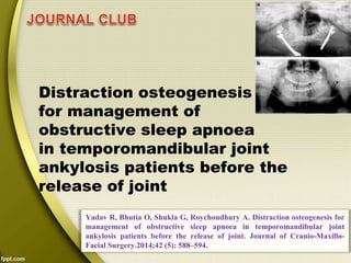
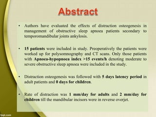
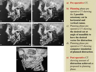
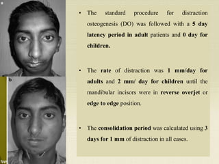
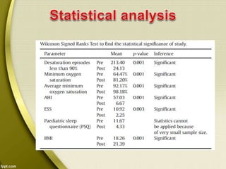
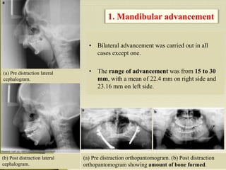
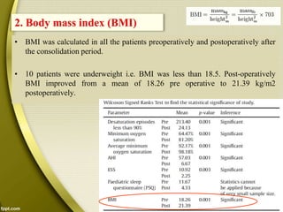
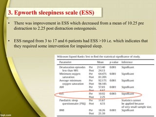
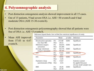
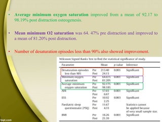
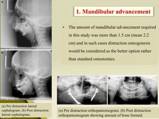
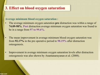
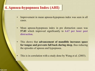
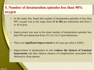
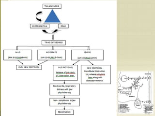
PRESENTED BY –
DR. SHEETAL KAPSE
3rd YEAR, P.G. STUDENT
MODERATORS -
DR. D. A. DARAWADE
DR. M. SATISH
DR. MANISH PANDIT
DR. DEEPAK THAKUR
Distraction osteogenesis
for management of
obstructive sleep apnoea
Yadav R, Bhutia O, Shukla G, Roychoudhury A. Distraction osteogenesis for
management of obstructive sleep apnoea in temporomandibular joint
ankylosis patients before the release of joint. Journal of Cranio-Maxillo-
Facial Surgery.2014;42 (5): 588–594.
in temporomandibular joint
ankylosis patients before the
release of joint
1. Dr. Rahul Yadav, Senior Resident, Department of Oral &
Maxillofacial Surgery, AIIMS, New Delhi, India.
2. Dr. Ongkila Bhutia, Associate Professor, Department of
Oral & Maxillofacial Surgery, AIIMS, New Delhi, India.
3. Dr. Garima Shukla, Department of Neurology, AIIMS,
New Delhi, India.
4. Dr. Ajoy Roychoudhury, BDS, MDS, Prof & Head,
Department of Oral & Maxillofacial Surgery, AIIMS, New
Delhi, India.
• Obstructive sleep apnoea
• Temporomandibular joint
ankylosis
• Blood oxygen saturation
• Retrognathia
Abstract
Introduction
Material and methods
Results
Discussion
Conclusion
References
• Authors have evaluated the effects of distraction osteogenesis in
management of obstructive sleep apnoea patients secondary to
temporomandibular joints ankylosis.
• 15 patients were included in study. Preoperatively the patients were
worked up for polysomnography and CT scans. Only those patients
with Apnoea-hypopnoea index >15 events/h denoting moderate to
severe obstructive sleep apnoea were included in the study.
• Distraction osteogenesis was followed with 5 days latency period in
adult patients and 0 days for children.
• Rate of distraction was 1 mm/day for adults and 2 mm/day for
children till the mandibular incisors were in reverse overjet.
• After 3 months post distraction assessment was done using
polysomnography and CT scan.
• TMJ ankylosis was released by doing gap arthroplasty after
distraction osteogenesis.
• Post distraction improvement was seen in clinical features of OSA
like daytime sleepiness and snoring.
• Epworth sleepiness scale improved from a mean of 10.25 to 2.25.
Polysomnographic analysis also showed improvement in all cases with
apnoea-hypopnoea index from 57.03 to 6.67 per hour.
• Lowest oxygen saturation improved from 64.47% to 81.20% and
average minimum oxygen saturation improved from 92.17% to
98.19%.
• Body mass index improved from a mean of 18.26 to 21.39 kg/m2.
• By this they concluded that Distraction osteogenesis is a stable and
beneficial treatment option for temporomandibular joint ankylosis
patients with obstructive sleep apnoea.
• Retrognathia whether acquired or congenital leads to reduced
posterior airway space and may cause obstructive sleep apnoea.
• Acquired retrognathia may occur due to temporomandibular joint
ankylosis.
• Obstructive sleep apnoea is a sleep-related disorder defined as
absence of breathing for 10 or more seconds despite the effort
to breathe (Kushida et al).
• Besides having immediate effects of hypoxia, arousal during
sleep, snoring and daytime sleepiness, long term repetitive
nocturnal upper airway obstruction has a risk of the
development of hypertension, stroke and myocardial
infarction.
Kushida CA, Littner MR, Morgenthaler T, Alessi CA, Bailey D, Coleman Jr J, et al: Practice parameters for the
indications for polysomnography and related procedures: an update for 2005. Sleep 28(4): 499e521, 2005 Apri1
• Exposure to intermittent hypoxia leads to oxidative stress,
inflammation, atherosclerosis and endothelial dysfunction
(Prabhakar).
• Intermittent hypercapnia and hypoxia may be the mechanism
responsible for cardiovascular effects of obstructive sleep apnoea
due to sustained activation of the sympathetic nervous system
(Patel et al).
Prabhakar NR: Physiological and genomic consequences of intermittent hypoxia:
invited review: oxygen sensing during intermittent hypoxia: cellular and molecular
mechanisms. J Appl Physiol 90(5): 1986e1994, 2001 May
Patel SR, White DP, Malhotra A, Stanchina ML, Ayas NT: Continuous positive airway
pressure therapy for treating sleepiness in a diverse population with obstructive
sleep apnoea: results of meta-analysis. Arch Intern Med 163(5): 565e571,2003 Mar 10
• Obstructive sleep apnoea in retrognathic patients can be treated
successfully by mandibular advancement procedures.
• The mandible can be advanced up to 10-12 mm by
orthognathic surgery. This advancement is useful only for
mild to moderate obstructive sleep apnoea.
• Obstructive sleep apnoea secondary to severe acquired
retrognathia usually requires extensive advancement.
• This is easily achieved by using distraction osteogenesis.
• Distraction induces histogenesis of blood vessels, muscles,
nerves, cartilages, ligaments, skin and mucosa.
• Mandibular lengthening by distraction osteogenesis is now a
commonly used technique to correct congenital and acquired
retrognathia (Rao et al., 2004; Shang et al., 2012).
• Distraction osteogenesis is less invasive, can be applied to
children and due to histogenesis there is less chance of relapse
(Iatrou et al., 2010; Miloro, 2010).
• This study reports the experience in the treatment of obstructive
sleep apnoea with distraction osteogenesis in severely
retrognathic patients’ secondary temporomandibular joint
ankylosis.
• A prospective study including 15 patients with retrognathia were
enrolled in the study.
• Retrognathia followed by release of temporomandibular joint
ankylosis was done in all 15 cases.
• Mean age was 18.2 years with a range of 2-46 years.
• Male: female ratio was 3:2.
• Institutional review board approval was obtained prior to
commencement of the study.
• Preoperatively the patients underwent polysomnography and CT
scans.
• Only those patients with an apnoea-hypopnoea index (AHI) >15
events/h, denoting moderate to severe obstructive sleep apnoea
(OSA),were included in the study.
• The distraction vector was planned by computer simulation.
Distraction was done using a stainless steel linear distractor
manufactured by Synthes (GmBh Oberdorf, Switzerland.) in all 15
cases.
• The distractors were placed using a submandibular incision and the
activation arm was taken out through a stab incision in the mental
foramen region.
a) Pre operative CT.
b) Planning phase pre
operative CT showing
the 2 possible
osteotomy cut in
horizontal and
vertical ramus.
c) Planning phase pre
operative CT showing
the desired cut at
angle of mandible to
have the desired
vector for distraction.
d) Planning phase pre
operative CT showing
computer simulation
of planned distraction.
e) Post operative CT
showing amount of
distraction achieved as
proposed in planning
phase.
• The standard procedure for distraction
osteogenesis (DO) was followed with a 5 day
latency period in adult patients and 0 day for
children.
• The rate of distraction was 1 mm/day for
adults and 2 mm/ day for children until the
mandibular incisors were in reverse overjet or
edge to edge position.
• The consolidation period was calculated using 3
days for 1 mm of distraction in all cases.
• Patients had polysomnography and CT scan 3 months after
distraction osteogenesis. Further surgery to remove the distractor
was performed after the completion of consolidation period.
• TMJ ankylosis was released 6 months after the removal of
distractors.
• Bilateral advancement was carried out in all
cases except one.
• The range of advancement was from 15 to 30
mm, with a mean of 22.4 mm on right side and
23.16 mm on left side.
1. Mandibular advancement
(a) Pre distraction orthopantomogram. (b) Post distraction
orthopantomogram showing amount of bone formed.
(b) Post distraction lateral
cephalogram.
(a) Pre distraction lateral
cephalogram.
• BMI was calculated in all the patients preoperatively and postoperatively after
the consolidation period.
• 10 patients were underweight i.e. BMI was less than 18.5. Post-operatively
BMI improved from a mean of 18.26 pre operative to 21.39 kg/m2
postoperatively.
2. Body mass index (BMI)
• There was improvement in ESS which decreased from a mean of 10.25 pre
distraction to 2.25 post distraction osteogenesis.
• ESS ranged from 3 to 17 and 6 patients had ESS >10 i.e. which indicates that
they required some intervention for impaired sleep.
3. Epworth sleepiness scale (ESS)
• Post distraction osteogenesis analysis showed improvement in all 15 cases.
• Out of 15 patients, 9 had severe OSA i.e. AHI >30 events/h and 6 had
moderate OSA (AHI 15-30 events/h).
• Post distraction osteogenesis polysomnography showed that all patients were
free of OSA i.e. AHI <5 events/h.
4. Polysomnographic analysis
• Mean AHI improved
from 57.03 to 6.67
events/h.
• Average minimum oxygen saturation improved from a mean of 92.17 to
98.19% post distraction osteogenesis.
• Mean minimum O2 saturation was 64. 47% pre distraction and improved to
a mean of 81.20% post distraction.
• Number of desaturation episodes less than 90% also showed improvement.
• All the patients who were subjected to distraction
osteogenesis had a skeletal class II malocclusion.
Post distraction osteogenesis all patients were
brought to an edge to edge position or even class III.
• After release of ankylosis by gap arthroplasty there
was backward positioning of mandible leading to an
overjet and overbite of around 2-4 mm.
(a) Pre distraction lateral profile.
(b) Post distraction lateral profile.
(a) Occlusion pre distraction.
(b) Occlusion post distraction.
• Infection at rod/pin site was noted in 4 patients, infection was managed by
oral antibiotics. There was no need for premature removal of distractor in any
case.
• In 2 children a tracheostomy was done. One patient had difficult intubation
and thus tracheostomy was done as an emergency procedure at the operation
table. Another patient had severe hypoxic episode while admitted in ward.
Patient aspirated and cyanosed as oxygen saturation fell to 59%. Immediate
needle cricothyroidotomy was done to maintain oxygen saturation. This was
followed by tracheostomy and distractor placement.
 Aspiration pneumonitis was managed by intravenous antibiotics and chest
physiotherapy. In both the patients bilateral distraction osteogenesis was
done and they were decannulated successfully after the consolidation period
and removal of distractors.
• Non-union was encountered in one patient after consolidation period. This
was managed with an iliac crest bone graft.
5. Complications
• In this study the cause of the retrognathic mandible in all patients
was post traumatic, long standing, temporomandibular joint (TMJ)
ankylosis. In this situation due to fusion of the condyle to the temporal
bone the growth of the mandible was retarded.
• In severe retrognathia the space available for the tongue is
diminished and as the tonicity of muscles decreases during sleep the
tongue falls back which causes obstruction of the upper airway,
resulting in episodes of apnoea.
• Repeated bouts of transient hypoxaemia occur leading to high
sympathetic nervous system activity which results in complications
like hypertension, angina, stroke, myocardial infarction and
cardiac failure (Somers et al., 2008).
• Distraction osteogenesis was performed in all patients before the
release of the TMJ ankylosis, because if TMJ ankylosis is released
before doing distraction osteogenesis then the already diminished
posterior airway space will be further compromised and will pose
problems in the post operative period (tracheostomy may be
needed).
• In all the cases a linear distractor was used for lengthening the
mandible as the only movement required was in the horizontal
ramus.
• The amount of mandibular advancement required
in this study was more than 1.5 cm (mean 2.2
cm) and in such cases distraction osteogenesis
would be considered as the better option rather
than standard osteotomies.
1. Mandibular advancement
(a) Pre distraction orthopantomogram. (b) Post distraction
orthopantomogram showing amount of bone formed.
(a) Pre distraction lateral
cephalogram. (b) Post distraction
lateral cephalogram.
• In all the adult patients ESS, and in 3 children pediatric sleep
questionnaires, were used (Chervin et al., 2000).
• In all adult patients pre operative ESS values ranged from 3 to 17,
6 patients had an ESS score >10, suggesting that these patients require
some intervention for impaired sleep.
2. Epworth sleepiness scale (ESS)
Johns, M.W. (1991). A new method for measuring daytime sleepiness: The Epworth sleepiness scale. Sleep, 14, 540-545.
The Epworth sleepiness
scale has been
frequently
recommended as a
standard measure of
daytime sleepiness in
the clinical evaluation
of obstructive sleep
apnoea (Pouliot et al.,
1997; Johns, 2000).
The proposed range for
normal sleep
propensity is 0-10.
(ESS)
• All these patients had apnoea-hypopnoea index 22 on
polysomnography, suggesting that all of them had moderate to severe
obstructive sleep apnoea.
• After distraction there was significant improvement in Epworth
sleepiness scale readings which ranged from 1 to 3.
• This correlates with the study done by Ishikawa et al. (2006).
• In children pre distraction, the pediatric sleep questionnaire score was
more than 10 in all patients and showed significant improvement post
distraction with a maximum score of 5.
Average minimum blood oxygen saturation :
• The average minimum oxygen saturation pre distraction was within a range of
74.09-98%. Post distraction average minimum oxygen saturation was found to
be in a range from 97 to 99.6%.
• The mean improvement in average minimum blood oxygen saturation was
from 92.17% in the pre operative period to 98.19% after distraction
osteogenesis.
• Improvement in average minimum oxygen saturation levels after distraction
osteogenesis was also shown by Anantanarayanan et al. (2008).
3. Effect on blood oxygen saturation
Minimum blood oxygen saturation:
• The improvement in blood oxygen saturation is because of reduced episodes of
transient hypoxaemia because of the increased space for the tongue, with no
fall back of the tongue to cause obstruction of airway.
• Improvement in mean apnoea-hypopnoea index was seen in all
cases.
• Mean apnoea-hypopnoea index in pre distraction cases was
57.03 which improved significantly to 6.67 per hour post
distraction.
• This shows that advancement of mandible increases space
for tongue and prevents fall back during sleep, thus reducing
the episodes of apnoea and hypopnoea.
• This is in correlation with a study done by Wang et al. (2003).
4. Apnoea-hypopnoea Index (AHI)
• In this study they found that number of desaturation episodes of less than
90% oxygen was in the range from 13 to 682 pre distraction and from 2
to 48 in post.
• Improvement was seen in the mean number of desaturation episodes less
than 90% pre distraction from 213.4 to 24.13 post distraction.
• There was significant improvement in all cases (p-value is 0.001).
• Improvement in desaturation in turn reduces the chances of transient
hypoxaemia and thus reduces chances of complications associated with
obstructive sleep apnoea.
5. Number of desaturation episodes less than 90%
oxygen
• According to World Health Organization classification any person with
a body mass index less than 18.5 is underweight.
• In the present study 10 patients were underweight. Post distraction
osteogenesis there was significant improvement in body mass index in
these patients (p-value is 0.001), with a mean body mass index pre
distraction of 18.26 to a post distraction of 21.39.
• This could be because as obstructive sleep apnoea was relieved after
distraction osteogenesis, there was increased delivery of oxygen to
tissues, which leads to improved metabolism in all patients, hence
improved body mass index.
6. Body mass index
• They noted that improvement in facial profile, snoring and daytime
sleepiness can be achieved by distraction osteogenesis in retrognathic
patients and in patients like those in the present study, where the
amount of advancement needed is more than 10 mm, distraction
osteogenesis can be considered as it causes tension across the
osteotomy and induces bone formation and histogenesis of blood
vessels, muscles, nerves, cartilages, ligaments, skin and mucosa
(Aronson et al., 1990, 1988, 1989). Thus there is less chance of
relapse as compared to orthognathic surgery.
7. Clinical features
• In the study 4 patients had infection at rod/pin site and this was managed by
daily dressings and oral antibiotics. This may be due to the long consolidation
period and less compliance on the patient’s part.
• In 2 children tracheostomy was done prior to distraction osteogenesis and post
distraction osteogenesis both patients were decannulated successfully.
• This again was due to reduced airway space and frequent upper respiratory
tract infections in the children, further compromising the already diminished
airway space leading to severe apnoea and drastic falls in blood oxygen
saturation, requiring emergency tracheostomy to improve blood oxygen
saturation by bypassing the upper airway.
8. Complications
• Mandibular distraction osteogenesis increases the airway space, allowing
successful removal of tracheostomy (Iatrou et al., 2010).
• Non-union was seen in one of the patient even after the completion of
consolidation period this may be due to poor patient compliance because of
lengthy treatment period.
2510 North Frontage RoadDarien, IL
60561(630) 737-9700
www.aasmnet.org ©AASM 2008
Types
• Mild OSA: AHI of 5-15
Involuntary sleepiness during activities that require little
attention, such as watching TV or reading
• Moderate OSA: AHI of 15-30
Involuntary sleepiness during activities that require some
attention, such as meetings or presentations
• Severe OSA: AHI of more than 30
Involuntary sleepiness during activities that require more
active attention, such as talking or driving
A. Moderate to severe obstructive sleep apnoea secondary to
temporomandibular joint ankylosis can be successfully treated by
distraction osteogenesis of the mandible followed by the release of
temporomandibular joint ankylosis after 6 months of distraction
osteogenesis.
B. Improvement in facial profile, BMI and ESS scores can be achieved by
doing distraction osteogenesis in retrognathic patient secondary to
temporomandibular joint ankylosis associated with obstructive sleep
apnoea.
C. Drawbacks of distraction osteogenesis include the need more patient
compliance, second surgery for removal of the distractors and frequent
visits to hospital. Noncompliant patients result in unfavourable
outcomes.
DO for osa

DO for osa

  • 1.
    PRESENTED BY – DR.SHEETAL KAPSE 3rd YEAR, P.G. STUDENT MODERATORS - DR. D. A. DARAWADE DR. M. SATISH DR. MANISH PANDIT DR. DEEPAK THAKUR
  • 2.
    Distraction osteogenesis for managementof obstructive sleep apnoea Yadav R, Bhutia O, Shukla G, Roychoudhury A. Distraction osteogenesis for management of obstructive sleep apnoea in temporomandibular joint ankylosis patients before the release of joint. Journal of Cranio-Maxillo- Facial Surgery.2014;42 (5): 588–594. in temporomandibular joint ankylosis patients before the release of joint
  • 3.
    1. Dr. RahulYadav, Senior Resident, Department of Oral & Maxillofacial Surgery, AIIMS, New Delhi, India. 2. Dr. Ongkila Bhutia, Associate Professor, Department of Oral & Maxillofacial Surgery, AIIMS, New Delhi, India. 3. Dr. Garima Shukla, Department of Neurology, AIIMS, New Delhi, India. 4. Dr. Ajoy Roychoudhury, BDS, MDS, Prof & Head, Department of Oral & Maxillofacial Surgery, AIIMS, New Delhi, India.
  • 4.
    • Obstructive sleepapnoea • Temporomandibular joint ankylosis • Blood oxygen saturation • Retrognathia
  • 5.
  • 6.
    • Authors haveevaluated the effects of distraction osteogenesis in management of obstructive sleep apnoea patients secondary to temporomandibular joints ankylosis. • 15 patients were included in study. Preoperatively the patients were worked up for polysomnography and CT scans. Only those patients with Apnoea-hypopnoea index >15 events/h denoting moderate to severe obstructive sleep apnoea were included in the study. • Distraction osteogenesis was followed with 5 days latency period in adult patients and 0 days for children. • Rate of distraction was 1 mm/day for adults and 2 mm/day for children till the mandibular incisors were in reverse overjet.
  • 7.
    • After 3months post distraction assessment was done using polysomnography and CT scan. • TMJ ankylosis was released by doing gap arthroplasty after distraction osteogenesis. • Post distraction improvement was seen in clinical features of OSA like daytime sleepiness and snoring. • Epworth sleepiness scale improved from a mean of 10.25 to 2.25. Polysomnographic analysis also showed improvement in all cases with apnoea-hypopnoea index from 57.03 to 6.67 per hour. • Lowest oxygen saturation improved from 64.47% to 81.20% and average minimum oxygen saturation improved from 92.17% to 98.19%.
  • 8.
    • Body massindex improved from a mean of 18.26 to 21.39 kg/m2. • By this they concluded that Distraction osteogenesis is a stable and beneficial treatment option for temporomandibular joint ankylosis patients with obstructive sleep apnoea.
  • 9.
    • Retrognathia whetheracquired or congenital leads to reduced posterior airway space and may cause obstructive sleep apnoea. • Acquired retrognathia may occur due to temporomandibular joint ankylosis. • Obstructive sleep apnoea is a sleep-related disorder defined as absence of breathing for 10 or more seconds despite the effort to breathe (Kushida et al). • Besides having immediate effects of hypoxia, arousal during sleep, snoring and daytime sleepiness, long term repetitive nocturnal upper airway obstruction has a risk of the development of hypertension, stroke and myocardial infarction. Kushida CA, Littner MR, Morgenthaler T, Alessi CA, Bailey D, Coleman Jr J, et al: Practice parameters for the indications for polysomnography and related procedures: an update for 2005. Sleep 28(4): 499e521, 2005 Apri1
  • 10.
    • Exposure tointermittent hypoxia leads to oxidative stress, inflammation, atherosclerosis and endothelial dysfunction (Prabhakar). • Intermittent hypercapnia and hypoxia may be the mechanism responsible for cardiovascular effects of obstructive sleep apnoea due to sustained activation of the sympathetic nervous system (Patel et al). Prabhakar NR: Physiological and genomic consequences of intermittent hypoxia: invited review: oxygen sensing during intermittent hypoxia: cellular and molecular mechanisms. J Appl Physiol 90(5): 1986e1994, 2001 May Patel SR, White DP, Malhotra A, Stanchina ML, Ayas NT: Continuous positive airway pressure therapy for treating sleepiness in a diverse population with obstructive sleep apnoea: results of meta-analysis. Arch Intern Med 163(5): 565e571,2003 Mar 10
  • 11.
    • Obstructive sleepapnoea in retrognathic patients can be treated successfully by mandibular advancement procedures. • The mandible can be advanced up to 10-12 mm by orthognathic surgery. This advancement is useful only for mild to moderate obstructive sleep apnoea. • Obstructive sleep apnoea secondary to severe acquired retrognathia usually requires extensive advancement. • This is easily achieved by using distraction osteogenesis. • Distraction induces histogenesis of blood vessels, muscles, nerves, cartilages, ligaments, skin and mucosa.
  • 12.
    • Mandibular lengtheningby distraction osteogenesis is now a commonly used technique to correct congenital and acquired retrognathia (Rao et al., 2004; Shang et al., 2012). • Distraction osteogenesis is less invasive, can be applied to children and due to histogenesis there is less chance of relapse (Iatrou et al., 2010; Miloro, 2010). • This study reports the experience in the treatment of obstructive sleep apnoea with distraction osteogenesis in severely retrognathic patients’ secondary temporomandibular joint ankylosis.
  • 14.
    • A prospectivestudy including 15 patients with retrognathia were enrolled in the study. • Retrognathia followed by release of temporomandibular joint ankylosis was done in all 15 cases. • Mean age was 18.2 years with a range of 2-46 years. • Male: female ratio was 3:2. • Institutional review board approval was obtained prior to commencement of the study. • Preoperatively the patients underwent polysomnography and CT scans.
  • 15.
    • Only thosepatients with an apnoea-hypopnoea index (AHI) >15 events/h, denoting moderate to severe obstructive sleep apnoea (OSA),were included in the study. • The distraction vector was planned by computer simulation. Distraction was done using a stainless steel linear distractor manufactured by Synthes (GmBh Oberdorf, Switzerland.) in all 15 cases. • The distractors were placed using a submandibular incision and the activation arm was taken out through a stab incision in the mental foramen region.
  • 16.
    a) Pre operativeCT. b) Planning phase pre operative CT showing the 2 possible osteotomy cut in horizontal and vertical ramus. c) Planning phase pre operative CT showing the desired cut at angle of mandible to have the desired vector for distraction. d) Planning phase pre operative CT showing computer simulation of planned distraction. e) Post operative CT showing amount of distraction achieved as proposed in planning phase.
  • 17.
    • The standardprocedure for distraction osteogenesis (DO) was followed with a 5 day latency period in adult patients and 0 day for children. • The rate of distraction was 1 mm/day for adults and 2 mm/ day for children until the mandibular incisors were in reverse overjet or edge to edge position. • The consolidation period was calculated using 3 days for 1 mm of distraction in all cases.
  • 18.
    • Patients hadpolysomnography and CT scan 3 months after distraction osteogenesis. Further surgery to remove the distractor was performed after the completion of consolidation period. • TMJ ankylosis was released 6 months after the removal of distractors.
  • 21.
    • Bilateral advancementwas carried out in all cases except one. • The range of advancement was from 15 to 30 mm, with a mean of 22.4 mm on right side and 23.16 mm on left side. 1. Mandibular advancement (a) Pre distraction orthopantomogram. (b) Post distraction orthopantomogram showing amount of bone formed. (b) Post distraction lateral cephalogram. (a) Pre distraction lateral cephalogram.
  • 22.
    • BMI wascalculated in all the patients preoperatively and postoperatively after the consolidation period. • 10 patients were underweight i.e. BMI was less than 18.5. Post-operatively BMI improved from a mean of 18.26 pre operative to 21.39 kg/m2 postoperatively. 2. Body mass index (BMI)
  • 23.
    • There wasimprovement in ESS which decreased from a mean of 10.25 pre distraction to 2.25 post distraction osteogenesis. • ESS ranged from 3 to 17 and 6 patients had ESS >10 i.e. which indicates that they required some intervention for impaired sleep. 3. Epworth sleepiness scale (ESS)
  • 24.
    • Post distractionosteogenesis analysis showed improvement in all 15 cases. • Out of 15 patients, 9 had severe OSA i.e. AHI >30 events/h and 6 had moderate OSA (AHI 15-30 events/h). • Post distraction osteogenesis polysomnography showed that all patients were free of OSA i.e. AHI <5 events/h. 4. Polysomnographic analysis • Mean AHI improved from 57.03 to 6.67 events/h.
  • 25.
    • Average minimumoxygen saturation improved from a mean of 92.17 to 98.19% post distraction osteogenesis. • Mean minimum O2 saturation was 64. 47% pre distraction and improved to a mean of 81.20% post distraction. • Number of desaturation episodes less than 90% also showed improvement.
  • 26.
    • All thepatients who were subjected to distraction osteogenesis had a skeletal class II malocclusion. Post distraction osteogenesis all patients were brought to an edge to edge position or even class III. • After release of ankylosis by gap arthroplasty there was backward positioning of mandible leading to an overjet and overbite of around 2-4 mm. (a) Pre distraction lateral profile. (b) Post distraction lateral profile. (a) Occlusion pre distraction. (b) Occlusion post distraction.
  • 27.
    • Infection atrod/pin site was noted in 4 patients, infection was managed by oral antibiotics. There was no need for premature removal of distractor in any case. • In 2 children a tracheostomy was done. One patient had difficult intubation and thus tracheostomy was done as an emergency procedure at the operation table. Another patient had severe hypoxic episode while admitted in ward. Patient aspirated and cyanosed as oxygen saturation fell to 59%. Immediate needle cricothyroidotomy was done to maintain oxygen saturation. This was followed by tracheostomy and distractor placement.  Aspiration pneumonitis was managed by intravenous antibiotics and chest physiotherapy. In both the patients bilateral distraction osteogenesis was done and they were decannulated successfully after the consolidation period and removal of distractors. • Non-union was encountered in one patient after consolidation period. This was managed with an iliac crest bone graft. 5. Complications
  • 29.
    • In thisstudy the cause of the retrognathic mandible in all patients was post traumatic, long standing, temporomandibular joint (TMJ) ankylosis. In this situation due to fusion of the condyle to the temporal bone the growth of the mandible was retarded. • In severe retrognathia the space available for the tongue is diminished and as the tonicity of muscles decreases during sleep the tongue falls back which causes obstruction of the upper airway, resulting in episodes of apnoea. • Repeated bouts of transient hypoxaemia occur leading to high sympathetic nervous system activity which results in complications like hypertension, angina, stroke, myocardial infarction and cardiac failure (Somers et al., 2008).
  • 30.
    • Distraction osteogenesiswas performed in all patients before the release of the TMJ ankylosis, because if TMJ ankylosis is released before doing distraction osteogenesis then the already diminished posterior airway space will be further compromised and will pose problems in the post operative period (tracheostomy may be needed). • In all the cases a linear distractor was used for lengthening the mandible as the only movement required was in the horizontal ramus.
  • 31.
    • The amountof mandibular advancement required in this study was more than 1.5 cm (mean 2.2 cm) and in such cases distraction osteogenesis would be considered as the better option rather than standard osteotomies. 1. Mandibular advancement (a) Pre distraction orthopantomogram. (b) Post distraction orthopantomogram showing amount of bone formed. (a) Pre distraction lateral cephalogram. (b) Post distraction lateral cephalogram.
  • 32.
    • In allthe adult patients ESS, and in 3 children pediatric sleep questionnaires, were used (Chervin et al., 2000). • In all adult patients pre operative ESS values ranged from 3 to 17, 6 patients had an ESS score >10, suggesting that these patients require some intervention for impaired sleep. 2. Epworth sleepiness scale (ESS)
  • 33.
    Johns, M.W. (1991).A new method for measuring daytime sleepiness: The Epworth sleepiness scale. Sleep, 14, 540-545. The Epworth sleepiness scale has been frequently recommended as a standard measure of daytime sleepiness in the clinical evaluation of obstructive sleep apnoea (Pouliot et al., 1997; Johns, 2000). The proposed range for normal sleep propensity is 0-10. (ESS)
  • 34.
    • All thesepatients had apnoea-hypopnoea index 22 on polysomnography, suggesting that all of them had moderate to severe obstructive sleep apnoea. • After distraction there was significant improvement in Epworth sleepiness scale readings which ranged from 1 to 3. • This correlates with the study done by Ishikawa et al. (2006). • In children pre distraction, the pediatric sleep questionnaire score was more than 10 in all patients and showed significant improvement post distraction with a maximum score of 5.
  • 35.
    Average minimum bloodoxygen saturation : • The average minimum oxygen saturation pre distraction was within a range of 74.09-98%. Post distraction average minimum oxygen saturation was found to be in a range from 97 to 99.6%. • The mean improvement in average minimum blood oxygen saturation was from 92.17% in the pre operative period to 98.19% after distraction osteogenesis. • Improvement in average minimum oxygen saturation levels after distraction osteogenesis was also shown by Anantanarayanan et al. (2008). 3. Effect on blood oxygen saturation
  • 36.
    Minimum blood oxygensaturation: • The improvement in blood oxygen saturation is because of reduced episodes of transient hypoxaemia because of the increased space for the tongue, with no fall back of the tongue to cause obstruction of airway.
  • 37.
    • Improvement inmean apnoea-hypopnoea index was seen in all cases. • Mean apnoea-hypopnoea index in pre distraction cases was 57.03 which improved significantly to 6.67 per hour post distraction. • This shows that advancement of mandible increases space for tongue and prevents fall back during sleep, thus reducing the episodes of apnoea and hypopnoea. • This is in correlation with a study done by Wang et al. (2003). 4. Apnoea-hypopnoea Index (AHI)
  • 38.
    • In thisstudy they found that number of desaturation episodes of less than 90% oxygen was in the range from 13 to 682 pre distraction and from 2 to 48 in post. • Improvement was seen in the mean number of desaturation episodes less than 90% pre distraction from 213.4 to 24.13 post distraction. • There was significant improvement in all cases (p-value is 0.001). • Improvement in desaturation in turn reduces the chances of transient hypoxaemia and thus reduces chances of complications associated with obstructive sleep apnoea. 5. Number of desaturation episodes less than 90% oxygen
  • 39.
    • According toWorld Health Organization classification any person with a body mass index less than 18.5 is underweight. • In the present study 10 patients were underweight. Post distraction osteogenesis there was significant improvement in body mass index in these patients (p-value is 0.001), with a mean body mass index pre distraction of 18.26 to a post distraction of 21.39. • This could be because as obstructive sleep apnoea was relieved after distraction osteogenesis, there was increased delivery of oxygen to tissues, which leads to improved metabolism in all patients, hence improved body mass index. 6. Body mass index
  • 40.
    • They notedthat improvement in facial profile, snoring and daytime sleepiness can be achieved by distraction osteogenesis in retrognathic patients and in patients like those in the present study, where the amount of advancement needed is more than 10 mm, distraction osteogenesis can be considered as it causes tension across the osteotomy and induces bone formation and histogenesis of blood vessels, muscles, nerves, cartilages, ligaments, skin and mucosa (Aronson et al., 1990, 1988, 1989). Thus there is less chance of relapse as compared to orthognathic surgery. 7. Clinical features
  • 41.
    • In thestudy 4 patients had infection at rod/pin site and this was managed by daily dressings and oral antibiotics. This may be due to the long consolidation period and less compliance on the patient’s part. • In 2 children tracheostomy was done prior to distraction osteogenesis and post distraction osteogenesis both patients were decannulated successfully. • This again was due to reduced airway space and frequent upper respiratory tract infections in the children, further compromising the already diminished airway space leading to severe apnoea and drastic falls in blood oxygen saturation, requiring emergency tracheostomy to improve blood oxygen saturation by bypassing the upper airway. 8. Complications
  • 42.
    • Mandibular distractionosteogenesis increases the airway space, allowing successful removal of tracheostomy (Iatrou et al., 2010). • Non-union was seen in one of the patient even after the completion of consolidation period this may be due to poor patient compliance because of lengthy treatment period.
  • 46.
    2510 North FrontageRoadDarien, IL 60561(630) 737-9700 www.aasmnet.org ©AASM 2008
  • 47.
    Types • Mild OSA:AHI of 5-15 Involuntary sleepiness during activities that require little attention, such as watching TV or reading • Moderate OSA: AHI of 15-30 Involuntary sleepiness during activities that require some attention, such as meetings or presentations • Severe OSA: AHI of more than 30 Involuntary sleepiness during activities that require more active attention, such as talking or driving
  • 48.
    A. Moderate tosevere obstructive sleep apnoea secondary to temporomandibular joint ankylosis can be successfully treated by distraction osteogenesis of the mandible followed by the release of temporomandibular joint ankylosis after 6 months of distraction osteogenesis. B. Improvement in facial profile, BMI and ESS scores can be achieved by doing distraction osteogenesis in retrognathic patient secondary to temporomandibular joint ankylosis associated with obstructive sleep apnoea. C. Drawbacks of distraction osteogenesis include the need more patient compliance, second surgery for removal of the distractors and frequent visits to hospital. Noncompliant patients result in unfavourable outcomes.

Editor's Notes

  • #68 A polysomnogram will typically record a minimum of 12 channels requiring a minimum of 22 wire attachments to the patient. These channels vary in every lab and may be adapted to meet the doctor's requests. There is a minimum of three channels for the EEG, one or two measure airflow, one or two are for chin muscle tone, one or more for leg movements, two for eye movements (EOG), one or two for heart rate and rhythm, one for oxygen saturation and one each for the belts which measure chest wall movement and upper abdominal wall movement. The movement of the belts is typically measured with piezoelectric sensors or respiratory inductance plethysmography. This movement is equated to effort and produces a low-frequency sinusoidal waveform as the patient inhales and exhales. Because movement is equated to effort, this system of measurement can produce false positives. It is possible, especially during obstructive apneas, for effort to be made without measurable movement