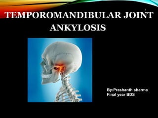
The presentation deals with the basics required for studying TMJ ankylosis. The text has been simplified and presented. It is well supported with illustrations.
Suggestions and feedback will be well appreciated. :)
The content covers majority of the aspect of immediate implant placement - why immediate implants?, case selection, decision making, classifications, surgical technique, healing following immediate implant placement, immediate implants in infected sockets/periapical infections, literature reviews and recommendations for clinical practice.
Dr. Ahmed M. Adawy, Professor Emeritus, Dep. Oral & Maxillofacial Surgery. Former Dean, Faculty of Dental Medicine, Al-Azhar University. Mandibular angle fractures account for 23% to 42% of all facial fractures. Fracture of mandibular angle can be classified as (A) Vertical favorable or unfavorable, (B) Horizontally favorable of unfavorable. Traditionally, mandibular angle fractures have been treated with either closed reduction and inter-maxillary fixation or open reduction and internal fixation with or without inter-maxillary fixation. Patients treated with inter-maxillary fixation have a restricted airway and loose excess weight. Rigid internal fixation and early return to function have eliminated the use of wire osteosenthysis and prolonged use of inter-maxillary fixation. The principal of rigid fixation, however, have inherent set of disadvantages including damage to the inferior alveolar nerve and the marginal mandibular branch of facial nerve. Postoperative malocclusion rates are also high. With the introduction of semi-rigid technique fracture of the mandibular angle could be treated according to Champy’s Ideal lines of osteosenthysis. The technique involves placement of a single monocortial miniplate on the superior border of the mandible. However, some studies suggested using a second miniplate along the inferior border. Wether one or two miniplates should be used is still debatable. The application of 3D plates may provide additional stability in 3 dimension and good resistance against torque forces.
Pterygoid implant insertion is an alternative to avoid sinus-lifting or other grafting procedures to treat the posterior maxilla.They are especially used in partial edentulism in order to avoid distal cantilevers.
The placement of a pterygoid implant requires surgical experience and expertise in the field of implantology.
Pterygoid implants have high success rates, to those of conventional implants, minimal complications and a good patient acceptance.
Dr Sachdeva's Facial Aesthetic and implant institute is one of the leading clinics in Delhi performing Pterygoid Implants in patients with bone resorption. So hurry up and book an appointment with us at Ashok Vihar, Delhi which has state of the art clinic and all the latest and advanced equipments.
To book an appointment contact:
Dr. Rajat Sachdeva
Director & Mentor
Dr Sachdeva’s Dental Aesthetic And Implant Institute
I 101, Ashok Vihar Phase 1, Delhi- 110052
Contact us at
Phone : +919818894041,01142464041
Our Websites:
www.sachdevadentalcare.com
www.dentalimplantindia.co.in
www.dentalclinicindelhi.com
www.dentalcoursesdelhi.com
Facebook- dentalcoursesdelhi
Youtube- drrajatsachdeva
Linkedin- drrajatsachdeva
Slideshare- Dr Rajat Sachdeva
Twitter Page- drrajatsachdeva
Instagram page- surgicalmasterrajat
Anatomical considerations for placing dental implants.
all the basic anatomical landmarks and considerations which are to be taken care off before and while placing a dental implant.
any type of implant it may be...wether endossous or subperiosteal or tranosteal.
lack of knowledge of basic anatomy will never lead to success of implant.
Pericoronitis is defined as inflammation of the oral soft tissues surrounding the crown of a partially erupted tooth. its treatment- operculectomy i.e. removal of the inflammed operculum
Naso-orbital-ethmoid (NOE) fractures: Management principles, options and rec...Dibya Falgoon Sarkar
Comprehensive discussion on diagnosis and management of NOE fractures. Surgical anatomy and approaches to NOE region is also discussed. Reconstruction of NOE complex is discussed. Recent advances in management of NOE fractures are also highlighted in this presentation
Dear Readers,
this is my ppt was made from a book of BAGHERI ( Current therapy in oral and maxillofacial surgery)- 2012 PLUS other sources.. hope you find it beneficial.
have a nice day,
hanan
This PowerPoint presentation provides a concise and technical exploration of NOE fractures, encompassing fracture classifications, diagnostic modalities, and treatment approaches. Delve into the intricacies of fracture pathology, radiological assessments, and surgical interventions
Couples presenting to the infertility clinic- Do they really have infertility...Sujoy Dasgupta
Dr Sujoy Dasgupta presented the study on "Couples presenting to the infertility clinic- Do they really have infertility? – The unexplored stories of non-consummation" in the 13th Congress of the Asia Pacific Initiative on Reproduction (ASPIRE 2024) at Manila on 24 May, 2024.
ARTIFICIAL INTELLIGENCE IN HEALTHCARE.pdfAnujkumaranit
Artificial intelligence (AI) refers to the simulation of human intelligence processes by machines, especially computer systems. It encompasses tasks such as learning, reasoning, problem-solving, perception, and language understanding. AI technologies are revolutionizing various fields, from healthcare to finance, by enabling machines to perform tasks that typically require human intelligence.
Anti ulcer drugs and their Advance pharmacology ||
Anti-ulcer drugs are medications used to prevent and treat ulcers in the stomach and upper part of the small intestine (duodenal ulcers). These ulcers are often caused by an imbalance between stomach acid and the mucosal lining, which protects the stomach lining.
||Scope: Overview of various classes of anti-ulcer drugs, their mechanisms of action, indications, side effects, and clinical considerations.
These lecture slides, by Dr Sidra Arshad, offer a quick overview of physiological basis of a normal electrocardiogram.
Learning objectives:
1. Define an electrocardiogram (ECG) and electrocardiography
2. Describe how dipoles generated by the heart produce the waveforms of the ECG
3. Describe the components of a normal electrocardiogram of a typical bipolar leads (limb II)
4. Differentiate between intervals and segments
5. Enlist some common indications for obtaining an ECG
Study Resources:
1. Chapter 11, Guyton and Hall Textbook of Medical Physiology, 14th edition
2. Chapter 9, Human Physiology - From Cells to Systems, Lauralee Sherwood, 9th edition
3. Chapter 29, Ganong’s Review of Medical Physiology, 26th edition
4. Electrocardiogram, StatPearls - https://www.ncbi.nlm.nih.gov/books/NBK549803/
5. ECG in Medical Practice by ABM Abdullah, 4th edition
6. ECG Basics, http://www.nataliescasebook.com/tag/e-c-g-basics
The prostate is an exocrine gland of the male mammalian reproductive system
It is a walnut-sized gland that forms part of the male reproductive system and is located in front of the rectum and just below the urinary bladder
Function is to store and secrete a clear, slightly alkaline fluid that constitutes 10-30% of the volume of the seminal fluid that along with the spermatozoa, constitutes semen
A healthy human prostate measures (4cm-vertical, by 3cm-horizontal, 2cm ant-post ).
It surrounds the urethra just below the urinary bladder. It has anterior, median, posterior and two lateral lobes
It’s work is regulated by androgens which are responsible for male sex characteristics
Generalised disease of the prostate due to hormonal derangement which leads to non malignant enlargement of the gland (increase in the number of epithelial cells and stromal tissue)to cause compression of the urethra leading to symptoms (LUTS
Ethanol (CH3CH2OH), or beverage alcohol, is a two-carbon alcohol
that is rapidly distributed in the body and brain. Ethanol alters many
neurochemical systems and has rewarding and addictive properties. It
is the oldest recreational drug and likely contributes to more morbidity,
mortality, and public health costs than all illicit drugs combined. The
5th edition of the Diagnostic and Statistical Manual of Mental Disorders
(DSM-5) integrates alcohol abuse and alcohol dependence into a single
disorder called alcohol use disorder (AUD), with mild, moderate,
and severe subclassifications (American Psychiatric Association, 2013).
In the DSM-5, all types of substance abuse and dependence have been
combined into a single substance use disorder (SUD) on a continuum
from mild to severe. A diagnosis of AUD requires that at least two of
the 11 DSM-5 behaviors be present within a 12-month period (mild
AUD: 2–3 criteria; moderate AUD: 4–5 criteria; severe AUD: 6–11 criteria).
The four main behavioral effects of AUD are impaired control over
drinking, negative social consequences, risky use, and altered physiological
effects (tolerance, withdrawal). This chapter presents an overview
of the prevalence and harmful consequences of AUD in the U.S.,
the systemic nature of the disease, neurocircuitry and stages of AUD,
comorbidities, fetal alcohol spectrum disorders, genetic risk factors, and
pharmacotherapies for AUD.
New Directions in Targeted Therapeutic Approaches for Older Adults With Mantl...i3 Health
i3 Health is pleased to make the speaker slides from this activity available for use as a non-accredited self-study or teaching resource.
This slide deck presented by Dr. Kami Maddocks, Professor-Clinical in the Division of Hematology and
Associate Division Director for Ambulatory Operations
The Ohio State University Comprehensive Cancer Center, will provide insight into new directions in targeted therapeutic approaches for older adults with mantle cell lymphoma.
STATEMENT OF NEED
Mantle cell lymphoma (MCL) is a rare, aggressive B-cell non-Hodgkin lymphoma (NHL) accounting for 5% to 7% of all lymphomas. Its prognosis ranges from indolent disease that does not require treatment for years to very aggressive disease, which is associated with poor survival (Silkenstedt et al, 2021). Typically, MCL is diagnosed at advanced stage and in older patients who cannot tolerate intensive therapy (NCCN, 2022). Although recent advances have slightly increased remission rates, recurrence and relapse remain very common, leading to a median overall survival between 3 and 6 years (LLS, 2021). Though there are several effective options, progress is still needed towards establishing an accepted frontline approach for MCL (Castellino et al, 2022). Treatment selection and management of MCL are complicated by the heterogeneity of prognosis, advanced age and comorbidities of patients, and lack of an established standard approach for treatment, making it vital that clinicians be familiar with the latest research and advances in this area. In this activity chaired by Michael Wang, MD, Professor in the Department of Lymphoma & Myeloma at MD Anderson Cancer Center, expert faculty will discuss prognostic factors informing treatment, the promising results of recent trials in new therapeutic approaches, and the implications of treatment resistance in therapeutic selection for MCL.
Target Audience
Hematology/oncology fellows, attending faculty, and other health care professionals involved in the treatment of patients with mantle cell lymphoma (MCL).
Learning Objectives
1.) Identify clinical and biological prognostic factors that can guide treatment decision making for older adults with MCL
2.) Evaluate emerging data on targeted therapeutic approaches for treatment-naive and relapsed/refractory MCL and their applicability to older adults
3.) Assess mechanisms of resistance to targeted therapies for MCL and their implications for treatment selection
2. • Greek terminology meaning ‘stiff joint’.
• Fusion between cranium and condyle.
• Jaw function is affected.
• Hypomobility or immobility of joint can lead to inability to open
mouth from partial to complete.
2
3. ETIOPATHOLOGY OF ANKYLOSIS OF TMJ
•
•
•
•
congenital
At birth (forceps delivery )
hemarthrosis
condylar # - intra / extra capsulra
Trauma
•
•
•
•
•
Parotitis
tonsilitis
Abscess around the joint
osteomyelitis of the jaw
actinomycosis
Infections
3
5. Pathophysiology
Trauma
↓
Extravasation of blood into the joint space
↓
Heamarthrosis
↓
Period of restricted mobility due to pain
↓
Fibrosis leading to further restriction
↓
Gradual bone formation
5
6. CLASSIFICATION
1. Based on the location:
-
-
Intra articular or true ankylosis
Extra articular or false ankylosis
2.
-
-
-
Based on the
Bony.
Fibrous.
Mixed.
type of tissue involved:
3. Based on the extent of fusion/severity of ankylosis:
-
-
Complete.
Incomplete.
4. Based on the side involved:
-
-
Unilateral.
Bilateral.
6
7. SAWHNEY CLASSIFICATION
1. Type I: Head of the condyle is flattened or deformed with close
approximation to the upper
make movement possible.
articular surface. Dense fibrous adhesions
2. Type II: Head misshapen or flattened but is distinguishable. Bony
fusion of head to outer edge of articular surface.
7
8. 3. Type III: Bony block seems to bridge across ramus and zygomatic
arch. Displaced condylar head. Elongation of coronoid process seen.
4. Type IV: Bony block is wide and deep and extends between ramus and
upper articular surface thereby completely replacing joint architecture.
8
9. CLINICAL MANIFESTATIONS
→ Unilateral Ankylosis:
• Facial asymmetry.
• Deviation of mandible and chin on affected side.
• Roundness and fullness of face on affected side.
• Cross bite maybe seen.
• Lower border of mandible has a concavity on affected side.
9
10. → Bilateral Ankylosis:
• Inability to open mouth progresses to decreased interincisal opening.
• Typical ‘bird face’ deformity with receding chin.
• Neck chin angle reduced or completely absent.
• Class II malocclusion.
• Protrusive upper incisors with anterior open bite.
• Multiple carious teeth with bad periodontal health.
10
11. DIAGNOSIS
Diagnosis is based on the following:
1.
2.
3.
a.
History of trauma, infection etc.
Clinical findings.
Radiographic findings:
OPG: Shows both joints picture which can be compared in unilateral
cases.
Lateral oblique view: Gives anteroposterior dimension of condylarb.
mass. Elongation of coronoid process seen.
c. Cephalometric radiograph: Taken to evaluate associated skeletal
deformities.
11
12. d. CT scan:
• Very helpful guide for surgery.
• Relation to middle cranial fossa, anteroposterior width can be
assessed.
• Any presence of fractured condylar head can be located.
12
13. MANAGEMENT OF TMJ ANKYLOSIS
Aims and Objectives of Surgery:
1. Release the ankylosed mass and creation of a gap to mobilize
joint.
Creation of a functional joint.
the
2.
3.
4.
5.
6.
To
To
To
To
reconstruct the joint and restore vertical height
prevent recurrence.
restore normal facial growth pattern.
of ramus.
improve esthetics and rehabilitate the patient.
Surgical Techniques:
I: Condylectomy.
II: Gap Arthroplasty.
III: Interpositional Arthroplasty.
13
14. Management:
Adult
• Cause:
• Cause:
• Trauma
• Aim:
• Restoration of
satisfactory
movement
• Trauma
•Infection
• Aim:
•Restoring
function and
movement
• Bony
replacement
with CCG
• Correction of
occlusal and
cosmetic
deformity
Childhood
14
15. SURGICALAPPROACHES
Blair
inverted hockey stick vertical incision
Dingman
question markAl-Kayat & Bramley in 1979-
modified preauricular approach
and Ivy in 1936- Thoma in 1958- angulated Preauricular incision-
Popowich and Crane in 1982-
15
17. Condylectomy:
Advocated in cases of fibrous ankylosis, where joint space is
obilterated with deposition of fibrous bands but there is not much
deformity of condylar head.
• Preauricular approach used commonly, others include Al Kayat
Bramley, inverted hockey stick.
17
18. Gap Arthroplasty:
• Section consists of two horizontal osteotomy cuts and removal of
bony wedge for creation of a gap.
• No substance is interposed between the two cut bony surfaces.
• Minimum gap of 1 cm to prevent reankylosis.
18
19. Interpositional Arthroplasty:
• Involves creation of a gap, but in addition a barrier is inserted between
the cut bony surfaces to minimize risk of recurrence and to maintain
vertical height of ramus.
18
19
22. KABAN’S PROTOCOL FOR MANAGEMENT OF TMJ ANKYLOSIS
1. Early surgical intervention.
2.
-
Aggressive resection:
Gap of at least 1 – 1.5 cm should be created.
3.
-
Ipsilateral coronoidectomy and temporalis myotomy:
After gap arthroplasty, coronoidectomy on the same side
carried out.
should be
- Temporalis muscle attachments are severed by carrying out temporalis
myotomy.
4. Contralateral coronoidectomy and temporalis myotomy.
22
23. 5. Lining of glenoid fossa region with temporalis fascia.
6. Reconstruction of ramus with costochondral graft.
7. Early mobilization and aggressive physiotherapy for
months postoperatively.
at least six
8. Regular long term follow up.
9. To carry out cosmetic surgery at later date, when growth of patient is
completed.
23
24. COMPLICATIONS DURING SURGERY
During Anesthesia:
a. As the patient cannot open the mouth, awake blind intubation has to
be done where co – operation is required which is difficult to achieve
sometimes.
b. Because of small mandible and altered position of larynx, intubation
poses a problem.
c. Aspiration of blood clot, tooth or foreign body during extubation.
d. Danger of falling back of tongue and obstructing airway is always
there after extubation.
24
26. FREY SYNDROME:
1st described by frey.
It is localised gustatory sweating in the area supplied by
auriculotemporal nerve.
Cause:
Congenital or acquired
Surgery of parotid gland, TMJ , parotid abscess, facial wound.
Clinical feature:
Pain in area supplied by ATN
Gustatory sweating
Erythema & flushing
Positive iodine starch test
1.
2.
3.
4.
26
28. RECURRENCE OFANKYLOSIS
Several factors said to be responsible:
1.
2.
3.
Inadequate gap created between fragments.
Fracture of costochondral graft.
Loosening of costochondral graft due to inadequate
ramus.
Inadequate postoperative physiotherapy.
Inadequate coverage of glenoid fossa surface.
fixation to
4.
5.
6. Higher osteogenic potential and periosteal osteogenic
responsible for high rate of recurrence in children.
power maybe
28