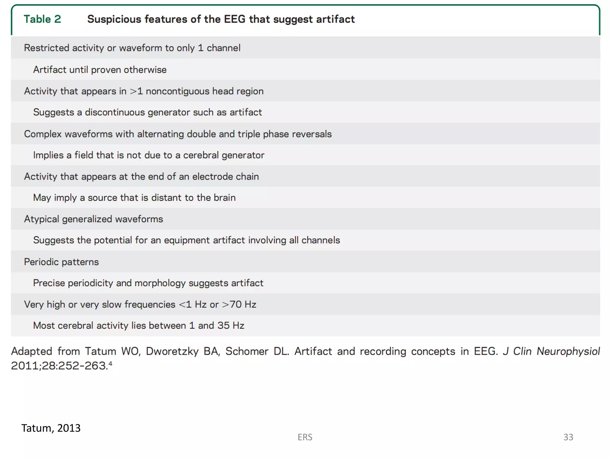
The Philosophy of
EEG & Epilepsy
• In 29% - 55% of patients with epilepsy, the initial EEG will
show EDs  approximately 15% of these patients have
repeatedly “negative” studies or normal-appearing EEG.
• Approximately 30% of patients seen at epilepsy centers for
refractory seizures do not have seizures and have been
misdiagnosed
– many of them have histories not in the least suggestive of seizures
– many of them have their diagnosis based largely (and sometimes
solely) on an ‘abnormal’ EEG
ERS 2Miller & Henry, 2013; Tatum, 2013; Benbadis, 2010
Bad EEG may be harmful
Good EEG is invaluable
• The main consequence of EEG misinterpretation is that false-positive
reports lead to inappropriate treatments.
• Without recording a seizure, the EEG is only supportive when diagnosing
epilepsy. However, the supportive nature of an interictal EEG is only valid if
clear interictal epileptiform discharges (IED) are encountered.
• When over-identified IED are reported, the misinterpretation of the EEG
often goes unchallenged until ictal recordings are performed. A repeat
normal recording does not resolve the issue of a prior false-positive
interpretation.
Miller & Henry, 2013; Tatum, 2013 ERS 3
Vague Nonspecific Symptoms + an Overread EEG =
Incorrect Diagnosis of Seizure(s)
“..Routine interictal EEG recording is one of the most abused
investigations in clinical medicine and is unquestionably
responsible for great human suffering.”
Benbadis, 2013 ERS 4
The essence of EEG reading:
a pattern recognition skill
• EEG interpretation is a different sort of mental skill—
it takes viewing of many studies, with a variety of
findings, to begin to reliably recognize common
abnormalities.
Miller & Henry, 2013
ERS 5
To accurately interpret an
abnormal EEG,
one must first have the
ability to identify normal
patterns
Miller & Henry, 2013 ; Tatum, 2013; Noachtar, 2018 ERS 6
The Stages of EEG Analysis
Stage 1
- Nothing makes sense
Stage 2
- You think you understand but you see abnormalities everywhere
Stage 3
- You gain more hindsight, you recognize a spike but wonder if it is
actually significant
Stage 4
- You are finally able to form your own opinion, even if it is different
from your teacher’s, this last stage is the sign you have matured, you
have acquired enough experience to have your own opinion and to
discuss an EEG.
ERS 7Gelisse & Crespel, 2016
A group of clinical neurophysiologists..
60% of the respondents were certified in clinical
neurophysiology by a national board of examiners..
>90% had encountered misread EEGs..
>95% of the misinterpretations were due to
overinterpretation:
– Of normal findings
– Of artifact
Tatum, 2013
ERS 8
ERS 9
A major contributor to the misdiagnosis of epilepsy is
the tendency to overread normal EEGs as abnormal
• The wrong diagnosis of seizures is sometimes based solely on
the “abnormal” EEG.
• The vast majority of overread patterns are wicket rhythms or
“nameless variants”.
• The common and unfortunate misconception is that phase
reversals are somehow indicative of abnormalities
 Phase reversals do not indicate epileptogenicity or even abnormality,
they only indicate location.
Benbadis, 2013 ERS 10
Reasons for the overreading
• The “looking too hard” syndrome
the reader is “trying too hard” to find abnormalities because the patient
had a “seizure” (history bias)
• Lack of training & inexperience
i.e., not seeing enough normal tracings and the range of normal variations
• Not applying strict criteria to make sharply contoured
waveforms epileptiform
Benbadis, 2013 ERS 11
Differentiating normal variants from meaningful spikes
and sharp waves can at times be challenging
Guidelines for EEG interpretation available for defining and
identifying EDs do not exist
but helpful rules have been described..
Miller & Henry, 2013; Benbadis, 2013; Tatum, 2013 ERS 12
Epileptiform Pattern
= Interictal epileptiform discharge, epileptiform activity
: Transients distinguishable from background activity with a characteristic morphology typically,
but neither exclusively nor invariably, found in interictal EEGs of people with epilepsy.
EPs have to fulfill at least 4 of the following 6 criteria:
1. Di- or tri-phasic waves with sharp or spiky morphology (i.e. pointed peak)
2. Different wave-duration than the ongoing background activity, either
shorter or longer.
3. Asymmetry of the waveform: a sharply rising ascending phase and a more
slowly decaying descending phase, or vice versa.
4. The transient is followed by an associated slow after-wave.
5. The background activity surrounding epileptiform discharges is disrupted
by the presence of the epileptiform discharges.
6. Distribution of the negative and positive potentials on the scalp suggests a
source of the signal in the brain, corresponding to a radial, oblique or
tangential orientation of the source (see dipole). This is best assessed by
inspecting voltage maps constructed using common-average reference.
ERS 13Kane et al, 2017
EEG patterns which may be mistaken as epileptiform
Noachtar, 2018 ERS 14
Tips for diagnostic purpose when identifying EDs
2 Minutes Rule
• If 2minutes after review of the EEG, a
“discharge” is unable to be clearly categorized
as an ED, a conservative interpretation should
apply, and the waveform interpreted as
nonepileptiform.
Tatum, 2013
ERS 15
ERS 16
Artifacts are intertwined with epilepsy
• Artifacts may beguile the interpreter into
misidentifying wave forms (false-positive) that
simulate ED.
• Artifacts may obscure the recording during ED or
seizures to eliminate EEG detection (false-negative)
from a diagnostic equation.
Tatum, 2013
ERS 17
Removing Artifacts
• Artifact recognition is the essential first step
• Despite computerization of EEG, artifact
identification, recognition, and elimination
will still be essential human tasks of EEG
interpretation
• The easiest means of achieving artifact
reduction is to avoid them
Tatum, 2013
ERS 18
Tatum, 2013
Artifacts are present in virtually every EEG and may
arise from a variety of extracerebral sources.
• Some artifact is crucial
to identify stages of
sleep and level of
consciousness.
• Some areas in the hos-
pital are electrically
complex/hostile to
recording & predispose
to artifacts.
ERS 19
A quiet patient, controlled setting, and a qualified
technologist are the foundation to minimizing the
amount of artifact.
• The responsibility of the technologist during the recording is
to prove whether a wave-form is artifact or not, and act to
identify or eliminate it from the recording  Technologist
monitors, eliminates, and camouflages extracerebral sources
bioelectric fields introduce artifact.
• Electrode contact with the scalp, maintenance of a quiet
environment, and troubleshooting are keys to minimize
artifact-related diagnoses of epilepsy.
• Troubleshooting artifact must be done at the time of the
recording. Post hoc filtering and montage manipulation may
help, but unless a noncephalic source is identified, the
electrocerebral field may appear real.
Tatum, 2013
ERS 20
ERS 21
Systematic approach to EEG abnormalities
• The interpretation of EEG is associated with a poor inter-
oberserver reliability.
• Pattern recognition is inherently prone to pitfalls when rules
[of polarity] and convention are ignored.
• Problems of fluctuation in the accuracy of EEG interpretation
may vary from person to person and even in the same person
over time.
• There are guidelines available for conducting EEG studies, but
those available for defining and identifying EDs do not exist
for EEG interpretation.
• It is important to follow a systematic approach to the
classification of EEG abnormalities.
Miller & Henry, 2013; Tatum, 2013; Noachtar, 2018 ERS 22
EEG interpretation & report [1]
• Requires knowledge of the patient’s age, past medical and
medication history, their clinical condition during the EEG,
particularly level of consciousness and responsiveness.
• Should follow a standard format that includes a factual
description, a classification and a clinical interpretation of the
EEG record.
• EEG interpretation summarizes the results of the EEG and
gives a clinical interpretation in light of the diagnosis and the
questions posed by the referring physician.
• Terminology of the EEG interpretation should follow common
neurological and clinical practice and use terms
understandable to other physicians not specialized in EEG
Noachtar, 2018 ERS 23
EEG interpretation & report [2]
• All EEG phenomena should be described as precisely as
possible in terms of frequency, amplitude, phase relation,
waveform, localization, quantity, and variability of these
parameters.
• The terminology used should follow international standards
and recommendations
Noachtar, 2018 ERS 24
Kaplan & Benbadis, 2013 ERS 25
There are only few normal individuals
with epileptiform dicharges in the EEG
• Epileptiform discharges in the EEG without having
seizures:
– Children: 2-3%
– Adults: 0.5%
the epileptifom discharges occur only during photic
stimulation in half of these individuals
Noachtar, 2018 ERS 26
“We do not treat the EEG”
• A normal EEG does not exclude a clinical
diagnosis of epilepsy
• An abnormal EEG finding may not be related to
the provisional diagnosis or presenting
symptoms
Miller & Henry, 2013; Tatum, 2013 ERS 27
• The diagnosis of seizures relies mainly on a good history,
which requires skills and time.
• The importance of the EEG is [often] overemphasized, and it is
especially detrimental when it is interpreted out of clinical
context.
• Overreading is more harmful than underreading.
• Every EEG should be interpreted with care and caution to
avoid pitfalls.
• Proper training is a crucial aspect of minimizing as many of
the errors as possible.
ERS 28
References
• Benbadis, S.R. (2010) 'The tragedy of over-read EEGs and wrong diagnoses of epilepsy', Expert
Review of Neurotherapeutics, vol. 10, no. 3, pp. 343-346.
• Gaspard, N. and Hirsch, L.J. (2013) 'Pitfalls in ictal EEG interpretation: Critical care and intracranial
recordings', Neurology, vol. 80, p. S26.
• Gelisse, P. and Crespel, A. (2016) Atlas of Electroencephalography Volume I: Awake and Sleep EEG,
Activation Procedures and Artifact, Paris: John Libbey Eurotext.
• Kane, N., Acharya, J., Benickzy, S., Caboclo, L., Finnigan, S. and Kaplan, P.W. (2017) 'A revised glossary
of terms most commonly used by clinical electroencephalographers and updated proposal for the
report format of the EEG findings. Revision 2017', Clinical Neurophysiology Practice, vol. 2, pp. 170-
185.
• Kaplan, P.W. and Benbadis, S.R. (2013) 'How to write an EEG report: Dos and don'ts', Neurology, vol.
80, p. S43.
• Miller, J.W. and Henry, J.C. (2013) 'Solving the dilemma of EEG misinterpretation', Neurology, vol. 80,
pp. 13-14.
• Noachtar, S. (2018) 'EAN/ILAE-CEA: How to approach EEG and avoid overreading in epilepsy - Level
1: Systematic approach to EEG abnormalities', 4th Congress of the European Academy of Neurology
, Lisbon.
• Tatum, W.O. (2013) 'Artifact-related epilepsy', Neurology, vol. 80, p. S12.
• Tatum, W.O. (2013) 'How not to read an EEG: Introductory statements', Neurology, vol. 80, p. S1.
• Tatum, W.O. (2013) 'Normal "suspicious" EEG', Neurology, vol. 80, p. S4.
ERS 29
ERS 30
How to identify artifacts?
ERS 31
Gaspard, 2013
ERS 32
Tatum, 2013
ERS 33
Gaspard, 2013 ERS 34
The role of EEG for vague cases suspected non-convulsive seizure..
ERS 35
Gaspard, 2013 ERS 36
Gaspard, 2013 ERS 37
ICU: the most hostile environment for EEG recording..
Tips n trick?
ERS 38
Gaspard, 2013ERS 39
How to write an EEG report
ERS 40
Items to include
in EEG report
Kaplan & Benbadis, 2013ERS 41
Guideline..
ERS 42
..Updated Proposal for the Report Format
of the EEG Findings, Revision 2017
Kane et al, 2017 ERS 43
..Updated Proposal for the Report Format
of the EEG Findings, Revision 2017
Kane et al, 2017
ERS 44
..Updated Proposal for the
Report Format
of the EEG Findings,
Revision 2017
Kane et al, 2017
ERS 45
..Updated Proposal for the Report Format
of the EEG Findings, Revision 2017
Kane et al, 2017
ERS 46
Thank You
ERS 47

The Philosophy of EEG Interpretation

  • 1.
  • 2.
    EEG & Epilepsy •In 29% - 55% of patients with epilepsy, the initial EEG will show EDs  approximately 15% of these patients have repeatedly “negative” studies or normal-appearing EEG. • Approximately 30% of patients seen at epilepsy centers for refractory seizures do not have seizures and have been misdiagnosed – many of them have histories not in the least suggestive of seizures – many of them have their diagnosis based largely (and sometimes solely) on an ‘abnormal’ EEG ERS 2Miller & Henry, 2013; Tatum, 2013; Benbadis, 2010
  • 3.
    Bad EEG maybe harmful Good EEG is invaluable • The main consequence of EEG misinterpretation is that false-positive reports lead to inappropriate treatments. • Without recording a seizure, the EEG is only supportive when diagnosing epilepsy. However, the supportive nature of an interictal EEG is only valid if clear interictal epileptiform discharges (IED) are encountered. • When over-identified IED are reported, the misinterpretation of the EEG often goes unchallenged until ictal recordings are performed. A repeat normal recording does not resolve the issue of a prior false-positive interpretation. Miller & Henry, 2013; Tatum, 2013 ERS 3
  • 4.
    Vague Nonspecific Symptoms+ an Overread EEG = Incorrect Diagnosis of Seizure(s) “..Routine interictal EEG recording is one of the most abused investigations in clinical medicine and is unquestionably responsible for great human suffering.” Benbadis, 2013 ERS 4
  • 5.
    The essence ofEEG reading: a pattern recognition skill • EEG interpretation is a different sort of mental skill— it takes viewing of many studies, with a variety of findings, to begin to reliably recognize common abnormalities. Miller & Henry, 2013 ERS 5
  • 6.
    To accurately interpretan abnormal EEG, one must first have the ability to identify normal patterns Miller & Henry, 2013 ; Tatum, 2013; Noachtar, 2018 ERS 6
  • 7.
    The Stages ofEEG Analysis Stage 1 - Nothing makes sense Stage 2 - You think you understand but you see abnormalities everywhere Stage 3 - You gain more hindsight, you recognize a spike but wonder if it is actually significant Stage 4 - You are finally able to form your own opinion, even if it is different from your teacher’s, this last stage is the sign you have matured, you have acquired enough experience to have your own opinion and to discuss an EEG. ERS 7Gelisse & Crespel, 2016
  • 8.
    A group ofclinical neurophysiologists.. 60% of the respondents were certified in clinical neurophysiology by a national board of examiners.. >90% had encountered misread EEGs.. >95% of the misinterpretations were due to overinterpretation: – Of normal findings – Of artifact Tatum, 2013 ERS 8
  • 9.
  • 10.
    A major contributorto the misdiagnosis of epilepsy is the tendency to overread normal EEGs as abnormal • The wrong diagnosis of seizures is sometimes based solely on the “abnormal” EEG. • The vast majority of overread patterns are wicket rhythms or “nameless variants”. • The common and unfortunate misconception is that phase reversals are somehow indicative of abnormalities  Phase reversals do not indicate epileptogenicity or even abnormality, they only indicate location. Benbadis, 2013 ERS 10
  • 11.
    Reasons for theoverreading • The “looking too hard” syndrome the reader is “trying too hard” to find abnormalities because the patient had a “seizure” (history bias) • Lack of training & inexperience i.e., not seeing enough normal tracings and the range of normal variations • Not applying strict criteria to make sharply contoured waveforms epileptiform Benbadis, 2013 ERS 11
  • 12.
    Differentiating normal variantsfrom meaningful spikes and sharp waves can at times be challenging Guidelines for EEG interpretation available for defining and identifying EDs do not exist but helpful rules have been described.. Miller & Henry, 2013; Benbadis, 2013; Tatum, 2013 ERS 12
  • 13.
    Epileptiform Pattern = Interictalepileptiform discharge, epileptiform activity : Transients distinguishable from background activity with a characteristic morphology typically, but neither exclusively nor invariably, found in interictal EEGs of people with epilepsy. EPs have to fulfill at least 4 of the following 6 criteria: 1. Di- or tri-phasic waves with sharp or spiky morphology (i.e. pointed peak) 2. Different wave-duration than the ongoing background activity, either shorter or longer. 3. Asymmetry of the waveform: a sharply rising ascending phase and a more slowly decaying descending phase, or vice versa. 4. The transient is followed by an associated slow after-wave. 5. The background activity surrounding epileptiform discharges is disrupted by the presence of the epileptiform discharges. 6. Distribution of the negative and positive potentials on the scalp suggests a source of the signal in the brain, corresponding to a radial, oblique or tangential orientation of the source (see dipole). This is best assessed by inspecting voltage maps constructed using common-average reference. ERS 13Kane et al, 2017
  • 14.
    EEG patterns whichmay be mistaken as epileptiform Noachtar, 2018 ERS 14
  • 15.
    Tips for diagnosticpurpose when identifying EDs 2 Minutes Rule • If 2minutes after review of the EEG, a “discharge” is unable to be clearly categorized as an ED, a conservative interpretation should apply, and the waveform interpreted as nonepileptiform. Tatum, 2013 ERS 15
  • 16.
  • 17.
    Artifacts are intertwinedwith epilepsy • Artifacts may beguile the interpreter into misidentifying wave forms (false-positive) that simulate ED. • Artifacts may obscure the recording during ED or seizures to eliminate EEG detection (false-negative) from a diagnostic equation. Tatum, 2013 ERS 17
  • 18.
    Removing Artifacts • Artifactrecognition is the essential first step • Despite computerization of EEG, artifact identification, recognition, and elimination will still be essential human tasks of EEG interpretation • The easiest means of achieving artifact reduction is to avoid them Tatum, 2013 ERS 18
  • 19.
    Tatum, 2013 Artifacts arepresent in virtually every EEG and may arise from a variety of extracerebral sources. • Some artifact is crucial to identify stages of sleep and level of consciousness. • Some areas in the hos- pital are electrically complex/hostile to recording & predispose to artifacts. ERS 19
  • 20.
    A quiet patient,controlled setting, and a qualified technologist are the foundation to minimizing the amount of artifact. • The responsibility of the technologist during the recording is to prove whether a wave-form is artifact or not, and act to identify or eliminate it from the recording  Technologist monitors, eliminates, and camouflages extracerebral sources bioelectric fields introduce artifact. • Electrode contact with the scalp, maintenance of a quiet environment, and troubleshooting are keys to minimize artifact-related diagnoses of epilepsy. • Troubleshooting artifact must be done at the time of the recording. Post hoc filtering and montage manipulation may help, but unless a noncephalic source is identified, the electrocerebral field may appear real. Tatum, 2013 ERS 20
  • 21.
  • 22.
    Systematic approach toEEG abnormalities • The interpretation of EEG is associated with a poor inter- oberserver reliability. • Pattern recognition is inherently prone to pitfalls when rules [of polarity] and convention are ignored. • Problems of fluctuation in the accuracy of EEG interpretation may vary from person to person and even in the same person over time. • There are guidelines available for conducting EEG studies, but those available for defining and identifying EDs do not exist for EEG interpretation. • It is important to follow a systematic approach to the classification of EEG abnormalities. Miller & Henry, 2013; Tatum, 2013; Noachtar, 2018 ERS 22
  • 23.
    EEG interpretation &report [1] • Requires knowledge of the patient’s age, past medical and medication history, their clinical condition during the EEG, particularly level of consciousness and responsiveness. • Should follow a standard format that includes a factual description, a classification and a clinical interpretation of the EEG record. • EEG interpretation summarizes the results of the EEG and gives a clinical interpretation in light of the diagnosis and the questions posed by the referring physician. • Terminology of the EEG interpretation should follow common neurological and clinical practice and use terms understandable to other physicians not specialized in EEG Noachtar, 2018 ERS 23
  • 24.
    EEG interpretation &report [2] • All EEG phenomena should be described as precisely as possible in terms of frequency, amplitude, phase relation, waveform, localization, quantity, and variability of these parameters. • The terminology used should follow international standards and recommendations Noachtar, 2018 ERS 24
  • 25.
    Kaplan & Benbadis,2013 ERS 25
  • 26.
    There are onlyfew normal individuals with epileptiform dicharges in the EEG • Epileptiform discharges in the EEG without having seizures: – Children: 2-3% – Adults: 0.5% the epileptifom discharges occur only during photic stimulation in half of these individuals Noachtar, 2018 ERS 26
  • 27.
    “We do nottreat the EEG” • A normal EEG does not exclude a clinical diagnosis of epilepsy • An abnormal EEG finding may not be related to the provisional diagnosis or presenting symptoms Miller & Henry, 2013; Tatum, 2013 ERS 27
  • 28.
    • The diagnosisof seizures relies mainly on a good history, which requires skills and time. • The importance of the EEG is [often] overemphasized, and it is especially detrimental when it is interpreted out of clinical context. • Overreading is more harmful than underreading. • Every EEG should be interpreted with care and caution to avoid pitfalls. • Proper training is a crucial aspect of minimizing as many of the errors as possible. ERS 28
  • 29.
    References • Benbadis, S.R.(2010) 'The tragedy of over-read EEGs and wrong diagnoses of epilepsy', Expert Review of Neurotherapeutics, vol. 10, no. 3, pp. 343-346. • Gaspard, N. and Hirsch, L.J. (2013) 'Pitfalls in ictal EEG interpretation: Critical care and intracranial recordings', Neurology, vol. 80, p. S26. • Gelisse, P. and Crespel, A. (2016) Atlas of Electroencephalography Volume I: Awake and Sleep EEG, Activation Procedures and Artifact, Paris: John Libbey Eurotext. • Kane, N., Acharya, J., Benickzy, S., Caboclo, L., Finnigan, S. and Kaplan, P.W. (2017) 'A revised glossary of terms most commonly used by clinical electroencephalographers and updated proposal for the report format of the EEG findings. Revision 2017', Clinical Neurophysiology Practice, vol. 2, pp. 170- 185. • Kaplan, P.W. and Benbadis, S.R. (2013) 'How to write an EEG report: Dos and don'ts', Neurology, vol. 80, p. S43. • Miller, J.W. and Henry, J.C. (2013) 'Solving the dilemma of EEG misinterpretation', Neurology, vol. 80, pp. 13-14. • Noachtar, S. (2018) 'EAN/ILAE-CEA: How to approach EEG and avoid overreading in epilepsy - Level 1: Systematic approach to EEG abnormalities', 4th Congress of the European Academy of Neurology , Lisbon. • Tatum, W.O. (2013) 'Artifact-related epilepsy', Neurology, vol. 80, p. S12. • Tatum, W.O. (2013) 'How not to read an EEG: Introductory statements', Neurology, vol. 80, p. S1. • Tatum, W.O. (2013) 'Normal "suspicious" EEG', Neurology, vol. 80, p. S4. ERS 29
  • 30.
  • 31.
    How to identifyartifacts? ERS 31
  • 32.
  • 33.
  • 34.
  • 35.
    The role ofEEG for vague cases suspected non-convulsive seizure.. ERS 35
  • 36.
  • 37.
  • 38.
    ICU: the mosthostile environment for EEG recording.. Tips n trick? ERS 38
  • 39.
  • 40.
    How to writean EEG report ERS 40
  • 41.
    Items to include inEEG report Kaplan & Benbadis, 2013ERS 41
  • 42.
  • 43.
    ..Updated Proposal forthe Report Format of the EEG Findings, Revision 2017 Kane et al, 2017 ERS 43
  • 44.
    ..Updated Proposal forthe Report Format of the EEG Findings, Revision 2017 Kane et al, 2017 ERS 44
  • 45.
    ..Updated Proposal forthe Report Format of the EEG Findings, Revision 2017 Kane et al, 2017 ERS 45
  • 46.
    ..Updated Proposal forthe Report Format of the EEG Findings, Revision 2017 Kane et al, 2017 ERS 46
  • 47.