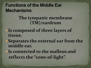
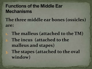
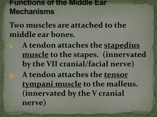
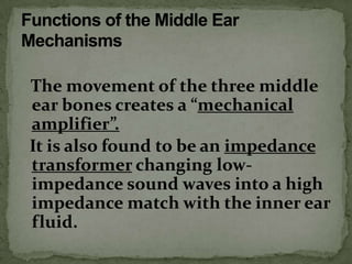
The tympanic membrane is the
receiver and collector of acoustic
pressure directed against the ear by
vibratory energy in the air around us.
The tympanic membrane (TM) is
conical, pointing medialward so that
more surface area of the membrane
can be available without having a
larger opening for a ear canal.
The TM is found to be at
approximately a fifty-five degree
angle (not perpendicular to the ear
canal) and has generally an elliptical
shape.
The tympanic membrane
           (TM)/eardrum
 Is composed of three layers of
  tissue.
 Separates the external ear from the
  middle ear.
 Is connected to the malleus and
  reflects the “cone-of-light”.
The three layers of tissue of the TM are:
1. The cutaneous layer (same layer as
   the EAM)
2. The fibrous layer (the “compliance”
   layer)
3. The mucous layer (same as the inner
   liner of the middle ear cavity)
The TM attachment at its superior
portion to the malleus creates a
movement which would more
resemble a swinging motion than a
piston (in and out) motion.
One tenth of one decibel of sound
pressure will create movement of a
healthy tympanic membrane by as
little as 1/100 the thickness of a
hydrogen molecule—it is very
sensitive to sound pressure—yes
even barometric pressure.
The TM moves much more easily at
its inferior portion.
Note: Due to gravity, this is the area
which may become most easily
compromised by ear canal debris or
middle ear fluid.
When the TM becomes too flaccid
(floppy) or too tense, the sound
transfer to the middle ear bones
(ossicles) becomes easily influenced.
The three middle ear bones (ossicles)
are:
1. The malleus (attached to the TM)
2. The incus (attached to the
   malleus and stapes)
3. The stapes (attached to the oval
   window)
Two muscles are attached to the
middle ear bones.
1. A tendon attaches the stapedius
   muscle to the stapes. (innervated
   by the VII cranial/facial nerve)
2. A tendon attaches the tensor
   tympani muscle to the malleus.
   (innervated by the V cranial
   nerve)
The movement of the three middle
ear bones creates a “mechanical
amplifier”.
It is also found to be an impedance
transformer changing low-
impedance sound waves into a high
impedance match with the inner ear
fluid.
As the movement of the malleus and
incus bones represent the to-and-fro
motion of a swing, they actually rock
the stapes in and out of the oval
window (do not push it in and out
like a plunger or piston).
This rocking action of the stapes
actually decreases once the brain
senses sound pressures greater than
seventy decibels due to the
contraction of the middle ear
muscles.
The most widely accepted theory
regarding this middle ear muscle
contraction is to protect the ear from
too much sound pressure—mostly
from low frequency energy (below
2000hz). In other words, their
contraction creates a high-pass filter.
The Eustachian tube
It equalizes the air pressure
between the middle ear cavity and
the external auditory meatus. It
usually opens and closes every time
we swallow.
The Eustachian Tube

It also serves as a drainage conduit
when any fluid has collected within
the middle ear such as the effects of a
head cold.

The middle ear mechanisms

  • 1.
    The tympanic membraneis the receiver and collector of acoustic pressure directed against the ear by vibratory energy in the air around us.
  • 2.
    The tympanic membrane(TM) is conical, pointing medialward so that more surface area of the membrane can be available without having a larger opening for a ear canal.
  • 3.
    The TM isfound to be at approximately a fifty-five degree angle (not perpendicular to the ear canal) and has generally an elliptical shape.
  • 4.
    The tympanic membrane (TM)/eardrum  Is composed of three layers of tissue.  Separates the external ear from the middle ear.  Is connected to the malleus and reflects the “cone-of-light”.
  • 5.
    The three layersof tissue of the TM are: 1. The cutaneous layer (same layer as the EAM) 2. The fibrous layer (the “compliance” layer) 3. The mucous layer (same as the inner liner of the middle ear cavity)
  • 6.
    The TM attachmentat its superior portion to the malleus creates a movement which would more resemble a swinging motion than a piston (in and out) motion.
  • 7.
    One tenth ofone decibel of sound pressure will create movement of a healthy tympanic membrane by as little as 1/100 the thickness of a hydrogen molecule—it is very sensitive to sound pressure—yes even barometric pressure.
  • 8.
    The TM movesmuch more easily at its inferior portion. Note: Due to gravity, this is the area which may become most easily compromised by ear canal debris or middle ear fluid.
  • 9.
    When the TMbecomes too flaccid (floppy) or too tense, the sound transfer to the middle ear bones (ossicles) becomes easily influenced.
  • 10.
    The three middleear bones (ossicles) are: 1. The malleus (attached to the TM) 2. The incus (attached to the malleus and stapes) 3. The stapes (attached to the oval window)
  • 11.
    Two muscles areattached to the middle ear bones. 1. A tendon attaches the stapedius muscle to the stapes. (innervated by the VII cranial/facial nerve) 2. A tendon attaches the tensor tympani muscle to the malleus. (innervated by the V cranial nerve)
  • 12.
    The movement ofthe three middle ear bones creates a “mechanical amplifier”. It is also found to be an impedance transformer changing low- impedance sound waves into a high impedance match with the inner ear fluid.
  • 13.
    As the movementof the malleus and incus bones represent the to-and-fro motion of a swing, they actually rock the stapes in and out of the oval window (do not push it in and out like a plunger or piston).
  • 14.
    This rocking actionof the stapes actually decreases once the brain senses sound pressures greater than seventy decibels due to the contraction of the middle ear muscles.
  • 15.
    The most widelyaccepted theory regarding this middle ear muscle contraction is to protect the ear from too much sound pressure—mostly from low frequency energy (below 2000hz). In other words, their contraction creates a high-pass filter.
  • 16.
    The Eustachian tube Itequalizes the air pressure between the middle ear cavity and the external auditory meatus. It usually opens and closes every time we swallow.
  • 17.
    The Eustachian Tube Italso serves as a drainage conduit when any fluid has collected within the middle ear such as the effects of a head cold.