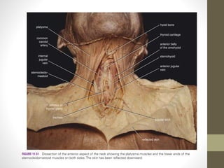
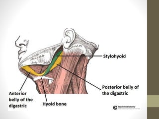
The hyoid bone 
Dr. Muhammad Mustafa 
Lectrure Anatomy NCS University System (DHS) , Peshawar 
DPT, KMU (IPMR), Peshawar 
facebook.com/ Muhammad Mutafa
• It is a u shaped bone 
• Situated in the anterior midline of the neck between the chin and the 
thyroid caartilage. 
• At rest it lies at the level of the 3rd cervical vertebra behind and the base of 
the mandible in front. 
• It is kept in suspended postion by muscles and ligaments 
• It provides attachment to the floor of the mouth and to the tongue above, 
to the larynx below and the epiglottis and the phraynx behind 
• It consists of central part called the body, and two pairs of cornua,greater 
and lesser.
The body 
• It has anterior and posterior surfaces ,and upper 
and lower borders. 
• The anterior surface is convex and is directed 
forwards and upwards.it is often divided by a 
median ridge into two halves. 
• The posterior surface in concave and is directed 
backwards and downwards. 
• Each lateral end is continous posteriorly with the 
greater horn or cornua. 
• Till middle life this connection between the body 
and greater cornua is fibrous.
The greater cornua 
• These are flatenned from above 
downwards.each cornua tapers posteriorly ,but 
ends in a tubercle. 
• it has two surfaces Upper and lower 
• Two borders medial and lateral and a tubercle.
The lesser cornua 
• These are small conical pieces of bone which 
project upwards from the junction of the body 
and greater cornua. 
• The lesser cornua are connected to the body of 
fibrous tissue . 
• Occasionally they are connected to the greater 
cornua by synovial joints which usually persist 
throughout life but may get ankylosed
attachments 
• Suprahyoid and infrahyoid muscles 
• Hypoglossus 
• Thyrohyoid membrane
• Anterior surface provides insertion to the geniohyoid and mylohyoid 
muscles 
• Gives origin to a part of the hypoglossus which extends to greater cornua 
• The upper border of body provides insertion to the lower fibres of the 
genioglossi and attachment to the thyrohyoid membrane 
• The lower border of the body provides attachment to the pretracheal fascia 
.in front of the fascia ,the sternohyoid is inserted medially and the 
omohyoid laterally 
• Below the omohyoid there is the linear attachment of the 
thyrohyoid,extedning back to the lower border of the greater cornua. 
• The medial border of the greater cornua provides attachment to they 
thyrohyoid membrane ,stylohyoid muscle and diagetric pulley 
• The lateral border of the greater cornua provides insertion to the 
thyrohyoid muscle anteriorly.the investing fascia is attached throughout 
length.
Clinical anatomy 
• In a suspected case of murder 
,fracture of the hyoid bone strongly 
indicates throtting and strangulation .
The hyoid bone

The hyoid bone

  • 1.
    The hyoid bone Dr. Muhammad Mustafa Lectrure Anatomy NCS University System (DHS) , Peshawar DPT, KMU (IPMR), Peshawar facebook.com/ Muhammad Mutafa
  • 3.
    • It isa u shaped bone • Situated in the anterior midline of the neck between the chin and the thyroid caartilage. • At rest it lies at the level of the 3rd cervical vertebra behind and the base of the mandible in front. • It is kept in suspended postion by muscles and ligaments • It provides attachment to the floor of the mouth and to the tongue above, to the larynx below and the epiglottis and the phraynx behind • It consists of central part called the body, and two pairs of cornua,greater and lesser.
  • 5.
    The body •It has anterior and posterior surfaces ,and upper and lower borders. • The anterior surface is convex and is directed forwards and upwards.it is often divided by a median ridge into two halves. • The posterior surface in concave and is directed backwards and downwards. • Each lateral end is continous posteriorly with the greater horn or cornua. • Till middle life this connection between the body and greater cornua is fibrous.
  • 6.
    The greater cornua • These are flatenned from above downwards.each cornua tapers posteriorly ,but ends in a tubercle. • it has two surfaces Upper and lower • Two borders medial and lateral and a tubercle.
  • 7.
    The lesser cornua • These are small conical pieces of bone which project upwards from the junction of the body and greater cornua. • The lesser cornua are connected to the body of fibrous tissue . • Occasionally they are connected to the greater cornua by synovial joints which usually persist throughout life but may get ankylosed
  • 10.
    attachments • Suprahyoidand infrahyoid muscles • Hypoglossus • Thyrohyoid membrane
  • 11.
    • Anterior surfaceprovides insertion to the geniohyoid and mylohyoid muscles • Gives origin to a part of the hypoglossus which extends to greater cornua • The upper border of body provides insertion to the lower fibres of the genioglossi and attachment to the thyrohyoid membrane • The lower border of the body provides attachment to the pretracheal fascia .in front of the fascia ,the sternohyoid is inserted medially and the omohyoid laterally • Below the omohyoid there is the linear attachment of the thyrohyoid,extedning back to the lower border of the greater cornua. • The medial border of the greater cornua provides attachment to they thyrohyoid membrane ,stylohyoid muscle and diagetric pulley • The lateral border of the greater cornua provides insertion to the thyrohyoid muscle anteriorly.the investing fascia is attached throughout length.
  • 16.
    Clinical anatomy •In a suspected case of murder ,fracture of the hyoid bone strongly indicates throtting and strangulation .