The epidemiology of schistosomiasis the Basrah study 2 serological profile.pdf
•
0 likes•8 views

This document summarizes a study on the serological profile and validity of ELISA testing for Schistosoma haematobium infection in Basrah, Iraq. Key findings include: - Antibody levels increased with age up to 15 years then plateaued, with no significant differences between males and females except in the 15-24 age group. - Those positive for current infection by urine examination had significantly higher antibody levels and seropositivity compared to those negative by urine examination. ELISA detected infection in 83% of urine-positive individuals. - For age groups 6-14 and 15-24, antibody levels and seropositivity were significantly higher in those reporting swimming compared to those who did not

Recommended
Recommended
More Related Content
Similar to The epidemiology of schistosomiasis the Basrah study 2 serological profile.pdf
Similar to The epidemiology of schistosomiasis the Basrah study 2 serological profile.pdf (20)
More from Alim A-H Yacoub Lovers
More from Alim A-H Yacoub Lovers (20)
Recently uploaded
Recently uploaded (20)
The epidemiology of schistosomiasis the Basrah study 2 serological profile.pdf
- 1. 460 TRANSACTIONS OFTHEROYAI. SOCIETY OFTROPICAL MEDICINE ANDHYGIENE (1987) 81, 460467 The epidemiology of schistosomiasis in the later stages of a control programme based on chemotherapy: the Basrah study. 2. The serological profile and the validity of the ELISA in seroepidemiological studies ALIM YACOUB,B. A. SOUTHGATE AND JANE E. LILLYWHITE Department of Tropical Hygiene, London School of Hygiene and Tropical Medicine, Keppel Street, London, WClE 7HT Abstract be An epidemiological study ofSchistosoma haematobium infection in Al-maadan locality, consideredto the main remaining endemic focus in Basrah, southern Iraq was carried out. The association between the serological profile of the population, measuredby the enzyme linked immunosorbent assay(ELISA), and various factors including current infection by urine examination, water contact pattern, past history of, and treatment for, schistosomiasis,and cercarial dermatitis wasinvestigated. Further study of the serological data by the relative operating characteristic (ROC) analysis to assess the validity of the ELISA in detecting schistosomalinfection, showed that current infection, past history of infection, age,cercarial dermatitis and categoryof household were significantly associated with the serological profile of the population. The analysisallowed quantification of the effectsof past history of infection and cercarial dermatitis on the validity of the ELISA in detecting schistosomiasis. Introduction The application of serological techniques in epidemiological surveys has contributed significantly to our understanding of the natural historv of many parasitic diseasesand their patterns of dis&bution in the communitv (LOBEL & KAGAN. 1978: LUCAS. 1976). Until aGo& 10 years ago, ser&pidemiological studies had a limited use in schistosomiasisbecause the serological testsemployed lacked sufficient sensi- tivity or specificity or were technically difficult to apply in the field (for critical reviews of the subjectsee KAGAN& PELLEGRINO, 1961;KAGAN,1982;HOUBA, 19801. -- --,. The development of the enzyme linked immuno- sorbent assav (ELISA) and its anolication for the serodiagnosig df schisiosomiasis (ENGVAL & PERL- MANN, 1972;HULDT et al., 1975),with the ability to processlarge numbers of sampleswith small amounts of reagents.,has stimulated researchto assess the test asa screenmgtool in epidemiological studies (FARAG & BARAKAT, 1978; SCHINSKIet al., 1978; JANITS- CHKEet al:, 1981;MCLARENet al., 1978, 1979).The use of purfied eggantigens hasenhanced further the validity of the ELISA in the detection of Schistosoma munsoni infection (MCLAREN et al., 1981). HILLYER et al. (19791 and LONG et al. (1981) carried out populati& based studies to evaluaie thi ELISA. in comoarison with other serodiaenostic tests,in detect&S. mansoniinfection in Puert’oRico and St Lucia respectively. YOGORE et al. (1983, 1984) and LEWERTet al. (1984) reported the results of an elegant seroepidemiological study of S. japonicum in the Philippines; they concluded that the ELISA could provide a more accurate measurement than stool Correspondence to be addressed to: Dr B. A. Southgate, Department of Tropical Hygiene, London School of Hygieneand Tropical Medicine, Keppel Street,London, WClE 7HT. examination of the prevalence and incidence of S. japonicum infection and recommended its use for monitoring the impact of the control programme already in operation. However, there are still the following important gaps in our knowledge of the seroepidemiology of schistosomiasis.(1) Studies should not be limited to obtaining serological and parasitological data but should collect information on other factorsrelevant to exposure to the infection (LOBEL & KAGAN, 1978). For example, although pasthistory of schistosomiasis and exposure to non-human schistosomeshave long been known to influence the interpretation of serolo- gical data, the extent of their effect has not been quantified mainly becauseof lack of epidemiological information on their distribution. (2) Evaluation of the validity of any serological test hasbeenbasedmainly on its sensitivity and specificity with respect to a reference parasitological test (the J index used in some studies is derived from these 2 measures). One of the main limitations of such an approach is the need to selecta cut-off point. Several methods have been suggested,none entirely satisfac- tory (DE SAVIGNY& VOLLER, 1980), and there is always an element of arbitrariness in such selection; there is a need to apply a statistical or mathematical technique which allows evaluation of the validity of the test across different cut-off points. (3) Seroepideniiological studies of S. haematobium infection in areas where this parasite is the only prevalent human schistosomehavenever beencarried out. The lack of a standardized high quality homolo- gous antigen which can be produced in sufficient amounts to be used in a field study has been an imnortant deterrent, significant crossreactionshavine: be& reported with het&ologous S. mansonicrude egg or worm antigens (ISMAIL, 1980; MCLAREN et al., 1978; TATICHEFF & MELAMED, 1983).
- 2. A. YACOUB et al. 461 To explore someof these issues, we carried out a seroepidemiological study of S. haematobium infection in Basrah, southern Iraq, with the following objec- tives. (1) Description of the serological profile of S. huemutobium infection in an endemic community in Basrah in relation to age and sex, water contact activities, current infection as detected by urine examination, cercarial dermatitis, category of the household, and evidence of previous schktosomiasis. (2) Assessment of the validitv of the ELISA in detecting S. haetnatobium infe&on in the field by using relative operating characteristic (ROC) analysis. ROC analysis allows assessmentto be carried out across different cut-off points. Materials and Methods Study population S. haemutobium is the only prevalent human schistosome in southern Iraq; a control programme based on screening primary schoolchildren by urine examination and treating those found infected, and the application of molluscicides, was launched in 1953. Reports of the Endemic Diseases Centre of Basrah indicated that a very low prevalence of infection had been attained among schoolchildren in the endemic areas. A more recent survey of schoolchildren in different areas of Basrah (YACOUB, 1985) showed that infection existed mainly in Al-maadan locality, 30 km north of Basrahcitv. Detailed descriotions of the studv nooulation and the house-to-housesurve);sare given in pa& i of this series(YACOUB & SOUTHGATE, 1987).After interviewing all members of a household, blood sampleswere collected by finger prick on Whatman No. 3 filter paper, dried, and kept in plastic bagsat -20°C during the period of the field work. The households were divided into Categories 1 and 2, describedby YACOUB & SOUTHGATE (1987).In this paper we report the effect of this categorization on the serological profile of the community. Laboratory procedures Urine sampleswere examined asdescribed by YACOUB & SOUTHGATE (1987). The ELISA test, employing S. munsoni soluble egg antigen, wasbasedon the method described by MCLARENet al. (1978) with some modifications. Bound antibody was measuredusing a peroxidase-labelled polyvalent anti-human IgG conjugate (Dako Ltd) and orthophenylene diamine/ HzOz assubstrate. The reaction wasstopped and read on a Multiskan photometer (Flow Laboratories) at 492 mn when the absorbanceof the reference positive reached a value of l%lO.A cut-off value of 0.40 wasselectedasgiving the best discrimination between positives and negatives based on testing random blood samplesfrom individuals with known narasitoloaical results. Statistical analyses Geometric mean absorbance values (A 492nm) and the proportions of seropositiveswere related to eachvariable in the study using the &i-squared test with continuity correc- tion, the calculation of standardized normal deviates(SND), or analysis of variance (F test). To evaluate the relative contribution of each variable taking into account the effect of others, multiple regression analysis was performed using the ELISA readings (log,, transformed) as the dependent variable while age, sex, current infection, water contact activities, history of haema- turia, treatment for schistosomiasis,cercarial dermatitis, and the category of the household were introduced as the independent variables. Age was introduced as a dummy variable; a computer program based on the statistical package for the social sciences(SPSS), using the stepwise method, was employed for the analysis. ROC analysis ROC analysis, originally developed in radar studiesto separate observer variability from innate detectability of a signal, wasapplied in the evaluation of screeningtestsfor the first ume by ERDREICH & LEE(1981).It allows assessment of the discriminating ability of a test across different cut-off points; we report its first application for the evaluation of the validity of a serodiagnostic test for parasitic infections. ROC analysis consists essentially of 2 components, the construction of the ROC curve and the calculation of various detectability measures. The ROC curve is constructed by plotting the proportion of true positives detected (i.e., sensitivity) on the Y axis of a graph, and the proportion of falsepositives (i.e., 1-specificity) on the X axis, for different cut-off points. The 45” slope diagonal, or ‘chance line’, results if the test is unable to discriminate between true and false positives. The higher the actual curve lies above the chanceline, the better is the test’s discriminant ability. The point on the curve at the maximum perpendicular distance from the chanceline representsthe optimum cut-off point. The areaunder the curve provides a quantitative measureof the discriminant ability of the test and can be estimated by the maximum likelihood method. A computer program developedby DORFMAN & ALF (1969)wasusedfor our data. The area under the curve ranges from 0.5 (for zero detectability) to 1.0 (for perfect detectability). ROC analysis was applied to evaluate the ELISA in detecting infection in the following groups of individuals. Group 1: thosewith current infection by urine examination; group 2: individuals with current infection and past history of schistosomiasis,or both; and group 3: individuals with current infection, past history of schistosomiasis,history of cercarial dermatitis, or any 2 or 3 of these features. Household visits Results 261 individuals were surveyed and have been described by YACOUB & SOUTHGATE (1987); 246 useable blood samples were collected. Serological profile and study variables The results of the detailed analysescarried out to explore the associationbetweenthe absorbancevalues and eachof the study variablesaregiven in Tables 1to 10. They are briefly summarized below. Serology, age and sex There was in general an increase in the level of antibodies up to 15yearsold; afterwards aplateauwas reached. There was no sirmificant different between malesand femalesexceptii the 15-24year agegroup. The samepattern was observedwhether the geomet- ric mean (Table 1) or the proportion of seropositives (Table 2) was used. Serology and current infection Table 3 shows that the geometric means of the absorbancevalues and the proportion of seropositives among those positive by urine examination were significantly higher than those found negative by urine examination. About 83% of those wsitive bv urine examination had values above the selected cut-off noint (i.e.. the sensitivitv of the ELISA) while 46% of those negative by urine examination had values below O-4(specificity of the test). The preva- lence ratesby both serology and parasitology for each age group are shown in Table 4. About 15% of the population were positive by parasitology comparedto 57% positive by serology. The greatest discrepancy wasobservedamongthe 25yearsand aboveagegroup of the population.
- 3. 462 EPIDEMIOLOGY OF SCHISTOSOMIASIS IN BASRAH. 2. Table l-Geometric means and 95% confidence intervals of the absorbance values with respect to age and sex Age (years) O-5 6-10 11-14 15-24 25-44 45+ 95% Geometric confidence Sex Number mean interval males 22 0.19 0.15-0.24 females 15 0.24 0.17-0.34 total 37 0.21 0.17-0.25 males 31 0.36 0.28-0.46 females 21 0.46 0.36-059 total 52 0.40 0.33-0.48 males 28 0.59 0.50-0.70 females 25 0.50 0.41-0.61 total 53 0.55 0.48-0.63 males 21 0.68 0.62-0.75 females 27 0.34 0.27-0.43 total 48 046 0.39-0.54 males 7 0.42 0.25-0.69 females 23 0.41 0.33-0.50 total 30 0.41 0.33-0.50 males 7 0.55 0.43-0.70 females 19 0.42 0.33-0.53 total 26 0.45 0.37-0.54 F=13.38, P<O.OOl Serology and water contact pattern Earlier analysis (not presented in this paper) showedthat swimming wasclosely associatedwith age and that it wasthe 6-14year agegroup which reported the maximum proportion of those who swam during the summer prior to the survey. The geometric mean of the absorbance values and the proportion of seropositives with respect to swimming for each age group areshown in Tables 5 and 6 respectively. It can be seenthat there were significant differences in both quantities betweenthosewho reported swimming and those who did not, for the 6-14 and 15-24year age groups. Since bathing, fetching water and washing clothes were reported mainly by individuals above 5 years old.,and sincethere wasno variation in reporting such activities within the remaining age groups, the analysiswith respectto serologywascarried out for all agegroups excluding the first 5years.Using either the geometric mean or the proportion of seropositives, there wasno significant difference betweenthosewho reported any of theseactivities and thosewho did not (Table 7). Serology and history of haematuria, treannent fm schisto- somiasis and cexarial dermatitis In general, those who reported a history of haema- turia or treatment for schistosomiasishad significantly Table 2-Distribution of seropositives with respect to age and sex Age (years) o-5 6-10 11-14 15-24 25-44 45+ Total Males Number Number tested positive (%) :: 3 (13.6) 16 (51.6) 2 tzl g:;i 7 ;t I;:::i 11: 72 (62.1) x*=39.7, P<O.OOl Females Total NUllher Number Number Number tested positive(%) tested positive (%) 15 3 (20.0) 6 (16.2) 2 :; ~$i:ij ;: :; t:;:ii I: :i i:::ij :i :: Ig:i 1:: 12 (63.0) :: 18 (69.2) 69 (53.1) 246 141 x*=16.09, (57.3) P<O.Ol x*=38.25, P<O.OOl Table 3-Geometric means (GM) of absorbance values and 95% confidence intervals, and the proportion of seropositives, with respect to current infection Urine examination Positive Negative Total Number GM 36 0.59 198 0.40 234 0.42 95% Confidence interval 0.50-0.70 0.37-044 0.39-0.45 % Sero- positive 83.3 54.5 59.0 Table 4-Degree of discrepancy between parasitological and serological results for different age groups of Al-maadan popula- tion Age % Positive by * Positive by Index of (y=rs) parasitology serology discrepancy* o-5 2.6 16.2 83.9% 6-14 24.8 67.6 63.3% 15-24 16.7 62.5 73.3% 25+ 3.7 60.7 93.9% Total 15.3 57.3 73.3% *Calculated from the formula (proportion positive by serology - proportion positive by parasitology) x lOO/proportion positive by serology higher levels of antibodies than those who did not (Table 8). The geometric mean of the absorbance values of thosewho reported shara (cercarial dermati- tis) was significantly higher than the geometric mean of those who did not (Table 9). However, among thosepositive by parasitology, there wasno significant difference between thosewho reported this condition and those who did not, while a significant difference occurred when the analysiswasconfined to thosewho were negative by urine examination. Serology and the category of the household The geometric mean of the absorbance values of membersof householdsof category 1wassignificantly lower than the geometric mean of members of households of category 2 (SND = 2.02, 0.01 <P<O*O5) (see Table 10). However, the difference between the two categories with respect to the proportion of seropositives was not significant (SND = 1.611, 0.10 <P<O*25). Results of the multiple regression analysis Sincemany of the study variables were interrelated, it was necessary to disentangle the association be-
- 4. A. YACOUB et cd. 463 Table S-Geometric means (GM) and 95%confidence intervals (CI) of the absorbance values with respect to swimming for each age group Age Swimming (years) No. GM (95% CI) No. Not swimming GM (95% CI) o-5 20 0.25 (O-19-0.33) 17 0.17 6-14* 0.51 (0*14-0.21) 80 (0.45-0.58) 0.34 E-24* (0.27-0.43) 19 0.70 (0.62-0.78) zz 0.35 (0.28-0.43) 25+ Total 12: 0.35 (0.21-0.60) 0.44 (O-38-0.51) 0.47 (0.42-0.52) 15: 0.35 *Significant difference at 0.05 level (0.31-0.39) Table bPropmtion of seropositives with respect to age among those who reported swimming and those who did not Age Swimming Not Swimming (years) No % Positives No. % Positives x2 P O-5 20 20.0 17 5.9 * 0.45 6-14 80 73.8 25 48.9 4.7 0.05 >P>O.O2 15-24 19 94.7 29 41.4 * >O,OOl 25+ 4 50.0 52 61.5 * 0.99 Total 123 68.3 123 46.3 11.2 >O.OOl *Fisher’s exact test (two-tailed) tween each of them and the serological data by controlling for the others. The significant results of the multiple regression analysis are shown in Table 11; history of haematuria, age, current infection, category of the household, and cercarial dermatitis were significantly associated with the serological profile of the Al-maadan population. These factors contributed 27% of the variance and the multiple correlation coefficient was 0.52. The negative sign attachedto the regressioncoefficient of the categoryof the household denoted that individuals in the second Table 7-Geometric means (GM) and 95% confidence intervals (CI) of absorbance values and proportions of seropositives with respect to washing clothes, bathing and fetching water Activity No. GM (95% CI) % Positive x2 P Bathing Yes No Total WY:? clothes No Total Fetching water Yes No Total 33 0.51 (0.41-O-63) 176 044 (0.41-0.48) 209 0.46 (O-42-0.49) 1:; 209 1:; 209 0.43 (0.38-O-50) 61.3 0.24 0~75>P>o*50 0.46 (0.42-0.51) 66.0 0.46 (0.42-O-49) 64.6 0.44 (0.37-0.52) 0.46 (0*42-0.50) 0.46 (O-43-0.49) 75.8 1.60 0.25>2'>0* 10 62.5 64.6 61.7 0.09 0.9O>P>O.75 65.4 64.6 Table g-Geometric means (GM) and 95% conhdence intervals (CI) and proportions of seropositives with respect to history of haematuria and treatment for schistosomiasis Category History of haematuria Yes No Total History of treatment Yes No Total No. GM 65 0.67 181 0.34 246 0.41 50 0.69 194 0.35 244 o-41 (95% CI) (O-61-0.73) (0.31-0.37) (0.38-0.44) (0.64-0.76) (0.32-0.39) (0.38-044) % Positive x2 P 93.8 46.2 <O*OOl 44.2 57.3 96.0 37.1 <o.oo 1 46.9 57.0
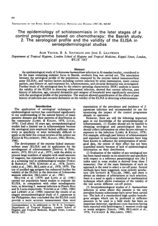
- 5. 464 EPIDEMIOLOGY OF SCHISTOSOMIASIS IN BASRAH. 2. Table 9-Serology and cetcarial dermatitis: geometric means (GM) of absorbance values of individuals with respect to current infection and cercarial dermatitis Category Number With dermatitis 1:; Without dermatitis Total 209 SNDt = 4.78, P<O*OOl For those infected with S. haematobium With dermatitis Without dermatitis :i Total SND = 1.61, P =3?l-11 For those not infected with S. haematobium With dermatitis ;; Without dermatitis Total 170 SND = 3.88, P<O*OOl Results of urine examination were not available from 4 individuals GM (95% CI)* 0.56 (O-SO-0.62) 0.39 (0-35-0-43) 0.46 (O-42-0.50) 0.68 (055-0.83) 0.49 (O-38-0.63) 0.59 (0~50-0~70) O-53 (0.47-0.60) 0.38 (0.34-0.43) 0.44 (0.40-0.48) *CI = confidence intervals j-SND = standardized normal deviate Table lO-Geome.tric means (GM) of absorbance values and the proportions of sempositives with respect to the category of the household significantly associatedwith the level of antibodies in the multiple regression analysis. Category NUdXr GM (95% Cl)* % seropositive The results of the ROC analysis The Figure showsthe ROC curves for the 3 groups 1 80 0.36 (0.31-0.42) 50.0 2 166 0.43 (0.39-0.47) 60.8 of individuals; closed circles represent the 0.4 cut-off Total 246 0.41 (0.38-044) 57.3 point, other cut-off points arelabelled. It can be seen that all 3 curves were above the chance line, i.e. the l CI = confidence intervals ELISA can discriminate between those with current Table 11-Significant results of multiple regression analysis using logre ELBA as the dependent variable and history of haematuria, treatment for schistosomiasis, age, sex, water contact activities, cercarial dermatitis, and the category of household as the independent variables Variable History of haematuria b* SE(b)* O-18 0.04 P* t P 0.33 4.8 <O*OOl Cercarial dermatitis 0.11 0.04 o-22 3.2 LO.01 Category of household -0.08 0.03 -0.15 -2.4 co.05 Current infection 0.11 0.04 0.16 2.5 <o-o5 Age 0.05 o-02 0.15 Constant -0.56 0.07 Multiple r = 0.52, SE(r) = 0.22, ?=0*27 *b=regression coefficient, SE = standard error, p = standardized b 2.2 <0*05 -8.1 <o-o01 category contributed positively to the antibody level relative to the first category. The standardized coef- ficient provides an estimate of the degree of the contribution of each independent variable to the explained variance. Sex, water contact activity, and history of treatment for schistosomiasis were not infection and those negative by urine examination; between thosewith current infection or apast history of haematuria, or both, and those who were negative by urine examination and did not report such history; and finally between those with current infection or a past history of either haematuria or cercarial dermati-
- 6. A. YACOUB t?Cal. 465 Table U-The area under the curve (Fig.) and its standard deviation for each category of individual tested by ELISA for S. hamurtobium infection Grout Standard Area deviation Individuals with current infection Individualswith current infection or pastexposure 0.70 00478 0.82 0.0302 Individualswith current infectionor pastexposureand cercarialdermatitis 0.88 0.0283 0.2 0.4 0.6 0.8 1 PROPORTION OF FALSE POSITIVES - Individuals with current lnfectlon - - - - lndlvldualr wlth current lnfectlon and past history of schlstosomlarls +e.*. Indlvlduals wlth current infectlon, past history and cercarlal dermatitis Fig. The applicationof the relativeoperatingcharacteristic (ROC) analysisto serologicaldata of Al-maadanpopulation. Theclosed circlesindicatetheselected cut-offpointusedin thestudy (W4).Opencirclesindicatetheothercut-offpointsinvestigated inthe study;valuesfrom abovedownwards followthe sameorderfor all three groupsof individuals. tis, or both, and those negative by urine examination who did not report a history of either condition. The heights of the 3 curves show that the ELISA has a better discriminating ability when those who gave a history of haematuria were considered positive in addition to thosewho were positive by urine examina- tion. Quantitatively, this observation is expressedby the values of the areaunder each of the 3 curves, as shown in Table 12. The Table alsoshowsestimatesof the standard deviations for the performanceof testsof signiiicance. There was a siani&ant difference be- t&en the areas under the &rves for the first and secondgroups (SND=2*12, O*Ol<P<O*OS).Howev- er, the diffecence between the second and the third groups was not significant (SND = 1.45, 0.10~ P<O*20). In summary, although the test discriminated be- tween those with current infection and those who were negative by urine examination (area=0*7), its discriminating ability wasfurther enhancedby giving consideration to the variable pasthistory of haematur- ia (area=0.82). Although the inclusion of those with cercarial dermatitis increased the area to O-88, this increase was not significant. Discussion In the present study S. mansoniegg antigen was used to screenfor S. haematobium. Since the latter is the only endemic human schistosome in southern Iraq, crossreactionsdue to other human schistosomes can be excluded. The sensitivity of the ELISA in detecting S. hematobium usink S. matsoni egg antigen was 83%. This firmre is comnarable to that rep&ted by MCLAREN e?al. (19783 using similar antigen to test Egyptian children and adults infected with S. haemutobium (83% and 76%). JANITSCHKE et al. (1981) reported a sensitivity of 86% when S. mansoni worm antigen was used to test for S. haematobium cases.ISMAIL(1980) tested sera from Egyptian casesof S. haemutobium and S. man.&. He showed that egg antigens of both parasites were equally reactive againstthe samesetof sera.Very few crossreactionswere reported from thesestudieswhen serafrom individuals with intestinal nematodeswere tested. Cross reactions were reported, however, by HILLYER & GOMEZ DE RIO (1979) with trichinosis. cysticercosisand fascioliasis. ‘k’hes; conditions could not affect our results since the first 2 do not exist in southern Iraq and human fascioliasis is very rare. The ROC analvsis revealed some interesting findings; it showed<thatthe ELISA was of adequati validity when used to discriminate between indi- viduals with respectto infection status asdetectedby urine examination. The discriminant ability of the test wasquantified by measuring the areaunder the ROC curve (O-7).This is a summary index of the perform- ance of the test across various cut-off points. The ROC curve also showed that the optimum cut-off point with respect to current infection was 0.4 (the point of the highest perpendicular distance from the chanceline). This wasin fact the cut-off point usedin this study to calculate the sensitivity iu;d specificity firmres. However. ROC analvsis allows flexibilitv in &e choiceof cut-hff points depending on the extent to which all positive casesneed to be identified and the implicatitins of such choicefor the health caresystem. ROC analysis can thus be considered as a powerful tool in seroepidemiologicalstudies of schistosomiasis. An attempt wasmadeto quantify the effect of apast history of infection and cercarial dermatitis, using ROC analysis, on the validity of the ELISA in detecting schlstosomiasis. By including those indi- viduals with a past history of infection as positive cases, the area under the curve was significantly increased to 0.82; when cercarial dermatitis was included asavariable, the increasein the discriminant ability of the test was not significant. The specificity of the ELISA in detecting current infection was 46%, calculated from those negative by parasitology in the Al-maadan population. This figure is low compared to those reported by LONG et al. (1981)and HILLYER etal. (1979)using the ELISA to detect S. man.& infection in St Lucia and Puerto Rico, who recorded specificity figures of 75% and 92%. MOTT & DIXON (1982)reported figures of 97%
- 7. 466 EPIDEMIOLOGY OF SCHISTOSOMIASISIN BASRAH. 2. and 90% using crude egg antigen to test European and Amazon Indian sera from persons who had never been to endemic areas. However, they reported a 47% specificity figure using the same antigen to test post-treatment sera from residents in S. mansoni areas. The variation in the reported specificity figures from different studies (including the present one) can be attributed to the following factors. (1) Variation in the choice of subjects to determine the specificity of the test. Higher figures are likely to be obtained when testing sera from individuals living in non-endemic areas. (2) The presence and nature of schistosomiasis control programmes in the endemic areas. Individuals who had had the infection and who had been treated were usually positive serologically even if they were parasitologically negative (see Table 8). Since a control programme based on selective chemotherapy has been in operation in Basrah for many years, a significant proportion of positive reactions could be attributed to the persistence of antibodies after successful cure. (3) The sensitivity of the parasitological test used as the reference test for calculating specificity. For example, YOGORE et al. (1983) showed that a single stool examination reduced the underesti- mate to 29%. This factor might have operated in the present study, and is discussed by YACOUB & SOUTHGATE (1987). (4) Variation in the frequency of cercarial dermatitis (whether caused by human or non-human schisto- somes), and of unisexual and unmated infections. Since serological data reflect the accumulated experience of a parasitic infection by individuals in a community, the value of such data can be enhanced if they are age related (DRAPER et al., 1972; LOBEL & KAGAN, 1978). Our study showed that age was one of the significant factors associated with the serological profile of schistosomiasis in the Al-maadan commun- ity (Table 11). However, it has also been shown that factors apart from current infection and age might be equally or more important when attempting to interpret the antibody profile of this community; past history of infection and cercarial dermatitis were significantly associated with the profile. In endemic areas of Basrah the causative organism of cercarial dermatitis (shara) is not known, and it may be caused by cercariae of S. haematobium. The only investiga- tion in these areas was made by WATSON & NAJIM (1956), who concluded that true cercarial dermatitis from non-human schistosomes was rare in southern Iraq and attributed the cases they examined to S. haematobium. The finding that category of household was a significant factor associated with the serological pro- file indicates that the distribution of antibodies is non-random and that there is a degree of aggregation in the 2 categories of households studied. The degree of clustering”or non-random distribution can also’ be investigated using such parameters as the distance of the household from the source of water or the distributian of infection at individual household level. Studies carried out by KLUMPP (1983), using para- sitoloeical data. and NOSENAS et al. (1975). using both -serological and parasitological data, ‘providi examples. Swimming was the only water contact activity associated with positive serology (Tables 5, 6, 7); however, none of tiiese activities contributed signi- ficantly to the antibody level when other factors were considered in the multiple regression analysis. This might be explained by the fact that serology measures the life-time experience of an individual, while infection detected by parasitology reflects variation in recent exposure through water contact. In conclusion, the results of this seroepidemiologic- al survey demonstrate the relative importance of variables, in addition to parasitological data, when attempting to interpret the serological profile of a community exposed to schistosomal infection. Acknowledgements We thank all staff at the Endemic DiseasesCentre. Basrah for their help during the field work. We are very grateful to all schoolchildren and inhabitants of Al-maadan locality, for without their cooperation this study would not have been possible. Thanks also go to Dr C. C. Draper for allowing us io process the blood samples at the -Seroepidemioibgy Laboratory of the London School of Hygiene and Tropical Medicine and for his advice on certain technical issues.The computer program for the ROC analysiswaskindly provided by the Institute of Psychiatry, London. Part of the material in this paper wasincluded in a thesis by A. Yacoub acceptedfor the award of aPhD degreeby the University of London. References De Savigny, D. & Voller, A. (1980).The communication of ELISA data from laboratory to clinician. Journal of Immunoassay, 1, 105-128. Dorfman. D. D. & Alf. E. (1969X Maximum-likelihood e&n&ion of parameters oi sign&detection theory and determination of confidence intervals*-*rating [sic] method data. Journal of Mathematical Psychology, 6, 487-496. Draper, C. C., Voller, A. & Carpenter, R. G. (1972). The epidemiologic interpretation of serologic datain malaria. ~4~47;~ Journal of Tropical Medicine and Hygiene, 21, El-Gindv. k. (1956). Monthlv orevalence rates of natural infei&on wiih S. ‘haematobiki cercariaein B. truncatus in central Iraq. Bulletin of Endemic Diseases, 7 (l/2), 1l-3 1. El-Gindv, M. & Radhawv, I. (1965). Viabilitv, incubation period and hatchability-of 3. trukatus eggsfrom central Iraq under outdoor conditions in different seasons. Bulletin of Endemic Diseases, 7 (3/4), 13-25. Engvall, E. & Perlmann, P. (1972).Enzyme linked immuno- sorbent assay (ELISA): III. Quantitation of specific antibodies by enzyme labelled anti-immunoglobulin in antigen coated plates. Journal of Immunology, 109, 129-135. Erdreich, L. & Lee, E. T. (1981). Use of relative operating characteristic analysisin epidemiology. Ammican3mrd of Epidemiology, 114, 649-662. Farag, H. F. & Barakat, R. M. R. (1978). The enzyme linked immunoassay in the diagnosis of bilharziasis. Tropenmedizin und Parasitologic, 29, 12-14. Hillyer, G. V. & Gomez de Rio, I. (1979). The enzyme linked immunosorbent assay(ELISA) for the immuno- diagnosisof schistosomiasis.AmericanJournal of Tropical Medicine and Hygiene, 28, 237-241. Hillyer, G. V., Ruiz-Tiben, E., Knight, W. B., Gomez de Rio, I. & Pelley, R. P. (1979). Immunodiagnosis of infection with S. mansoni: comparison of ELISA, radioimmunoassay and precipitation tests performed with antigens from eggs. American Journal of Tropical Medicine and Hygiene, 28, 661-669. Houba, V. (1980).Schistosomiasis.In: Immunological Znves- tigation of Tropical Parasitic Diseases, Houba, V. (editor).
- 8. A. YACOUB et al. 467 Edinburgh, London and New York: Churchill Living- of the Royal Societyof Tropical Medicine and Hygiene, 73, stone, pp. 130-147. 636-639. Huldt, G., Lagerquist, B., Phillips, T., Draper, C. C. & Voller, A. (1975). Detection of antibodies in schistoso- miasis by enzymelinked immunosorbent assay(ELISA). Annals of Tropical Medicine and Parasitology, 69, 483- 488. Ismail. M. M. (1980).Observations on someseroloeical testsin Schistosoma haematobium infections in n&t and ex- perimental animals. Ph.D. thesis, University of London, pp. 105122. McLaren, M. L., Lillywhite, J. E., Dunne, D. W. & Doenhoff, M. J. (1981). Serodiagnosisof human Schisto- soma mansoni infections: enhanced sensitivity and spe- cificity in ELISA using a fraction containing S. mansoni antigens w, and LX,. Transactions of the Royal Societyof Tropical Medicine and Hygiene, 75, 72-79. Mott, K. E. & Dixon, H. (1982). Collaborative study on antigens for immunodiagnosis of schistosomiasis.Bulle- tin of the World Health Organization, 60, 729-753. Nosenas, J. S., Matsuda, H., Blas, B. L., Tanaka, H. & Santos, A. T., Jr (1975). Evaluation of the circumoval precipitation test using dried blood on filter paper as a diagnostic tool in epidemiological survey for schistoso- miasis. Japanese 3oumal of Experimental Medicine, 45, 367-375. Janitschke, K., El-Kalouby, A. H., Braun-Munzinger, R. A., El-Baz, H. & Mahmoud, M. (1981). Evaluation of the ELISA test as an epidemiological tool in schistoso- miasis. Journal of Tropical Medicine and Hygiene, 84, 147-154. Kagan, I. G. (1982). Serodiagnosisof schistosomiasis.In: Immunoparasitologv: Principles and Methods in Malaria and Schistosomiasis Research. Strickland, G. T. & Hun- fi’~ri-l,&, Jr (editors). USA: Praeger Publications, pp. Kagan, I. G. & Pellegrino, J. (1961). A critical review of immunological methods for the diagnosisof bilharziasis. Bulletin of the World Health Organization, 25, 61l-674. Klumpp, R. K. (1983). A study of the transmission of Schistosomahaematobium in Volta Lake, Ghana. Ph.D. thesis, University of London, pp. 238-247and 302-306. Lewert, R. M., Yogore, M. G., Jr & Bias, B. L. (1984). Seroepidemiology of schistosomiasisjaponica by ELISA in the Philinuines. II. Unreliabilitv of stool examination in the mea’surementof incidence: American Journal of Tropical Medicine and Hygiene, 33, 872-881. Lobe& H. 0. & Kagan, I. G. (1978). Seroepidemiology of parasitic diseases.Annual Reviews of Microbiology, 32, 329-347. Long, E. G., McLaren, M., Goddard, M. J., Bartholomew, R. K., Peters, P. & Goodgame, R. (1981). Comparison of ELISA, radioimmunoassayand stool examination for Schistosoma mansoni infection. Transactions of the Royal Society of Tropical Medicine and Hygiene, 75, 365-371. Lucas, A. 0. (1976). Surveillance of communicable diseases in tropical Africa. International Journal of Epidemiology, 5, 39-43. McLaren, M., Draper, C. C., Roberts, J. M., Minter- Goedbloed, E., Ligthart, G. S., Teesdale, G. H., Amin, M. A., Omer, A. H. S., Bartlett, A. & Voller, A. (1978). Studies on the enzyme linked immunosorbent assay (ELISA) test for Schistosoma mansoni infections. Annals of Tropical Medicine and Parasitology, 72, 243-253. McLaren, M., Long, E. G., Goodgame, R. W. & Lilly- white, J. E. (1979). Application of the enzyme linked immunosorbent assay(ELISA) for the serodiagnosisof Schistosoma mansoniinfections in St Lucia. Transactions Peters, P. A., Warren, K. S. & Mahmoud, A. A. F. (1976). Rapid accurate quantification of schistosome eggs via Nuclepore filters. Journal of Parasitology, 62, 154-155. Schinski, V. D., Clutter, W. C. & Murrell, K. D. (1976). Enzyme and ‘*sI-labelled anti-immunoglobulin assaysin the immunodiagnosis of schistosomiasis.American Jour- nal of Tropical Medicine and Hygiene, 25, 824-831. Taticheff, S. StMelamed, M. (1983).Evaluation of ELISA - with S. mansoni egg antigen - in the serodiagnosisof schistosomiasis.Ethiopian Medical 3ourna1, 21, 27-33. Watson, J. M. & Najim, A. T. (1956). Studies on bilharziasis in Iraq. Part 12.Observationson schistosome dermatitis. Journal of the Iraq Medical Professions, 4, 4-10. Yacoub, A. (1985). The epidemiology of Schistosomahaema- tobium infection in Basrah. southern Iraa: seroloeical. parasitological and behavioural studies. Ph.D. thesis; University of London. Yacoub, A. & Southgate,B. A. (1987).The euidemiologv of schistosomiasisin the later stagesof acontrol programme basedon chemotheraov: the Basrahstudv. 1.Descriotive epidemiology and pa%itological results. Transact& of the Royal Societyof Tropical Medicine and Hygiene, 81, 449~A<9 . ._ ,_.. Yogore, M. G., Jr, Lewert, R. M. & Blas, B. L. (1983). Seroepidemiology of schistosomiasisjaponica by ELISA in the Philippines. 1. Underestimation by stool examina- tion of the prevalence of infection in schoolchildren. American Journal of Tropical Medicine and Hygiene, 32, 1322-1334. Yogore, M. G.,, Jr, Lewert! R. M. & Bias, B. L. (1984). Seroepidermologyof schistosomiasisjaponica by ELISA in the Philippines. III. Selectivemasschemotherapy with praziquantel in a control programme. American Journal of Tropical Medicine and Hygiene, 33, 882-890. Accepted for publication 9 June 1986