
Recommended
More Related Content
What's hot
What's hot (20)
Similar to Tex Rad.Pps
Similar to Tex Rad.Pps (20)
Recently uploaded
Recently uploaded (20)
Tex Rad.Pps
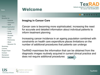
- 1. Home Welcome Imaging in Cancer Care Cancer care is becoming more sophisticated, increasing the need for accurate and detailed information about individual patients to inform treatment planning Increasing cancer incidence in an ageing population combined with constraints on health care expenditure places limitations on the number of additional procedures that patients can undergo TexRAD maximises the information that can be obtained from the diagnostic images routinely acquired in current clinical practice and does not require additional procedures HOW? 1 2 3 4 5 6 6 7 8
- 2. What is TexRAD? Measuring Tumour Complexity: TexRAD is a software application that analyses the textures in existing radiological scans to assist the clinician in assessing the prognosis of the cancer patient. This presentation will guide you through how TexRAD will improve patient care. Please use links on left hand side to go directly to particular sections. What is TexRAD? Screen shot of TexRAD software 1 2 3 4 5 6 6 7 8
- 3. Benefits Benefits Patients | Clinicians | OEM
- 7. Clinical Case Studies Clinical Application Colorectal cancer (Liver / Distant Metastatic Disease) Case Study | Clinical Demo | Evidence Lung cancer Case Study | Clinical Demo | Evidence Breast cancer Case Study | Clinical Demo | Evidence Prostate cancer Case Study | Clinical Demo | Evidence Renal cancer Case Study | Clinical Demo | Evidence Pulmonary Disorders | Schizophrenia | Dynamic Texture Analysis
- 8. Colorectal Homepage Colorectal Cancer (Liver / Distant Metastatic Disease) Case Study | Demo | Evidence PACS workstation
- 9. Colorectal Case Study Colorectal Case Study Typical example: A patient visits a clinic after curative colorectal cancer surgery Undergoes a routine follow-up CT scan The Radiologist considers that the CT looks normal with no focal abnormalities However, 18 months later the patient relapses with focal metastatic disease of the liver – fatal consequence TexRAD could have assisted the radiologist to improve this scenario as part of routine clinical procedure 1 2 3 4 5 6 HOW? 6 7 8 Case Study | Demo | Evidence
- 10. Colorectal CS 2 Colorectal Case Study How TexRAD supports clinician & patient in case study From the routine CT scans taken in the clinic, TexRAD software uniquely extracts and measures fine , medium and coarse textures - in this example, from the Liver CT. TexRAD highlights texture anomalies which are not apparent to normal visual examination These texture anomalies can be used to predict the risk of metastatic disease From this additional information, radiologist may suggest alternative treatment pathways to the patient 1 2 3 4 5 6 6 7 8 Case Study | Demo | Evidence
- 11. Colorectal Demo 1 Colorectal - Demo 1 2 3 4 5 6 6 7 TexRAD screen shot of Liver Case Study | Demo | Evidence PACS workstation 8
- 12. Colorectal Demo 2 Screenshot - Liver TexRAD analysis of apparently normal appearing liver (after curative surgery of primary tumour) as seen on follow-up CT of a patient with colorectal cancer could predict the risk of metastatic disease Colorectal - Demo 1 2 3 4 5 6 6 7 Case Study | Demo | Evidence 8
- 13. Colorectal Demo 3 Colorectal - Demo 1 2 3 4 5 6 6 7 Screenshot - Work flow demonstration sequence for Liver Case Study | Demo | Evidence 8 Clinician’s Workflow
- 14. Colorectal Demo 4 STAGE 1 - Display the target clinical image of interest A TexRAD analysis is applied to the appropriate 2D CT image highlighting the liver (tissue of interest - TOI). The specialist clinical consultant (e.g. Radiologist) will select the image containing this TOI. Colorectal - Demo 1 2 3 4 5 6 6 7 Case Study | Demo | Evidence 8 Conventional Abdominal CT Image
- 15. Colorectal Demo 5 STAGE 2 – Draw region of interest (ROI) to be analysed Using TexRAD’s graphical user interface tools, image window level/width can be altered to clearly delineate this TOI, interactive magnification/panning/centring can be used for better visualization of this TOI. Clinician can choose an appropriate ROI tool (e.g. Polygon ROI) from a list of options based on the application. This ROI is super-imposed on the TOI within the original image. Colorectal - Demo 1 2 3 4 5 6 6 7 Case Study | Demo | Evidence 8
- 16. Colorectal Demo 6 STAGE 3 – Texture Analysis TexRAD employs a novel algorithm (patent applied for) primarily to extract subtle but prognostic metrics currently not available in clinic. The software also graphically displays clinically relevant fine, medium and coarse liver textures separately (below) in addition to their fusion with the original CT image. Colorectal - Demo 1 2 3 4 5 6 6 7 Case Study | Demo | Evidence 8
- 17. Colorectal Demo 7 Colorectal - Demo 1 2 3 4 5 6 6 7 Case Study | Demo | Evidence STAGE 4 – TexRAD Spectroscopy Summarises the entire texture results graphically for the colorectal cancer patient. Clinician can easily interpret the results for a quick assessment. 8
- 18. Colorectal Demo 8 STAGE 5 – Risk Stratification Report A risk stratification report specific to the colorectal cancer is generated, which should be used only to assist the clinician to make an accurate decision. The report contains patient ID and scan details, TexRAD analysis result, explanation and contact information. Colorectal - Demo 1 2 3 4 5 6 6 Case Study | Demo | Evidence 8 7 SAMPLE REPORT ONLY FOR ILLUSTRATION
- 19. Colorectal Background 1 Colorectal Background Facts about Colorectal Cancer Colorectal cancer is the second most common malignancy in Western societies [1]. 40% of the patients undergoing resection of the primary tumour will relapse and die of their disease, making colorectal cancer the second leading cause of death related to cancer [2]. The liver is the sole site of secondary tumour spread (i.e. metastasis) in 20-40% of patients [3] and therefore it is a common practice to follow-up patients after their curative resection [4]. There is an overall survival benefit for intensifying the follow-up of such patients with imaging of the liver being associated with reduced mortality (Odds ratio = 0.66, 95% confidence limits 0.46-0.95) [5]. 1 2 3 4 5 6 6 7 8 Case Study | Demo | Evidence
- 20. Colorectal Background 2 Colorectal Background Facts about Colorectal Cancer The American Society of Clinical Oncology (ASCO) now recommends annual CT of the chest and abdomen for 3 years after primary therapy for patients at higher risk of recurrence [6]. However, the risk of recurrence is not uniform for these patients and identification of predictive factors that are linked to outcomes may allow modification of surveillance strategies for particular sub-groups and provide effective and optimum patient care. ASCO has also highlighted the need for research in this area [6]. 1 2 3 4 5 6 6 7 8 Case Study | Demo | Evidence
- 21. Colorectal Background 3 Colorectal Background Comparison of Risk Stratification techniques Additional physiological imaging techniques such as Doppler ultrasound and quantitative analysis of liver contrast enhancement or perfusion on CT also have the potential to identify patients at higher risk of recurrence [7, 8] These techniques may reflect alterations of liver hemodynamics associated with occult metastases [9, 10]. However, the additional image acquisitions required by these techniques have been a barrier to their adoption into surveillance programs. (cost/additional radiation dosage) 1 2 3 4 5 6 6 Case Study | Demo | Evidence 7 8
- 22. Colorectal Evidence 1 Colorectal Evidence Why TexRAD is better - evidence TexRAD applied to routinely acquired CT images highlights subtle liver changes that may occur in association with alterations in liver physiology. Furthermore computer simulations and clinical studies have suggested that measurements of liver texture on CT may reflect liver vascularity [11-13]. The ability of Liver TexRAD to detect occult tumour on CT is further supported by studies demonstrating textural differences between normal livers and apparently normal areas of tissue within livers bearing tumours, areas also known to exhibit alterations in blood flow [13-15]. 1 2 3 4 5 6 6 Case Study | Demo | Evidence 7 8
- 23. Colorectal Evidence 2 Colorectal Evidence Why TexRAD is better - evidence Preliminary results of Liver TexRAD demonstrate the potential for liver texture on contrast-enhanced CT to provide a novel parameter that is not only insensitive to variations in acquisition parameters but can also act as a marker of survival for patients following resection of colorectal cancer [16]. 1 2 3 4 5 6 6 Case Study | Demo | Evidence 7 8
- 27. Lung Homepage Lung Cancer Case Study | Demo | Evidence PACS workstation TexRAD screen shot of Lung lesion
- 28. Lung Case Study Lung Case Study Typical example: A patient diagnosed with lung cancer requires an accurate staging of the tumour and lymph node disease. Radiologists are unable to obtain a confident risk stratification from CT alone, which is critical for early prognosis and favourable patient outcome. The patient undergoes FDG PET-CT imaging, which overcomes the limitations of CT alone. However, this is an additional and expensive procedure. TexRAD can improve the performance of CT and potentially be employed for selection of patients for PET. 1 2 3 4 5 6 6 7 8 HOW? Case Study | Demo | Evidence
- 29. Lung CS 2 Lung Case Study How TexRAD supports clinician & patient in case study From routine CT scans taken in the clinic TexRAD software uniquely extracts and measures fine , medium and coarse textures - in this example, from the Lung lesion on CT - and classifies degree of adverse tumour biology These texture gradation can be used to predict tumour stage and the risk of metastatic and lymph node disease Based on this additional information, radiologist may optimally select patients who will benefit from FDG PET 1 2 3 4 5 6 6 7 8 Case Study | Demo | Evidence
- 30. Lung Demo 1 Lung TexRAD Demo 1 2 3 4 5 6 6 7 Case Study | Demo | Evidence PACS workstation TexRAD screen shot of Lung lesion 8
- 31. Lung Demo 2 Lung TexRAD analyses a focal cancerous lesion as seen on the conventional CT image of a patient with NSCLC could predict tumour stage, metabolism and lymph node disease involvement Lung - Demo 1 2 3 4 5 6 6 7 Case Study | Demo | Evidence 8
- 32. Lung Demo 3 Lung - Demo 1 2 3 4 5 6 6 7 Case Study | Demo | Evidence 8 Clinician’s Workflow
- 33. Lung Demo 4 STAGE 1 - Display the target clinical image of interest A TexRAD analysis is applied to the appropriate 2D CT image highlighting the lung lesion (tissue of interest - TOI). The specialist clinical consultant (e.g. Radiologist) will select the image containing this TOI. Lung - Demo 1 2 3 4 5 6 6 7 Case Study | Demo | Evidence 8 Conventional Lung CT Image
- 34. Lung Demo 5 STAGE 2 – Draw region of interest (ROI) to be analysed Using TexRAD’s graphical user interface tools, image window level/width can be altered to clearly delineate this TOI, interactive magnification/panning/centring can be used for better visualisation of this TOI. Clinician can choose an appropriate ROI tool (e.g. Elliptical ROI, which encloses the TOI in an automated fashion) from a list of options based on the application. This ROI is super-imposed on the TOI within the original image. Lung - Demo 1 2 3 4 5 6 6 7 Case Study | Demo | Evidence 8 Magnified CT Image For Drawing ROI
- 35. Lung Demo 6 Lung - Demo STAGE 3 – Texture Analysis TexRAD employs a novel algorithm (patent applied for) primarily to extracts subtle but prognostic metrics currently not available in clinic. The software also graphically displays clinically relevant fine, medium and coarse lung lesion textures separately (below) in addition to their fusion with the original CT image. 1 2 3 4 5 6 6 7 Case Study | Demo | Evidence 8 Fine Lung Lesion Texture Medium Lung Lesion Texture Coarse Lung Lesion Texture
- 36. Lung Demo 7 Lung - Demo STAGE 4 – TexRAD Spectroscopy Summarises the entire texture results graphically for the lung cancer patient. Clinician can easily interpret the results for a quick assessment. 1 2 3 4 5 6 6 7 Case Study | Demo | Evidence 8
- 37. Lung Demo 8 Lung - Demo STAGE 5 – Risk Stratification Report A risk stratification report specific to the lung cancer is generated, which should be used only to assist the clinician to make an accurate decision. The report contains patient ID and scan details, TexRAD analysis result, explanation and contact information. 1 2 3 4 5 6 6 Case Study | Demo | Evidence 8 7 SAMPLE REPORT ONLY FOR ILLUSTRATION
- 38. Lung Background Lung - Background Facts about lung Cancer Lung cancer is the most common form of death related to cancer in men and second most common in woman [1, 2], accounting for 1.3 million deaths worldwide annually [3]. Non-small cell lung carcinoma (NSCLC) is the most common form of lung cancer prevalent in 80% of all cases [4]. Following initial diagnosis, patients with NSCLC undergo staging. The most common imaging staging procedure has been CT. However, due to low accuracy for CT staging, clinical guidelines now recommend Fluoro-deoxy-glucose (FDG) PET-CT unless the initial CT imaging shows evidence of inoperable disease. An improvement in the accuracy of CT could improve the selection of patients for FDG-PET. Case Study | Demo | Evidence 5 6 6 7 8 1 2 3 4
- 39. Lung Evidence 1 Lung Evidence Clinical Evidence – ICIS 2008 [5] 75 patients, PET identified tumour and nodal stages I, II, III & IV PET glucose uptake (SUV) was also measured Non-contrast CT images were employed for texture analysis Case Study | Demo | Evidence 5 6 6 7 8 1 2 3 4
- 40. Lung Evidence 2 Lung Evidence Case Study | Demo | Evidence Texture-Tumour Stage Association MGI (fine) vs Stage: rs =0.77, p=0.0002 Entropy (fine) vs Stage: rs =0.51, p=0.03 SUV vs Stage: rs =0.50, p=0.04 Texture-Nodal Stage Prediction Uniformity (fine) predicted node positive disease - area under the ROC curve = 0.7, p < 0.005, sensitivity=65%, specificity=76% In comparison with CT alone, sensitivity=43%, specificity=85% Combining CT and Texture, sensitivity=87%. Specificity=67% Texture-Metabolic Association Entropy (coarse) vs SUV: r=0.51, p=0.03 Uniformity (coarse) vs SUV: r=-0.52, p=0.03 5 6 6 7 8 1 2 3 4
- 41. Lung Evidence 3 Lung Evidence Clinical Evidence - Recent Findings accepted in ECR’10 [6] Case Study | Demo | Evidence Kaplan-Meier survival curves for NSCLC patients with lung lesions separated by (A) Texture analysis on CT and (B) PET glucose uptake (SUV). Survival curves were significantly different for Texture (p<0.001)]. 5 6 6 7 8 1 2 3 4
- 42. Breast Homepage Breast Cancer Case Study | Demo | Evidence TexRAD screen shot of Breast lesion
- 43. Breast Case Study Breast Case Study Typical example: A female participant within a breast screening program undergoes an initial mammogram examination. Further diagnostic mammogram confirms presence of cancerous tissue. Core biopsy identified non-invasive ductal carcinoma in situ (DCIS) and the standard breast conservation excision was performed. Surprisingly, the final excised specimen provided evidence of invasive focus, routinely underestimated by core biopsy, resulting in additional surgical procedure involving the axilla TexRAD could have predicted the risk of invasive disease preoperatively from mammographic lesions assisting in treatment planning and optimal selection of biopsy or surgery 1 2 3 4 5 6 6 7 8 HOW? Case Study | Demo | Evidence
- 44. Breast CS 2 Breast Case Study How TexRAD supports clinician & patient in case study From routine mammographic images taken in the clinic TexRAD software uniquely extracts and measures fine , medium and coarse textures - in this example, from the breast lesion - and characterises differences in calcification architecture These texture differences can be used to preoperatively estimate the likelihood of invasive focus from among patients with DCIS This additional information may assist the clinician in better treatment planning and optimal selection of sentinel node biopsy or axillary surgery 1 2 3 4 5 6 6 7 8 Case Study | Demo | Evidence
- 45. Breast Demo 1 Breast - Demo 1 2 3 4 5 6 6 7 Case Study | Demo | Evidence TexRAD screen shot of Breast lesion 8
- 46. Breast Demo 2 Breast - Demo Breast TexRAD analysis of an obvious stellate mass as seen on a mammogram of a patient with core-biopsy proven breast cancer could estimate the risk of invasive disease 1 2 3 4 5 6 6 7 Case Study | Demo | Evidence 8
- 47. Breast Demo 3 Breast - Demo 1 2 3 4 5 6 6 7 Case Study | Demo | Evidence 8 Clinician’s Workflow
- 48. Breast Demo 4 STAGE 1 - Display the target clinical image of interest A TexRAD analysis is applied to the appropriate 2D mammographic image highlighting the breast lesion (tissue of interest - TOI). The specialist clinical consultant (e.g. Radiologist) will select the image containing this TOI. Breast - Demo 1 2 3 4 5 6 6 7 Case Study | Demo | Evidence 8
- 49. Breast Demo 5 STAGE 2 – Draw region of interest (ROI) to be analysed Using TexRAD’s graphical user interface tools, image window level/width can be altered to clearly delineate this TOI, interactive magnification/panning/centring can be used for better visualisation of this TOI. Clinician can choose an appropriate ROI tool (e.g. Polygon ROI) from a list of options based on the application. This ROI is super-imposed on the TOI within the original image. Breast - Demo 1 2 3 4 5 6 6 7 Case Study | Demo | Evidence 8
- 50. Breast Demo 6 Breast - Demo STAGE 3 – Texture Analysis TexRAD employs a novel algorithm (patent applied for) primarily to extracts subtle but prognostic metrics currently not available in clinic. The software also graphically displays clinically relevant fine, medium and coarse breast lesion textures separately (below) in addition to their fusion with the original mammographic image. 1 2 3 4 5 6 6 7 Case Study | Demo | Evidence 8
- 51. Breast Demo 7 STAGE 4 – TexRAD Spectroscopy Summarises the entire texture results graphically for the breast cancer patient. Clinician can easily interpret the results for a quick assessment. Breast - Demo 1 2 3 4 5 6 6 7 Case Study | Demo | Evidence 8
- 52. Breast Demo 8 STAGE 5 – Risk Stratification Report A risk stratification report specific to the breast cancer is generated, which should be used only to assist the clinician to make an accurate decision. The report contains patient ID and scan details, TexRAD analysis result, explanation and contact information. Breast - Demo SAMPLE REPORT ONLY FOR ILLUSTRATION 1 2 3 4 5 6 6 Case Study | Demo | Evidence 8 7
- 53. Breast Background 1 Breast - Background Facts about Breast Cancer Invasive cancer of the breast and DCIS can both present as focal lesions on mammography. Pure DCIS is not an invasive process and rarely metastasises to regional lymph nodes [1]. The introduction of mammographic breast screening has resulted in a dramatic increase in the diagnosis of DCIS and its detection rate has reached 15-20% of all mammographically detected cancers [2-4]. Based on core biopsy specimens, the standard surgical planning for DCIS is to offer breast conservation excision. 1 2 3 7 8 Case Study | Demo | Evidence 4 5 6
- 54. Breast Background 2 Breast - Background Facts about Breast Cancer However, the detection of DCIS on core biopsy is quite frequently followed by evidence of invasion within the final excision specimen. This occurs in 11-44% of patients and results in the need of second operative procedure involving the axilla [4, 5]. Therefore the main concern is whether any kind of axillary staging is indicated in patients preoperatively diagnosed with DCIS only. 1 2 3 7 8 Case Study | Demo | Evidence 4 5 6
- 55. Breast Background 3 Breast - Background Facts about Breast Cancer The most often employed axillary treatment option for patients with DCIS is to perform a sentinel node biopsy at the time of initial excision. Nevertheless it may lead to reoperation in cases of proven invasive focus on excision. Therefore an effective way of estimating the likelihood of an invasive focus preoperatively in patients diagnosed with DCIS would assist in better treatment planning and optimal use of sentinel node biopsy or axillary surgery. 1 2 3 7 8 Case Study | Demo | Evidence 4 5 6
- 56. Breast Evidence 1 Breast Evidence Clinical Evidence – ICIS 2007 [6] 1 2 7 8 Case Study | Demo | Evidence Graph indicates the relationship between relative fine to medium uniformity value and extent of disease (tumour type) from mammographic focal region of interest 3 4 5 6
- 57. Breast Evidence 2 Breast Evidence Clinical Evidence – ICIS 2007 [6] 1 2 7 8 Case Study | Demo | Evidence Graph indicates the inverse relationship between relative fine to coarse mean grey-level intensity value and oestrogen receptor (ER) status from mammographic focal region of interest 3 4 5 6
- 58. Breast Evidence 3 Breast Evidence Clinical Evidence – ICIS 2007 [6] 1 2 7 8 Case Study | Demo | Evidence Graph indicates the inverse relationship between relative medium to coarse mean grey-level intensity value and progesterone receptor (PR) status from mammographic focal region of interest 3 4 5 6
- 59. Prostate Demo 1 Prostate Demo Case Study | Demo | Evidence Screenshot of the Prostate TexRAD highlighting the conventional CT image of the prostate (top-left), followed by the derived texture maps superimposed on the conventional CT image – fine (red), medium (green) and coarse (blue) texture 5 6 6 7 8 1 2 3 4
- 60. Prostate Evidence 1 Prostate Early Evidence Clinical Evidence – Submitted an abstract UKRC 2010 [1] Case Study | Demo | Evidence A medium texture (mean grey-level intensity) below 2.4 predicted the presence of tumour with an area under the ROC curve of 0.909, p = 0.0001, sensitivity = 84% and specificity = 84% Texture was significantly different between tumour & normal prostate tissue 5 6 6 7 8 1 2 3 4
- 61. Prostate Evidence 2 Prostate Early Evidence Clinical Evidence – Submitted an abstract UKRC 2010 [1] Case Study | Demo | Evidence Medium mean grey-level intensity correlates inversely with Prognostic risk factors (determined on the basis of gleason score, PSA level and clinical T stage) Texture correlates with prognostic risk factor (adverse tumour biology and/or patient outcome) 5 6 6 7 8 1 2 3 4
- 62. Renal Demo 1 Renal Metastases Demo TexRAD: Potential predictive imaging biomarker of response to treatment in metastatic renal cancer TexRAD PRE-TREATMENT Case Study | Demo | Evidence Screenshot of the Renal TexRAD highlighting ‘Right lobe liver’ metastases (pre-treatment) on the conventional CT image (top-left), followed by the derived texture maps superimposed on the conventional CT image – fine (red), medium (green) and coarse (blue) texture 5 6 6 7 8 1 2 3 4
- 63. Renal Demo 2 Renal Metastases Demo TexRAD: Potential predictive imaging biomarker of response to treatment in metastatic renal cancer TexRAD POST-TREATMENT Case Study | Demo | Evidence Screenshot of the Renal TexRAD highlighting ‘Right lobe liver’ metastases (post-treatment) on the conventional CT image (top-left), followed by the derived texture maps superimposed on the conventional CT image – fine (red), medium (green) and coarse (blue) texture 5 6 6 7 8 1 2 3 4
- 64. Renal Evidence 1 Renal Early Evidence Clinical Evidence – Recent findings a ccepted in ECR 2010 [1] Case Study | Demo | Evidence CT Texture significantly predicts patients with poor prognosis (treatment response) 5 6 6 7 8 1 2 3 4 p < 0.005
- 65. Renal Evidence 2 Renal Early Evidence Clinical Evidence – Recent findings a ccepted in ECR 2010 [1] Case Study | Demo | Evidence Existing anatomical imaging response criteria (RECIST) did not significantly predict patients with poor prognosis 5 6 6 7 8 1 2 3 4
- 66. Pulmonary Disorders 1 Pulmonary Disorders Clinical Evidence – Investigative Radiology 2008 [1] 5 6 6 7 8 3D volume rendered coronal, left and right sagittal views of (A) airway lung (fine) texture, (B) vascular lung (coarse) texture, and (C) the fusion of airway and vascular lung texture (intensity- and gradient-based) in a patient with no pulmonary disorders. 1 2 3 4
- 67. Pulmonary Disorders 2 Pulmonary Disorders Clinical Evidence – Investigative Radiology 2008 [1] 5 6 6 7 8 3D volume rendered coronal, left and right sagittal views of (A) airway lung (fine) texture, (B) vascular lung (coarse) texture, and (C) the fusion of airway and vascular lung texture (intensity- and gradient-based) in a patient with only pulmonary embolism. 1 2 3 4
- 68. Pulmonary Disorders 3 Pulmonary Disorders Clinical Evidence – Investigative Radiology 2008 [1] 5 6 6 7 8 3D volume rendered coronal, left and right sagittal views of (A) airway lung (fine) texture, (B) vascular lung (coarse) texture, and (C) the fusion of airway and vascular lung texture (intensity- and gradient-based) in a patient with pulmonary embolism and emphysema. 1 2 3 4
- 69. Pulmonary Disorders 4 Pulmonary Disorders Clinical Evidence – Investigative Radiology 2008 [1] The best distinction between emphysematous and non-emphysematous lung was observed for filtered fine airway texture quantified as MGI The best distinction between normal and patients with pulmonary disorders was observed for filtered coarse vascular texture quantified as entropy and uniformity A progressive percentage change from normal was observed for coarse vascular texture within PE and emphysematous lung Airway and vascular texture metrics demonstrated postural gradient consistent with known physiology 5 6 6 7 8 1 2 3 4
- 70. Brain Disorder 1 Brain Disorder-Schizophrenia Preliminary Clinical Evidence [1] 1 2 3 4 5 6 6 7 8 3D volume rendered left sagittal view of conventional MR whole brain GM (top row) and WM (bottom row) and corresponding textures (intensity- and gradient-based) respectively in a patient with schizophrenia. RAW GREY MATTER FINE GM TEXTURE MEDIUM GM TEXTURE COARSE GM TEXTURE RAW WHITE MATTER FINE WM TEXTURE MEDIUM WM TEXTURE COARSE WM TEXTURE
- 71. Brain Disorder 2 Brain Disorder-Schizophrenia Preliminary Clinical Evidence [1] 1 2 3 4 5 6 6 7 8 FILTERED MEDIUM-TO-COARSE TEXTURE RATIO – GM
- 72. Ongoing Research 1 Ongoing Research Ongoing Research includes: Oesophageal carcinoma (PET-CT) Lymphoma (PET-CT) Dynamic Texture Analysis 5 6 7 8 1 2 3 4
- 73. Ongoing Research 2 Dynamic Texture Analysis Dynamic CE-CT: Dukes B Colorectal Cancer 6 6 7 8 1 2 3 4 Arterial Perfusion = 0.7 ml/min/ml Portal Perfusion = 0.45 ml/min/ml HPI = 60.96 ml/min/ml 5 0s 3s 6s 9s 12s 15s 18s 21s 24s 27s 30s 33s 36s Perfusion
- 74. Ongoing Research 3 Dynamic Texture Analysis Arterial Perfusion = 0.2 ml/min/ml Portal Perfusion = 0.06 ml/min/ml HPI = 75.76 ml/min/ml Dynamic Fine CE-CT Texture: Dukes B Colorectal Cancer [1] 6 6 7 8 1 5 2 3 4 0s 3s 6s 9s 12s 15s 18s 21s 24s 27s 30s 33s 36s Perfusion
- 75. Ongoing Research 4 Dynamic Texture Analysis Dynamic Medium CE-CT Texture: Dukes B Colorectal Cancer [1] 0s 3s 6s 9s 12s 15s 18s 21s 24s 27s 30s 33s 36s Perfusion Arterial Perfusion = 0.05 ml/min/ml Portal Perfusion = 0.03 ml/min/ml HPI = 59.11 ml/min/ml 6 6 7 8 1 2 3 4 5
- 76. Ongoing Research 5 Dynamic Texture Analysis Dynamic Coarse CE-CT Texture: Dukes B Colorectal Cancer [1] 0s 3s 6s 9s 12s 15s 18s 21s 24s 27s 30s 33s 36s Perfusion Arterial Perfusion = 0.61 ml/min/ml Portal Perfusion = 0.13 ml/min/ml HPI = 82.42 ml/min/ml 6 6 7 8 1 2 3 4 5 0s 3s 6s 9s 12s 15s 18s 21s 24s 27s 30s 33s 36s Perfusion
- 79. Project Team Principal Developer Dr. Balaji Ganeshan Clinical Collaborators Prof. Kenneth Miles Dr. Olga Strukowska Prof. Hugo Critchley Image Processing Collaborators Dr. Rupert Young Prof. Chris Chatwin Business Development Manager Mike Herd Mike Wylde Intellectual Property & Technology Transfer Office Russell Nicholls Medical Students & F2 Doctors Sandra Abaleke Elleny Panayiotou Ian Pressney Kate Burnand Mansi Rajpopat People
- 80. Contact Dr Balaji Ganeshan TexRAD Project Manager Research and Enterprise University of Sussex Falmer, Brighton UK BN1 9SB Tel: +44 (0) 7727228107 Email: TexRAD@sussex.ac.uk Contact
- 81. Disclaimer Notice TexRAD is meant to be used as an assistive risk stratification tool in clinical practice and should not form the basis for clinical decision making. The results obtained from TexRAD system should only be interpreted by a health care professional. IMPORTANT: TexRAD is being further evaluated and not yet available for routine clinical practice. Disclaimer
- 82. References References Supporting evidence and academic references Colorectal | Lung | Breast | Prostate | Renal | Lung | Brain | DTA