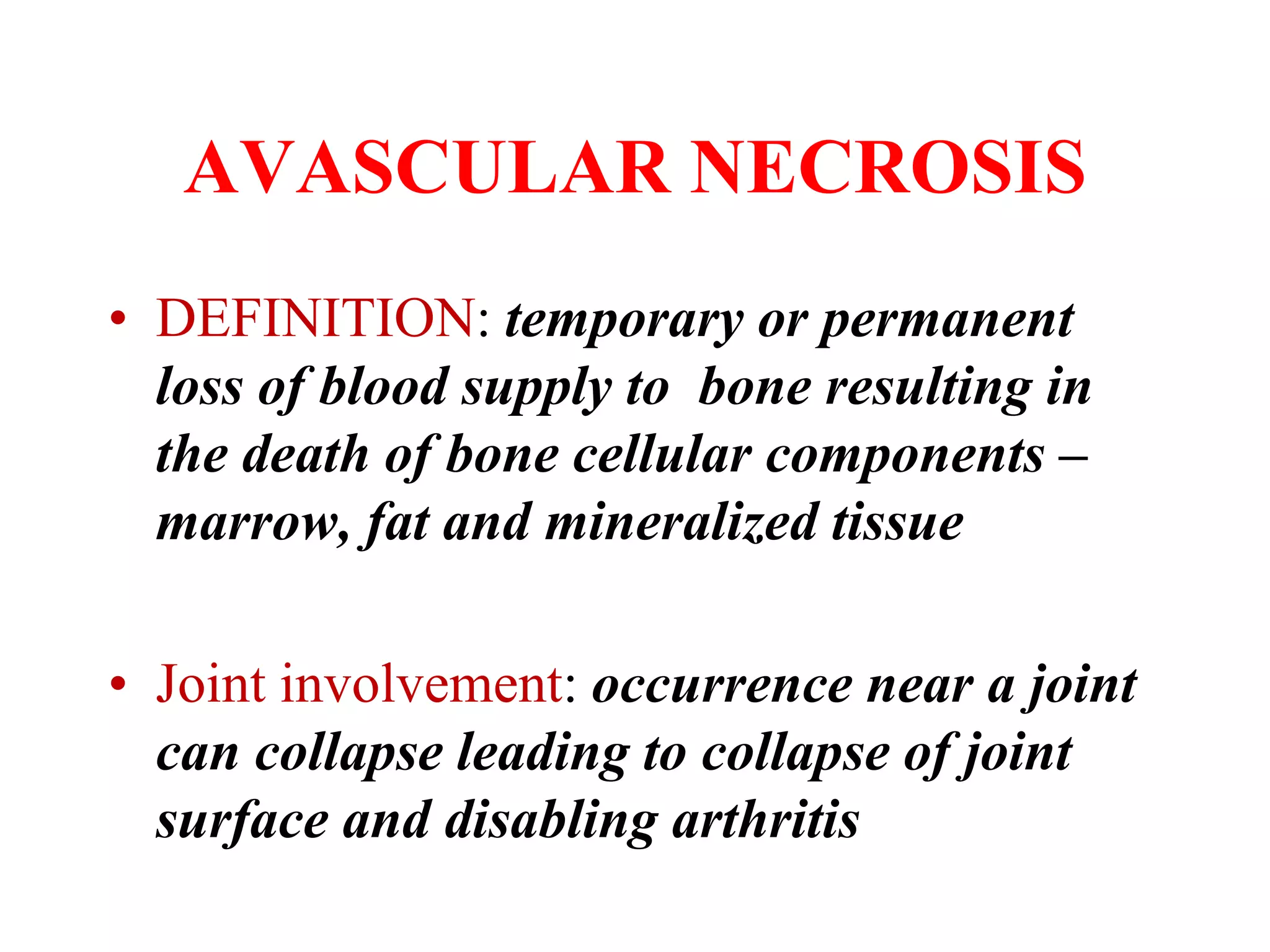
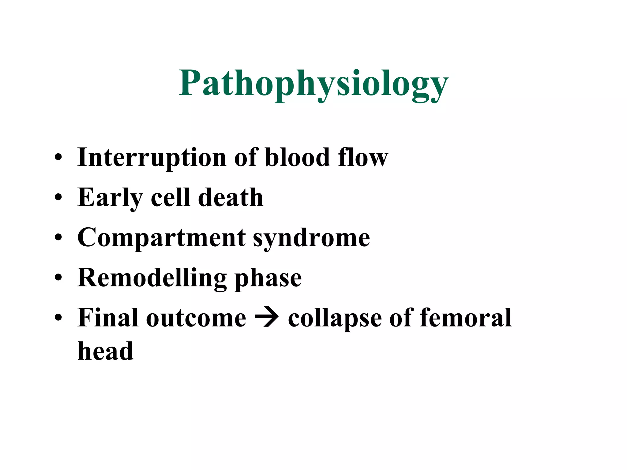
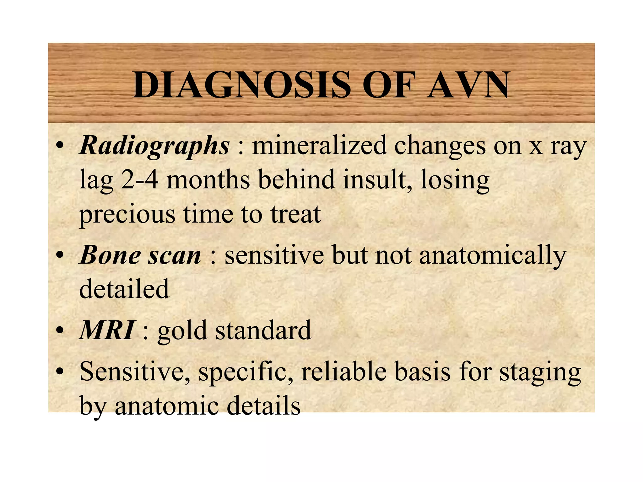
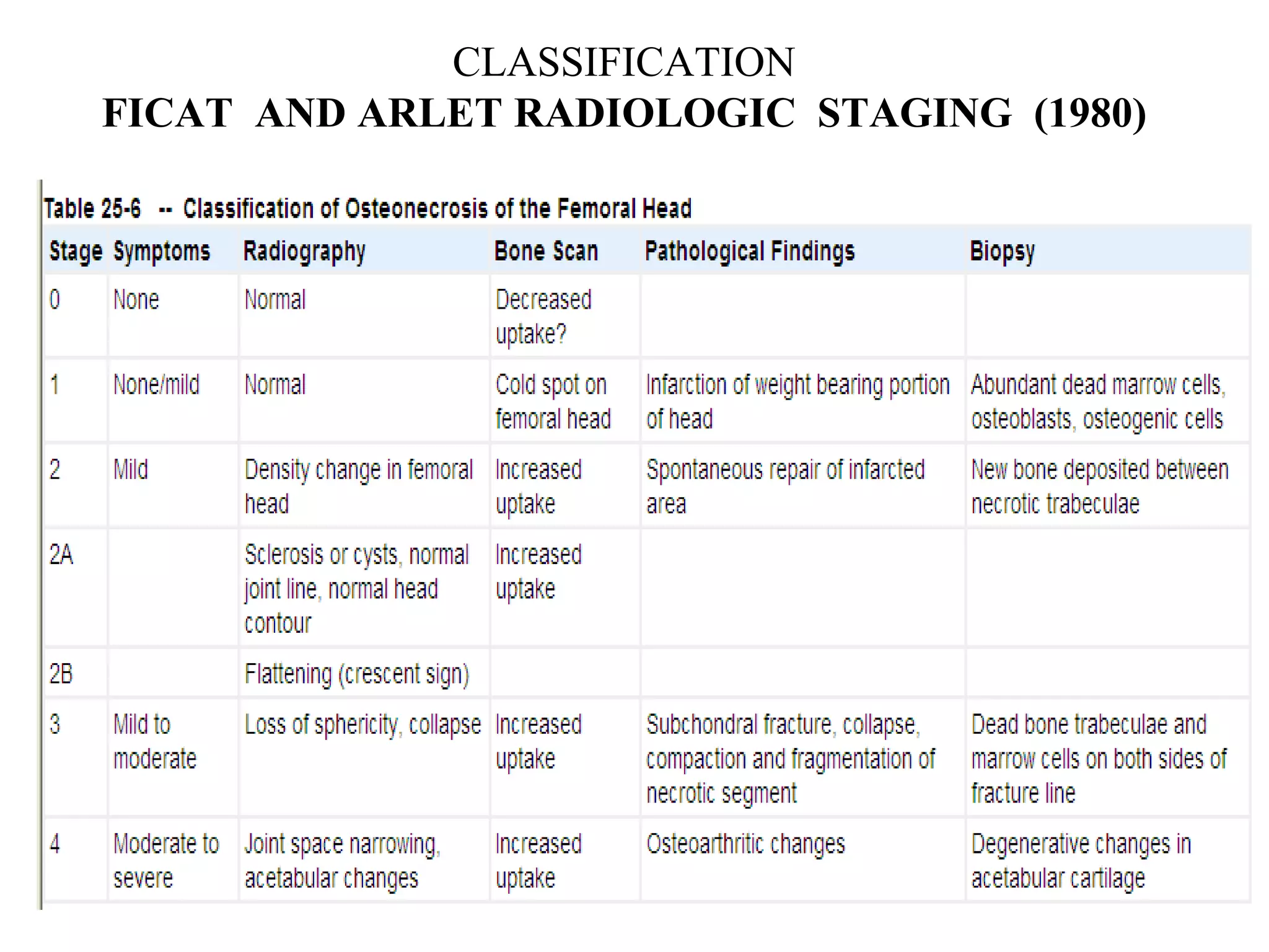
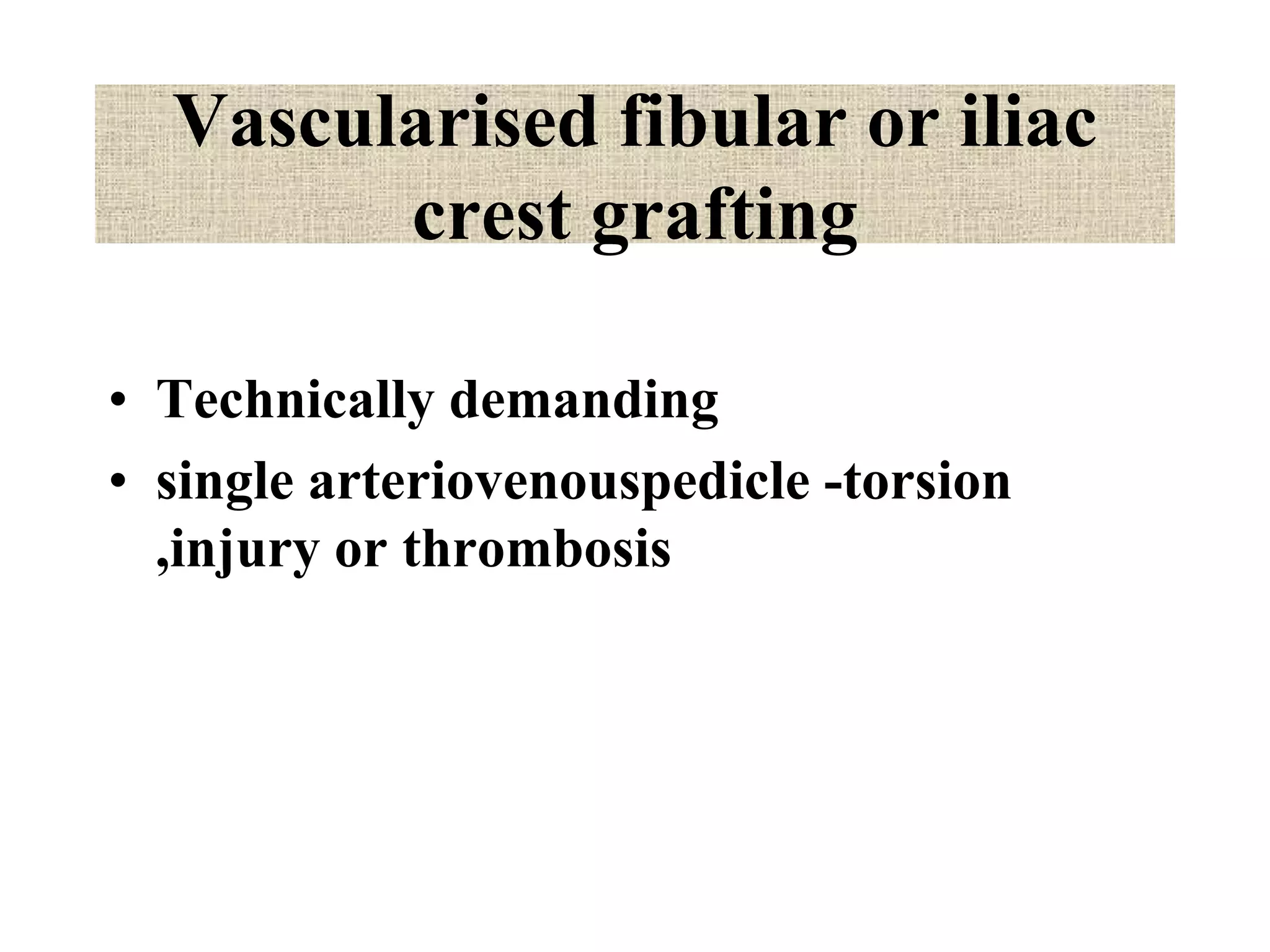
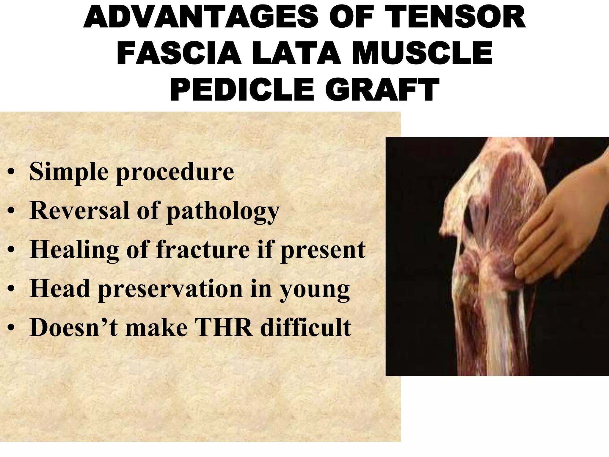
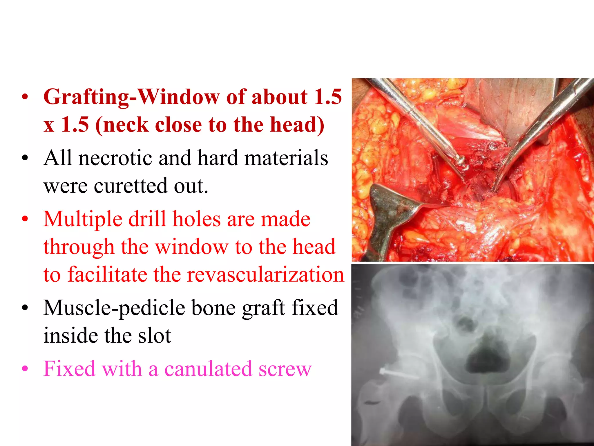
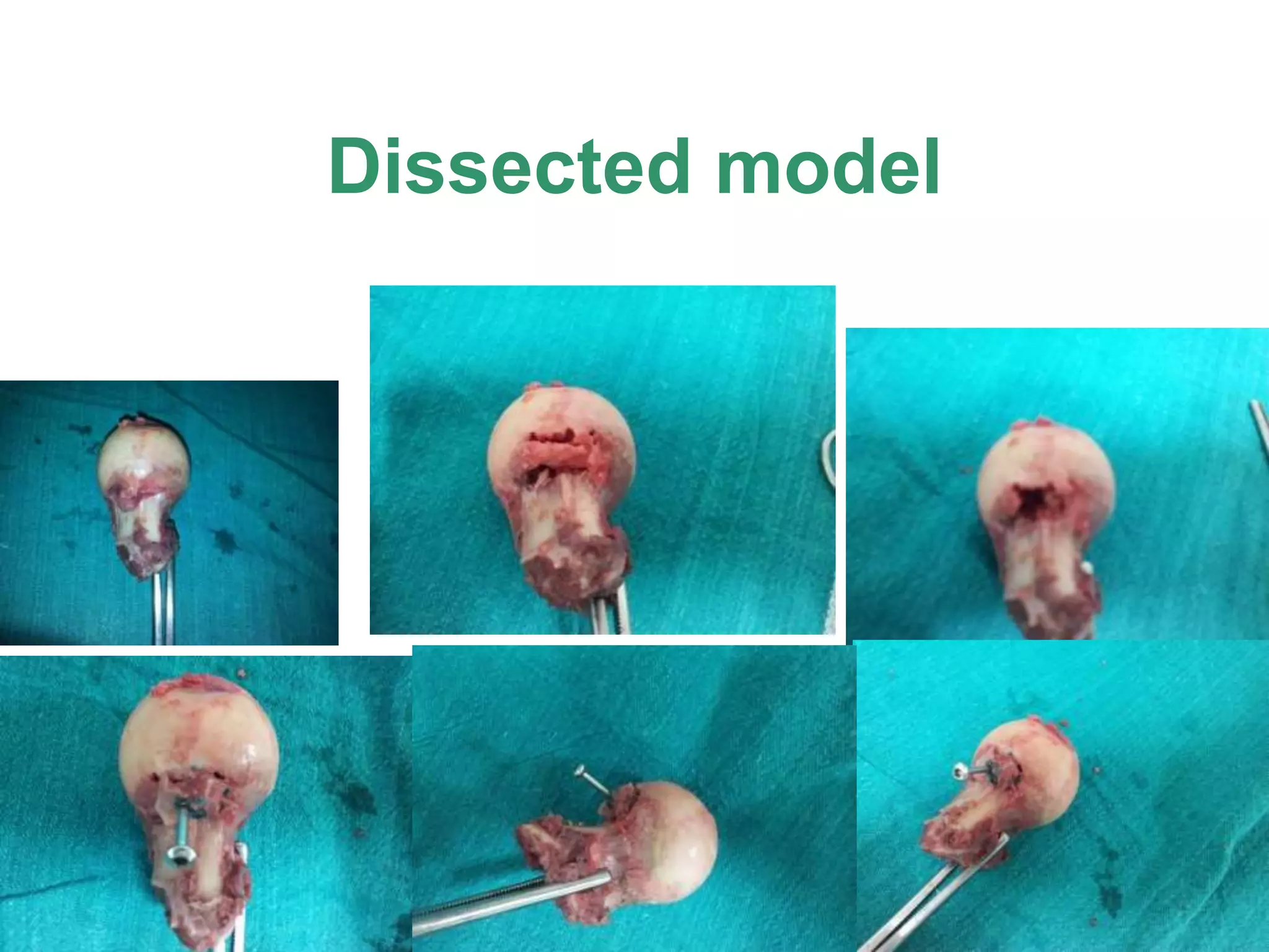
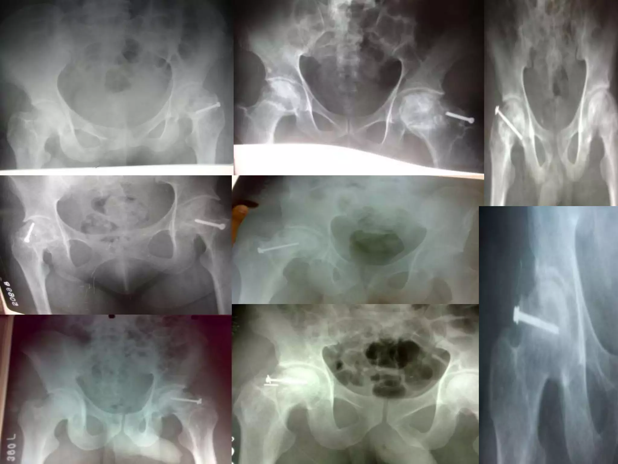
TENSOR FASCIA LATA
MUSCLE PEDICLE
GRAFT[TFL MPG] IN AVN hip
DR MOHAMED ASHRAF. Professor and head
drashraf369@gmail.com
DR Narayanan SK . Assistant professor
Govt TD medical college,alleppey,kerala,india
• From here to here painful
• active young man photo
• crippled wheelchaired man
PAINFUL
FROM HERE TO HERE
FROM HERE TO HERE TO HERE
CATASTROPHIC
• Presenting our long term
results of:
Modified TFL MPG
[tensor fascia lata
muscle pedicle graft]
AVASCULAR NECROSIS
• DEFINITION: temporary or permanent
loss of blood supply to bone resulting in
the death of bone cellular components –
marrow, fat and mineralized tissue
• Joint involvement: occurrence near a joint
can collapse leading to collapse of joint
surface and disabling arthritis
Pathophysiology
• Interruption of blood flow
• Early cell death
• Compartment syndrome
• Remodelling phase
• Final outcome  collapse of femoral
head
Etiology
• Idiopathic:
– Any bone
– Bilateral
– Most common FH
– M:F 8:1
– 30-60 years
• Post traumatic
• Secondary : alcoholism, steroids, radiation,
gout, hemoglobinopathies etc.
WHY FEMORAL HEAD?
• Most vulnerable: most
remote area of skeleton’s
vascular tree
• No collateral back up :
end arterioles supply
subchondral bone
• Heavy load : subject to
repeated body weight
loading
DIAGNOSIS OF AVN
• Radiographs : mineralized changes on x ray
lag 2-4 months behind insult, losing
precious time to treat
• Bone scan : sensitive but not anatomically
detailed
• MRI : gold standard
• Sensitive, specific, reliable basis for staging
by anatomic details
CLASSIFICATION
FICAT AND ARLET RADIOLOGIC STAGING (1980)
A R C O
TREATMENT OF AVN
• Preservation : sphericity of femoral head
• Decrease marrow pressure  prevents
further necrosis
• Increase vascularity
Stage one disease natural history : 70- 80% progress to FH
collapse without intervention
TREATMENT OF AVN
CONSERVATIVE
• Bed rest
• Constant traction
• Analgesics
• Limited weight
bearing
• Electrical stimulation
• Bisphosphonates
SURGICAL
• Core decompression/
with bone grafting
• Osteochondral graft
• Muscle pedicle graft
• Free vascularised bone
graft
– Osteotomy
• Joint reconstruction
CORE DECOMPRESSION
• relieves the pain for short
term
• but wont stop disease
progression.
• Not useful for
revascularisation of femoral
head.
OSTEOTOMY IN AVN
• Technically very
difficult
• Further decreases
vascularity of head
• Subsequent total hip
replacement difficult
Vascularised fibular or iliac
crest grafting
• Technically demanding
• single arteriovenouspedicle -torsion
,injury or thrombosis
THR IN YOUNG
• Analysis of twenty-seven studies
• 25 revealed a higher rate of early failure of
THR than age-matched patients with other
diagnoses
• Mont et all , JBJS Am Volume 77-A(3),March 1995,pp 459-474
• IMPORTANCE
BEST THING ON YOUR NECK IS
YOUR OWN HEAD
ADVANTAGES OF TENSOR
FASCIA LATA MUSCLE
PEDICLE GRAFT
• Simple procedure
• Reversal of pathology
• Healing of fracture if present
• Head preservation in young
• Doesn’t make THR difficult
• To evaluate functional outcome of
securely fixed muscle pedicle graft in
AVN
• To popularise the method of muscle
pedicle bone grafting fixed with CCS for
treatment of osteonecrosis of femoral
head.
AIM OF THE STUDY
MATERIAL AND METHODS
• Retrospective study
• Between 2007 and 2010
• Fifty patients of AVN stage I, II and III
treated with core decompression and
tensor fascia lata muscle pedicle graft
• minimum follow up period of two years.
• Maximum follow up period 5 years
• Ficat and Arlet staging was used
• Preoperative and postoperative Harris
Hip score and Visual analog scale were
used.
TFL MPG Surgical technique
Standard Watson-Jones
approach
GRAFT MOBILISATION
• Middle third of TFL with an
overlying piece of fascial
sleeve + 2.5cm length and
height of the iliac graft
• Bone part predrilled and
tapped
• No stretching of the muscle
pedicle
• Grafting-Window of about 1.5
x 1.5 (neck close to the head)
• All necrotic and hard materials
were curetted out.
• Multiple drill holes are made
through the window to the head
to facilitate the revascularization
• Muscle-pedicle bone graft fixed
inside the slot
• Fixed with a canulated screw
position skin incision exposing anterolateral
aspect of femur head
A 1cm to 1.5cm
window made at head
neck junction
Curettage of necrotic
bone
Core decompression
extending skin
incision for harvesting
TFL MPG
Predrilling and
tapping for cannulated
screw
A 30 mm long 4mm
cannulated cancellous
screw used
Inspected for secure
fixation
closure
Dissected model
POST-OP PROTOCOL
• Sutures removed on 10th day
• Static exercises started as soon as the pain is
relieved
• ROM exercises after 2 weeks
• NON-weight bearing for 6 weeks
• Partial weight bearing up to 12 weeks
• Full weight bearing after 12 weeks
FOLLOW UP
• Clinical and Biologic improvement in
response to the treatment
• Clinical improvement —relates to pain relief
and function restoration and delay of total
joint arthroplasty
• Biologic improvement —decreasing lesion size
or increasing bone density or blood flow
RESULTS
• All fifty patients had pain relief and
improvement in range of movements at
the end of 10-12 weeks.
• Eight patients had residual low intensity
pain for a period of 24 weeks and 10
patients had painless limp for a period of
16-18 weeks.
• Eight patients with stage III had
improvement in flexion beyond 90°- 100°
till the last follow up of five years.
• Radiographs taken in the post operative
period showed good position of the graft
with canulated cancellous screw in situ.
• Subsequent radiographs at 4, 6, 12 and
24 months showed good union in forty
four patients.
six hips from stage III progressed to
further collapse , but without any
progression to arthrosis .
• Two patients had superficial infection at
the operative site and the wound
responded promptly.
• One patient developed screw loosening by
accidental fall but graft incorporated
• no other complications occurred in any
other patients.
• At the final follow up period of 2 years,
Haris hip score improved. 82% of
patients had improvement in Haris Hip
score of more than 22 points.
CONCLUSION
• TFL MPG gives predictably good results in
avn before major collapse and arthrosis
• TFL MPG is the biological option since it
revascularise and reverses the pathology
• Procedure can be performed by general
orthopaedician exposed to hip surgery
• Early collapse is not a contraindication
since cartilage remains less affected
Message is clear
this simple but excellent method
must be the procedure of choice
for AVN without major collapse
and arthrosis
MBBS [GMC CALICUT]
D ORTHO [GMCTRIVANDRUM]
MS ORTHO [MMC MADRAS]
DNB,MNAMS [NEW DELHI]
drashraf369@gmail.com

Tensor fascia lata[tfl] muscle pedicle grafting for avn hip dr mohamed ashraf.hod orthopaedics.govt td medical college hospital alleppey,kerala,india

  • 1.
    TENSOR FASCIA LATA MUSCLEPEDICLE GRAFT[TFL MPG] IN AVN hip DR MOHAMED ASHRAF. Professor and head drashraf369@gmail.com DR Narayanan SK . Assistant professor Govt TD medical college,alleppey,kerala,india
  • 2.
    • From hereto here painful • active young man photo • crippled wheelchaired man PAINFUL FROM HERE TO HERE
  • 3.
    FROM HERE TOHERE TO HERE CATASTROPHIC
  • 4.
    • Presenting ourlong term results of: Modified TFL MPG [tensor fascia lata muscle pedicle graft]
  • 5.
    AVASCULAR NECROSIS • DEFINITION:temporary or permanent loss of blood supply to bone resulting in the death of bone cellular components – marrow, fat and mineralized tissue • Joint involvement: occurrence near a joint can collapse leading to collapse of joint surface and disabling arthritis
  • 6.
    Pathophysiology • Interruption ofblood flow • Early cell death • Compartment syndrome • Remodelling phase • Final outcome  collapse of femoral head
  • 7.
    Etiology • Idiopathic: – Anybone – Bilateral – Most common FH – M:F 8:1 – 30-60 years • Post traumatic • Secondary : alcoholism, steroids, radiation, gout, hemoglobinopathies etc.
  • 8.
    WHY FEMORAL HEAD? •Most vulnerable: most remote area of skeleton’s vascular tree • No collateral back up : end arterioles supply subchondral bone • Heavy load : subject to repeated body weight loading
  • 9.
    DIAGNOSIS OF AVN •Radiographs : mineralized changes on x ray lag 2-4 months behind insult, losing precious time to treat • Bone scan : sensitive but not anatomically detailed • MRI : gold standard • Sensitive, specific, reliable basis for staging by anatomic details
  • 10.
    CLASSIFICATION FICAT AND ARLETRADIOLOGIC STAGING (1980)
  • 11.
  • 12.
    TREATMENT OF AVN •Preservation : sphericity of femoral head • Decrease marrow pressure  prevents further necrosis • Increase vascularity Stage one disease natural history : 70- 80% progress to FH collapse without intervention
  • 13.
    TREATMENT OF AVN CONSERVATIVE •Bed rest • Constant traction • Analgesics • Limited weight bearing • Electrical stimulation • Bisphosphonates SURGICAL • Core decompression/ with bone grafting • Osteochondral graft • Muscle pedicle graft • Free vascularised bone graft – Osteotomy • Joint reconstruction
  • 14.
    CORE DECOMPRESSION • relievesthe pain for short term • but wont stop disease progression. • Not useful for revascularisation of femoral head.
  • 15.
    OSTEOTOMY IN AVN •Technically very difficult • Further decreases vascularity of head • Subsequent total hip replacement difficult
  • 16.
    Vascularised fibular oriliac crest grafting • Technically demanding • single arteriovenouspedicle -torsion ,injury or thrombosis
  • 17.
    THR IN YOUNG •Analysis of twenty-seven studies • 25 revealed a higher rate of early failure of THR than age-matched patients with other diagnoses • Mont et all , JBJS Am Volume 77-A(3),March 1995,pp 459-474
  • 18.
    • IMPORTANCE BEST THINGON YOUR NECK IS YOUR OWN HEAD
  • 19.
    ADVANTAGES OF TENSOR FASCIALATA MUSCLE PEDICLE GRAFT • Simple procedure • Reversal of pathology • Healing of fracture if present • Head preservation in young • Doesn’t make THR difficult
  • 20.
    • To evaluatefunctional outcome of securely fixed muscle pedicle graft in AVN • To popularise the method of muscle pedicle bone grafting fixed with CCS for treatment of osteonecrosis of femoral head. AIM OF THE STUDY
  • 21.
    MATERIAL AND METHODS •Retrospective study • Between 2007 and 2010 • Fifty patients of AVN stage I, II and III treated with core decompression and tensor fascia lata muscle pedicle graft • minimum follow up period of two years. • Maximum follow up period 5 years
  • 22.
    • Ficat andArlet staging was used • Preoperative and postoperative Harris Hip score and Visual analog scale were used.
  • 23.
    TFL MPG Surgicaltechnique Standard Watson-Jones approach
  • 24.
    GRAFT MOBILISATION • Middlethird of TFL with an overlying piece of fascial sleeve + 2.5cm length and height of the iliac graft • Bone part predrilled and tapped • No stretching of the muscle pedicle
  • 25.
    • Grafting-Window ofabout 1.5 x 1.5 (neck close to the head) • All necrotic and hard materials were curetted out. • Multiple drill holes are made through the window to the head to facilitate the revascularization • Muscle-pedicle bone graft fixed inside the slot • Fixed with a canulated screw
  • 26.
    position skin incisionexposing anterolateral aspect of femur head A 1cm to 1.5cm window made at head neck junction Curettage of necrotic bone Core decompression
  • 27.
    extending skin incision forharvesting TFL MPG Predrilling and tapping for cannulated screw A 30 mm long 4mm cannulated cancellous screw used Inspected for secure fixation closure
  • 28.
  • 29.
    POST-OP PROTOCOL • Suturesremoved on 10th day • Static exercises started as soon as the pain is relieved • ROM exercises after 2 weeks • NON-weight bearing for 6 weeks • Partial weight bearing up to 12 weeks • Full weight bearing after 12 weeks
  • 30.
    FOLLOW UP • Clinicaland Biologic improvement in response to the treatment • Clinical improvement —relates to pain relief and function restoration and delay of total joint arthroplasty • Biologic improvement —decreasing lesion size or increasing bone density or blood flow
  • 31.
    RESULTS • All fiftypatients had pain relief and improvement in range of movements at the end of 10-12 weeks. • Eight patients had residual low intensity pain for a period of 24 weeks and 10 patients had painless limp for a period of 16-18 weeks. • Eight patients with stage III had improvement in flexion beyond 90°- 100° till the last follow up of five years.
  • 32.
    • Radiographs takenin the post operative period showed good position of the graft with canulated cancellous screw in situ. • Subsequent radiographs at 4, 6, 12 and 24 months showed good union in forty four patients.
  • 33.
    six hips fromstage III progressed to further collapse , but without any progression to arthrosis . • Two patients had superficial infection at the operative site and the wound responded promptly. • One patient developed screw loosening by accidental fall but graft incorporated
  • 34.
    • no othercomplications occurred in any other patients. • At the final follow up period of 2 years, Haris hip score improved. 82% of patients had improvement in Haris Hip score of more than 22 points.
  • 35.
    CONCLUSION • TFL MPGgives predictably good results in avn before major collapse and arthrosis • TFL MPG is the biological option since it revascularise and reverses the pathology • Procedure can be performed by general orthopaedician exposed to hip surgery • Early collapse is not a contraindication since cartilage remains less affected
  • 39.
    Message is clear thissimple but excellent method must be the procedure of choice for AVN without major collapse and arthrosis
  • 41.
    MBBS [GMC CALICUT] DORTHO [GMCTRIVANDRUM] MS ORTHO [MMC MADRAS] DNB,MNAMS [NEW DELHI] drashraf369@gmail.com