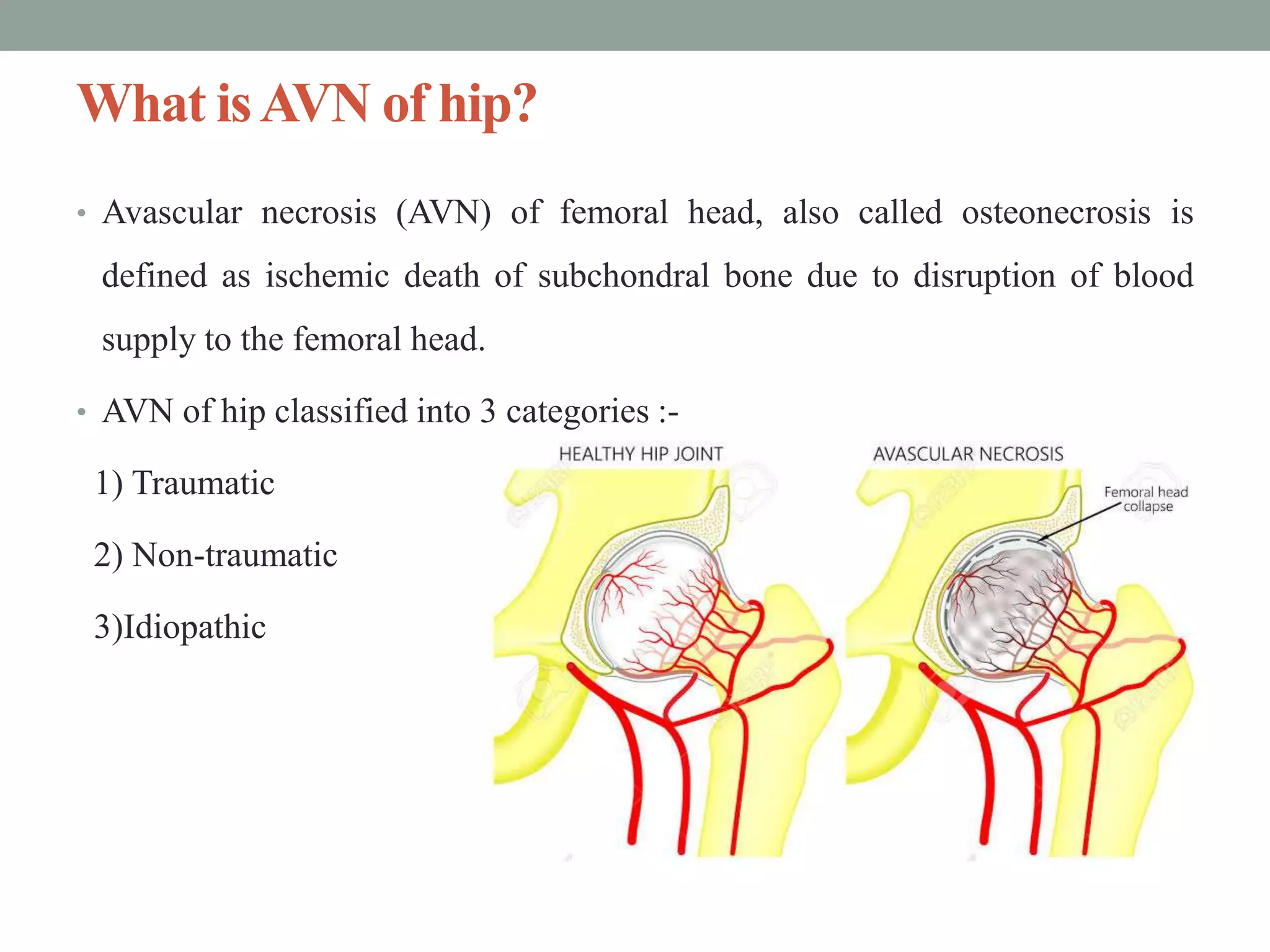
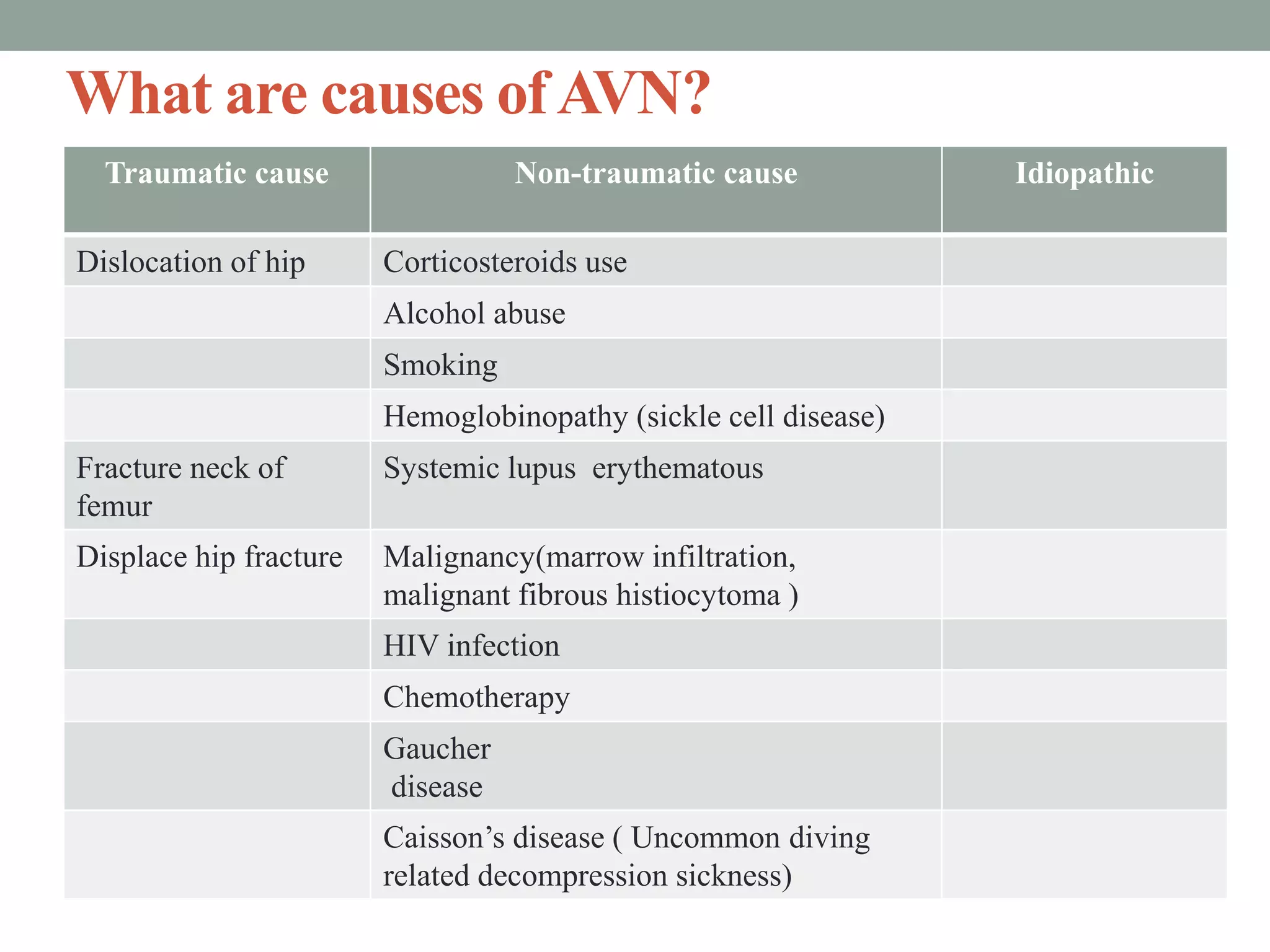
AVASCULAR NECROSIS (AVN)
OF HIP
Assistant Professor
Dr.Priyanka S.(PT)
Contents
• Introduction
• Causes
• Blood supply
• Mechanism
• Pathophysiology
• Investigation
• History
• Treatment
• References
What isAVN of hip?
• Avascular necrosis (AVN) of femoral head, also called osteonecrosis is
defined as ischemic death of subchondral bone due to disruption of blood
supply to the femoral head.
• AVN of hip classified into 3 categories :-
1) Traumatic
2) Non-traumatic
3)Idiopathic
What are causes ofAVN?
Traumatic cause Non-traumatic cause Idiopathic
Dislocation of hip Corticosteroids use
Alcohol abuse
Smoking
Hemoglobinopathy (sickle cell disease)
Fracture neck of
femur
Systemic lupus erythematous
Displace hip fracture Malignancy(marrow infiltration,
malignant fibrous histiocytoma )
HIV infection
Chemotherapy
Gaucher
disease
Caisson’s disease ( Uncommon diving
related decompression sickness)
Blood supply
• The capsule encloses most of the
femoral neck and the entire femoral
head.
• The primary blood supply to the femoral
head and neck arises from the medial
and lateral circumflex femoral arteries.
Mechanism of osteonecrosis
Pathophysiology
• Although the pathophysiology of AVN is not fully understood.
• The final common pathway is interruption of blood flow to the bone and
resultant necrosis of marrow, medullary bone, and cortex.
• Corticosteroids induced AVN- Fat accumulates in the liver of patients
treated where serum lipid concentration also increases. It give rise to fat
embolism and AVN.
• Alcohol induced AVN - Hyperlipidemia is the excess of fatty cells in the
body, the increased fat content of the blood causes blockage of the blood
vessels.
• Cigarette smoking : Due to changes in nitric oxide bioavailability, there is
an increased oxidative stress level and endothelial dysfunction.
• Sickle cell induced AVN : Sickle cell disease is a disorder that causes red
blood cells to become sickled (banana-shaped), as well as sticky and rigid,
which blocks blood flow in small blood vessels of the body. This loss of
blood causes narrowing of the joint and collapse of the bone.
• Chemotherapy -Multiple courses of chemotherapy, resulting in increased
production of cytokines, which caused the formation of thrombi and
decreased arterial blood supply. Blood disruption to the epiphyses of the
femoral head led to the development of AVN.
Investigations
• Radiographs (only after a few months to as long as 2 years, that one can
diagnose avascular necrosis on X-ray).
• MRI : Highest sensitivity (99%) and specificity (99%)
• The disease can be staged radio-graphically (Ficat & Arlet, 1964).
• Steinberg’s system of classification which is progression of Ficat &
Arlet.
Ficat and arlet classification
• Ficat & Arlet classification system of the femoral head
Classification Clinical Radiographs MRI
Stage 0 No symptoms; preclinical Normal Normal
Stage 1 Possible groin pain Normal or mild osteopenia Possible edema
Stage 2 Groin pain and stiffness; Osteopenia and/or subchondral Outlines area of
pain with activity cysts; diffuse porosis; involvement of FH
precollapse of joint space
Stage 3 Groin pain, stiffness, Crescent sign and/or subchondral
radiation of pain; collapse (flattening) of joint with
pain with activity secondary degenerative changes; Same as
radiographs
loss of sphericity of femoral head
Stage 4 Groin pain and limp; End-stage disease with collapse; Same as
radiographs ; Pain at rest extensive destruction of joint
Steinberg staging system
• Steinberg expands the Ficat system into six stages and includes :
• Stage Features
• 0 Normal radiograph, bone scan and magnetic resonance imaging
• I Normal radiograph, abnormal bone scan and or MRI
• IA Mild (involves < 15% of femoral head)
• IB Moderate (involves 15% to 30% of femoral head)
• IC Severe (involves > 30% of femoral head)
• II Cystic and sclerotic changes in the femoral head
• IIA Mild (involves < 15% of femoral head)
• IIB Moderate (involves 15% to 30% of femoral head)
• IIC Severe (involves > 30% of femoral head)
• III Subchondral collapse (crescent sign) without flattening of the FH
• IIIA Mild (involves < 15% of femoral head)
• IIIB Moderate (involves 15% to 30% of femoral head)
• IIIC Severe (involves > 30% of femoral head)
• IV Flattening of the femoral head/femoral head collapse
• IVA Mild (involves < 15% of femoral head)
• IVB Moderate (involves 15% to 30% of femoral head)
• IVC Severe (involves > 30% of femoral head)
• V Joint space narrowing and/or acetabular changes
• VA Mild
• VB Moderate
• VC Severe
• VI Advance degenerative joint disease
History
1.Age- Osteonecrosis can affect anyone, but is more common in people
between the ages of 40 and 65.
2.Sex- Men develop osteonecrosis of the hip more often than women (7:3).
3.Chief complaints – Asymptomatic early on in the disease process.
• In symptomatic, the stated history is usually of hip pain that may radiate to
the groin and/or thigh.
• Pain aggravating Factor- Walking and climbing stairs.
• Relieving Factor – Rest (Pain will be present in absence of movements)
History
4 .Past medical history- History of trauma
5. Drug history- Corticosteroids and steroids have high association of
AVN.
6. Personal history- Addictions like alcohol and cigarette smoking are risk
factors of AVN.
7. BMI- Obesity: osteonecrosis is positively associated with BMI. Overweight
and obesity are, just like steroid and alcohol use, often associated with
hyperlipidemia.
History
8. Observation - Trendlenburg’s sign may be positive..
9. Palpation- Tenderness over the groin region and tenderness to palpation of
the hip region.
10. Painful movements
11.Shortening of limb may be present.
Treatment
• Treatment decisions should be based on the staging of the lesions but mainly
on the presence or absence of collapse.
• Generally, non-operative treatments or core decompression can show value
in asymptomatic and symptomatic small to medium-sized pre-collapse
lesion.
• Medium to larger-sized lesions can have treatment with bone grafting
(vascularized or non-vascularized), or osteotomies.
• If femoral collapse has occurred or acetabular involvement is present,
arthroplasty is indicated.
• Conservative management spans a variety of non-operative treatments. These
may include physical therapy, restricted weight-bearing, alcohol cessation,
discontinuation of steroid therapy, pain control medication, and targeted
pharmacologic therapy, among others.
Surgical treatment
• Core decompression: (for stages 1
and 2 of the disease) creates a tract for
example in the femoral head that
decompresses the head. Hereby
facilitating increased blood flow,
which will then promote neo-
vascularization that could possibly
stimulate new bone growth.
Osteotomy
• It is an surgical operation whereby a
bone is cut to shorten or lengthen it or to
change its alignment.
• It decreases the load from the necrotic
bone either by rotation of the femoral
head and neck or by a varus or valgus
angulation of the proximal femur.
• This treatment is only for young patients
without co-morbidities interfering with
bone healing.
Joint replacement
• Total hip arthroplasty (commonly in stages 3 and 4) is the only option. In
this total hip replacement the damaged bone and cartilage are removed and
replaced with prosthetic components.
Physiotherapeutic Management
• Conservative management such as physiotherapy is necessary to prevent
further deterioration of the affected hip.
• while it has been shown to delay the disease progression, physiotherapy alone
cannot cure the disease – with 70-80% of clients requiring surgical treatment.
• Physiotherapy treatment aims include:
1. To reduce pain
2. Decrease weight bearing on affected extremity.
3. Patient education.
4.To improve joint mobility.
5.To strengthen the muscle of hip joint
1. Crutches or a walking aid - To decrease the weight-bearing load through
the head of the femur.
2. Patients education - Reduction of risk factors such as smoking, alcohol
abuse, obesity and corticosteroids.
3. To reduce pain - Modalities like ES, TENS and HCP is helpful to reduce
pain.
4. To improve mobility of joint - Passive ROM exercises (Flexion,extension
& Abduction) and Active ROM exercises in pain free range.
5. To prevent reflex inhibition of muscle – Activation exercises for
hamstrings, glutes and core muscle.
Do’s and Don’ts
Do’s
• Do keep the leg facing forward
• Do keep the affected leg in front as you
sit or stand
• Do use a high kitchen or barstool in the
kitchen
• Do kneel on the knee on the operated
leg (the bad side)
• Do use ice to reduce pain and swelling,
but remember that ice will diminish
sensation.
Don’ts
• Don't cross your legs at the knees
for at least 8 weeks.
• Don't bring your knee up higher
than your hip.
• Don't lean forward while sitting or
as you sit down.
• Don't try to pick up something on
the floor while you are sitting.
Do’s
• Do apply heat before exercising to
assist with range of motion.
• Use a heating pad or hot, damp
towel for 15 to 20 minutes.
• Do cut back on your exercises if
your muscles begin to ache, but
don't stop doing them.
Don’ts
• Don't turn your feet excessively
inward or outward when you bend
down.
• Don't bend at the waist beyond 90°
• Don't kneel on the knee on the
non-operated leg (the good side)
• Avoid toe-standing
References
1. S. Brent Broadzman &Robert C. Manske, PT “Clinical Orthopaedic
Rehabilitation”- 3rd edition.
2. Jayant Joshi & Prakash Kotwal “ Orthopaedics and applied
Physiotherapy”- 3rd edition.
3. Moya-Angeler J, Gianakos AL, Villa JC, Ni A, Lane JM. Current concepts
on osteonecrosis of the femoral head. World J Orthop 2015; 6(8): 590-601
[PMID: 26396935 DOI: 10.5312/wjo.v6.i8.590].
4. American Academy of Orthopedic Surgeons: http://orthoinfo.aaos.org
AVN Of Hip.pptx

AVN Of Hip.pptx

  • 1.
    AVASCULAR NECROSIS (AVN) OFHIP Assistant Professor Dr.Priyanka S.(PT)
  • 2.
    Contents • Introduction • Causes •Blood supply • Mechanism • Pathophysiology • Investigation • History • Treatment • References
  • 3.
    What isAVN ofhip? • Avascular necrosis (AVN) of femoral head, also called osteonecrosis is defined as ischemic death of subchondral bone due to disruption of blood supply to the femoral head. • AVN of hip classified into 3 categories :- 1) Traumatic 2) Non-traumatic 3)Idiopathic
  • 4.
    What are causesofAVN? Traumatic cause Non-traumatic cause Idiopathic Dislocation of hip Corticosteroids use Alcohol abuse Smoking Hemoglobinopathy (sickle cell disease) Fracture neck of femur Systemic lupus erythematous Displace hip fracture Malignancy(marrow infiltration, malignant fibrous histiocytoma ) HIV infection Chemotherapy Gaucher disease Caisson’s disease ( Uncommon diving related decompression sickness)
  • 5.
    Blood supply • Thecapsule encloses most of the femoral neck and the entire femoral head. • The primary blood supply to the femoral head and neck arises from the medial and lateral circumflex femoral arteries.
  • 6.
  • 7.
    Pathophysiology • Although thepathophysiology of AVN is not fully understood. • The final common pathway is interruption of blood flow to the bone and resultant necrosis of marrow, medullary bone, and cortex. • Corticosteroids induced AVN- Fat accumulates in the liver of patients treated where serum lipid concentration also increases. It give rise to fat embolism and AVN.
  • 8.
    • Alcohol inducedAVN - Hyperlipidemia is the excess of fatty cells in the body, the increased fat content of the blood causes blockage of the blood vessels. • Cigarette smoking : Due to changes in nitric oxide bioavailability, there is an increased oxidative stress level and endothelial dysfunction. • Sickle cell induced AVN : Sickle cell disease is a disorder that causes red blood cells to become sickled (banana-shaped), as well as sticky and rigid, which blocks blood flow in small blood vessels of the body. This loss of blood causes narrowing of the joint and collapse of the bone.
  • 9.
    • Chemotherapy -Multiplecourses of chemotherapy, resulting in increased production of cytokines, which caused the formation of thrombi and decreased arterial blood supply. Blood disruption to the epiphyses of the femoral head led to the development of AVN.
  • 10.
    Investigations • Radiographs (onlyafter a few months to as long as 2 years, that one can diagnose avascular necrosis on X-ray). • MRI : Highest sensitivity (99%) and specificity (99%) • The disease can be staged radio-graphically (Ficat & Arlet, 1964). • Steinberg’s system of classification which is progression of Ficat & Arlet.
  • 12.
    Ficat and arletclassification • Ficat & Arlet classification system of the femoral head Classification Clinical Radiographs MRI Stage 0 No symptoms; preclinical Normal Normal Stage 1 Possible groin pain Normal or mild osteopenia Possible edema Stage 2 Groin pain and stiffness; Osteopenia and/or subchondral Outlines area of pain with activity cysts; diffuse porosis; involvement of FH precollapse of joint space Stage 3 Groin pain, stiffness, Crescent sign and/or subchondral radiation of pain; collapse (flattening) of joint with pain with activity secondary degenerative changes; Same as radiographs loss of sphericity of femoral head Stage 4 Groin pain and limp; End-stage disease with collapse; Same as radiographs ; Pain at rest extensive destruction of joint
  • 14.
    Steinberg staging system •Steinberg expands the Ficat system into six stages and includes : • Stage Features • 0 Normal radiograph, bone scan and magnetic resonance imaging • I Normal radiograph, abnormal bone scan and or MRI • IA Mild (involves < 15% of femoral head) • IB Moderate (involves 15% to 30% of femoral head) • IC Severe (involves > 30% of femoral head) • II Cystic and sclerotic changes in the femoral head • IIA Mild (involves < 15% of femoral head) • IIB Moderate (involves 15% to 30% of femoral head) • IIC Severe (involves > 30% of femoral head) • III Subchondral collapse (crescent sign) without flattening of the FH
  • 15.
    • IIIA Mild(involves < 15% of femoral head) • IIIB Moderate (involves 15% to 30% of femoral head) • IIIC Severe (involves > 30% of femoral head) • IV Flattening of the femoral head/femoral head collapse • IVA Mild (involves < 15% of femoral head) • IVB Moderate (involves 15% to 30% of femoral head) • IVC Severe (involves > 30% of femoral head) • V Joint space narrowing and/or acetabular changes • VA Mild • VB Moderate • VC Severe • VI Advance degenerative joint disease
  • 16.
    History 1.Age- Osteonecrosis canaffect anyone, but is more common in people between the ages of 40 and 65. 2.Sex- Men develop osteonecrosis of the hip more often than women (7:3). 3.Chief complaints – Asymptomatic early on in the disease process. • In symptomatic, the stated history is usually of hip pain that may radiate to the groin and/or thigh. • Pain aggravating Factor- Walking and climbing stairs. • Relieving Factor – Rest (Pain will be present in absence of movements)
  • 17.
    History 4 .Past medicalhistory- History of trauma 5. Drug history- Corticosteroids and steroids have high association of AVN. 6. Personal history- Addictions like alcohol and cigarette smoking are risk factors of AVN. 7. BMI- Obesity: osteonecrosis is positively associated with BMI. Overweight and obesity are, just like steroid and alcohol use, often associated with hyperlipidemia.
  • 18.
    History 8. Observation -Trendlenburg’s sign may be positive.. 9. Palpation- Tenderness over the groin region and tenderness to palpation of the hip region. 10. Painful movements 11.Shortening of limb may be present.
  • 19.
    Treatment • Treatment decisionsshould be based on the staging of the lesions but mainly on the presence or absence of collapse. • Generally, non-operative treatments or core decompression can show value in asymptomatic and symptomatic small to medium-sized pre-collapse lesion. • Medium to larger-sized lesions can have treatment with bone grafting (vascularized or non-vascularized), or osteotomies.
  • 20.
    • If femoralcollapse has occurred or acetabular involvement is present, arthroplasty is indicated. • Conservative management spans a variety of non-operative treatments. These may include physical therapy, restricted weight-bearing, alcohol cessation, discontinuation of steroid therapy, pain control medication, and targeted pharmacologic therapy, among others.
  • 21.
    Surgical treatment • Coredecompression: (for stages 1 and 2 of the disease) creates a tract for example in the femoral head that decompresses the head. Hereby facilitating increased blood flow, which will then promote neo- vascularization that could possibly stimulate new bone growth.
  • 22.
    Osteotomy • It isan surgical operation whereby a bone is cut to shorten or lengthen it or to change its alignment. • It decreases the load from the necrotic bone either by rotation of the femoral head and neck or by a varus or valgus angulation of the proximal femur. • This treatment is only for young patients without co-morbidities interfering with bone healing.
  • 23.
    Joint replacement • Totalhip arthroplasty (commonly in stages 3 and 4) is the only option. In this total hip replacement the damaged bone and cartilage are removed and replaced with prosthetic components.
  • 24.
    Physiotherapeutic Management • Conservativemanagement such as physiotherapy is necessary to prevent further deterioration of the affected hip. • while it has been shown to delay the disease progression, physiotherapy alone cannot cure the disease – with 70-80% of clients requiring surgical treatment. • Physiotherapy treatment aims include: 1. To reduce pain 2. Decrease weight bearing on affected extremity. 3. Patient education. 4.To improve joint mobility. 5.To strengthen the muscle of hip joint
  • 25.
    1. Crutches ora walking aid - To decrease the weight-bearing load through the head of the femur. 2. Patients education - Reduction of risk factors such as smoking, alcohol abuse, obesity and corticosteroids. 3. To reduce pain - Modalities like ES, TENS and HCP is helpful to reduce pain. 4. To improve mobility of joint - Passive ROM exercises (Flexion,extension & Abduction) and Active ROM exercises in pain free range. 5. To prevent reflex inhibition of muscle – Activation exercises for hamstrings, glutes and core muscle.
  • 26.
    Do’s and Don’ts Do’s •Do keep the leg facing forward • Do keep the affected leg in front as you sit or stand • Do use a high kitchen or barstool in the kitchen • Do kneel on the knee on the operated leg (the bad side) • Do use ice to reduce pain and swelling, but remember that ice will diminish sensation. Don’ts • Don't cross your legs at the knees for at least 8 weeks. • Don't bring your knee up higher than your hip. • Don't lean forward while sitting or as you sit down. • Don't try to pick up something on the floor while you are sitting.
  • 27.
    Do’s • Do applyheat before exercising to assist with range of motion. • Use a heating pad or hot, damp towel for 15 to 20 minutes. • Do cut back on your exercises if your muscles begin to ache, but don't stop doing them. Don’ts • Don't turn your feet excessively inward or outward when you bend down. • Don't bend at the waist beyond 90° • Don't kneel on the knee on the non-operated leg (the good side) • Avoid toe-standing
  • 28.
    References 1. S. BrentBroadzman &Robert C. Manske, PT “Clinical Orthopaedic Rehabilitation”- 3rd edition. 2. Jayant Joshi & Prakash Kotwal “ Orthopaedics and applied Physiotherapy”- 3rd edition. 3. Moya-Angeler J, Gianakos AL, Villa JC, Ni A, Lane JM. Current concepts on osteonecrosis of the femoral head. World J Orthop 2015; 6(8): 590-601 [PMID: 26396935 DOI: 10.5312/wjo.v6.i8.590]. 4. American Academy of Orthopedic Surgeons: http://orthoinfo.aaos.org