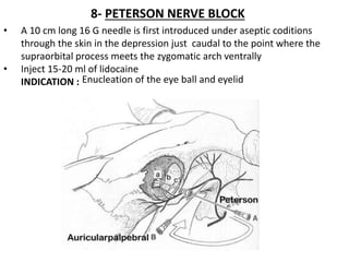
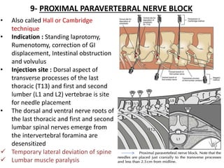
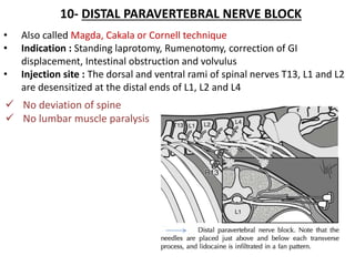
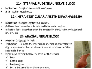
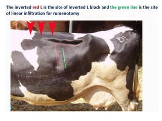
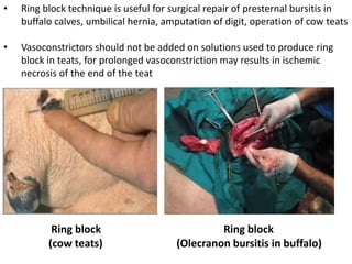
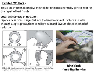
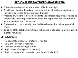
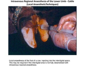
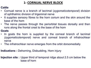
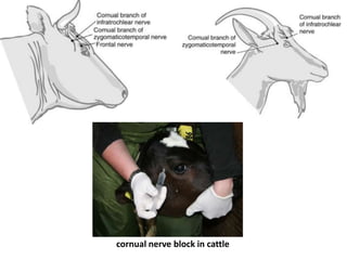
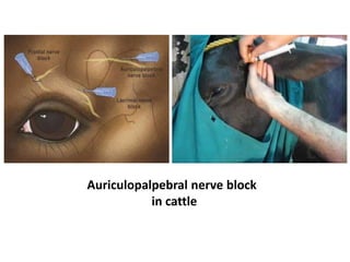
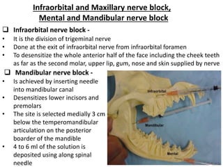
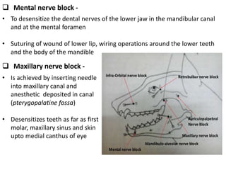
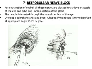
COLLEGE OF VETERINARY
AND ANIMAL SCIENCEs
S.V.P. UNIVERSITY OF AG. & TECH., MEERUT
Presented To:
Dr. Ashwani Arya
Course Advisor
Presented By:
Surya Kant Agarwal
ID. NO. V-3098/14
Various Methods Of Local Infiltration Anaesthesia And
Regional Block, For Surgical Procedures Of Different
Regions Of Body In Small And Large Animals
INFILTRATION ANAESTHESIA
 In this procedure the nerve ending is desensitized at the actual site of
operation
 Depending on the duration one can use procaine, lignocaine or bupivacaine
 Never inject local analgesic through infected and inflammed tissues
 Where this is used is on the teats of the cattle (do not use epinephrine
here, as vasoconstriction could lead to ischemic necrosis and sloughing of
tissue) or around the limb of cattle
 Following disinfection of the skin 0.5 to 1 ml of local anaesthetic is injected
intradermally before injecting into deeper tissues. This called as an
intradermal skin wheal
 Small circular wheals are created for catheterization of vessels
 A linear continuous wheal can be produced by the use of a longer needle
with single prick and it reduces the number of pricks in case of paravertibral
nerve block
 Advantages -
 Require no greater skill or the knowledge of the anatomy of the site
 Muscle relaxation and no interference to healing
 Disadvantages –
 Large volume of LAA in the tissue to be incised and sutured
 Epinephrine in the anesthetic solution may be interfering with the blood
supply and retard healing
 If the flank laparotomy is being done the LA must not only infiltrated SC but
into the fascia and the muscle as well
 Uses -
 To minimize or prevent pain
 To facilitate surgery
 Skin incision
 Surgical removal of superficial tumors
 Wound repair
 Types -
two types of infilteration anaesthesia
 Line block / infiltration
 Field block
A - Linear infiltration
• Preparation of the area to be anaesthetized for aseptic interference
• Define the intended line of the incision one of the following methods :
 Scratching of the skin by the tip of the injectable needle
 The needle is inserted parallel to the skin incision and for every 1 cm
area of incision 1 ml of the local anaesthetic solution is deposited
 The needle is withdrawn gently as the solution is deposited
 If the length of the incision is longer than the length of the needle the
needle can be inserted at the mid point of incision to preent multiple
pricks
 Using of surgical marker
Area blocked : skin and muscles layers of the flank and parietal peritoneum
along the line of incisions
Needle : 18 G, 7-10 cm.
Anesthetic : 10-100 ml of 2% lidocaine
Methods : make multiple SC injections of 0.5 to 1ml of AA, 1-2 cm apart then
infiltrate the muscle layer and the partial peritoneum through the
desensitized skin
 Although sensation is mainly confined to the skin, but in some
circumstances it is recommended to infiltrate the muscular layer beneath
the skin as sensory nerves pas through it and this will achieve better
analgesia
 Moreover, involvement of motor nerves that passses through the muscles
reduces movement of the muscles during incision
 Example of the linear infiltration of the left flank in cattle that involves
both subcutaneous and underlying muscles for induction of rumenotomy
or cesarean section
 Advantages -
 Simple and easy technique
 It consumes smaller amount of anesthetic and shorter time than inverted-
L technique
 Disadvantages –
A. Dealyed healing
B. Changes in the anatomical features
C. Consumption of large amount of drug than paravertebral
D. Lack of muscle relaxation
E. High cost
F. Formation of haematoma along the incision line
G. Toxicity after inj to peritoneal cavity (more than 250 ml (5g) of 2% lidocaine )
Linear infiltration analgesia in cow
B - Field block analgesia
1- Cup shaped field block –
It is an inverted pyramidal shape analgesic area that is created by two
punctures, and can be used when the pass of nerve supply is not exactly
known. Usually it is applied to an area of bulky musculature
 Advantages -
 Absence of anatomical distortion at seat of incision
 When the drug contains vasoconstrictor, it will produce efficient ischemia
 Complete muscular relaxation
 No retardation of healing
2- Inveted “L” block –
It is a field block technique through which only the dorsal and anterior aspects
of the flank region are injected subcutaneously with LA solution to produce
complete analgesia of the flank ( for rumenotomy or cesarean )
3- Ring block -
• Used in the extremities like limb for amputation of digit in cattle or in teat
for teat surgery
• Infiltration is done proximal to the site of operation
• Ring block technique is useful for surgical repair of presternal bursitis in
buffalo calves, umbilical hernia, amputation of digit, operation of cow teats
• Vasoconstrictors should not be added on solutions used to produce ring
block in teats, for prolonged vasoconstriction may results in ischemic
necrosis of the end of the teat
Ring block
(Olecranon bursitis in buffalo)
Ring block
(cow teats)
Ring block
(umbilical hernia)
Inverted “V” block -
This is an another alternative method for ring block normally done in teat for
the repair of teat fistula
Local anaesthesia of fracture -
Lignocaine is directly injected into the haematoma of fracture site with
through aseptic precautions to relieve pain and favours closed method of
reduction
REGIONAL INTRAVENOUS ANAESTHESIA
 This technique is used for amputation of limbs and digits
 A tight tourniquet or blood pressure measuring cuff is placed proximal to
the site of operation to block venous drainage
 Procaine or 2% lignocaine is injected into the engorged superficial vein so as
to facilitiate the retrograde flow of blood and deposition and infiltration of
local anaesthetic into the tissues
 Bupivaccaine is not normally used in this technique due to its myocardial
effects
 10-20 ml of the solution is sufficient in bovines while about 5 ml is required
in small ruminants
 Advantages -
 No special knowledge of anatomy is needed
 Only one injection is required
 Little risk of introducing bacteria
 Rapid onset of analgesia (5-10 mint.)
 Rapid recovery after removal of tourniquet (5-10 mint.)
Intravenous Regional Anaesthesia of the Lower Limb - Cattle
(Local AnaestheticTechniques)
NERVE BLOCKS
1- CORNUAL NERVE BLOCK
Cattle
• Cornual nerve is a branch of lacrimal (zygomaticotemporal) division
of ophthalmic division of trigeminal nerve
• It supplies sensory fibres to the horn corium and the skin around the
base of the horn
• The nerve passes through the periorbital tissues dorsally and then
runs along the frontal crest to the base of the horn
Goats
• In goats the horn is supplied by the corneal branch of lacrimal
(zygomaticotemporal) nerve and corneal branch of infratrochlear
nerve
• The infratrochlear nerve emerges from the orbit dorsomedially
Indications : Dehorning, Disbudding, Horn injury
Injection site : Upper third of temporal ridge about 2.5 cm below the
base of horn
cornual nerve block in cattle
2- AURICULOPALPEBRAL NERVE BLOCK
• This block can be used to prevent the eyelids moving during clinical
examination or surgery
• It blocks the orbicularis oculi muscle
• The nerve course runs from the base of the ear past the eye ventrally along
the facial crest
Purpose -
 Prevent eyelids closure during examination of the eyeball
Injection site -
 The needle is inserted in front of the base of the ear at the end of the
zygomatic arch and is introduced until its point lies at the dorsal border of
the arch
Caution -
 This block does not produce analgesia of the eye or the lids
 In conjunction with topic analgesia (2% lidocaine) it is useful for the
removal of foreign bodies form the cornea and conjunctival sac
Auriculopalpebral nerve block
in cattle
Infraorbital and Maxillary nerve block,
Mental and Mandibular nerve block
 Infraorbital nerve block -
• It is the division of trigeminal nerve
• Done at the exit of infraorbital nerve from infraorbital foramen
• To desensitize the whole anterior half of the face including the cheek teeth
as far as the second molar, upper lip, gum, nose and skin supplied by nerve
 Mandibular nerve block -
• Is achieved by inserting needle
into mandibular canal
• Desensitizes lower incisors and
premolars
• The site is selected medially 3 cm
below the temperomandibular
articulation on the posterior
boarder of the mandible
• 4 to 6 ml of the solution is
deposited using along spinal
needle
 Mental nerve block -
• To desensitize the dental nerves of the lower jaw in the mandibular canal
and at the mental foramen
• Suturing of wound of lower lip, wiring operations around the lower teeth
and the body of the mandible
 Maxillary nerve block -
• Is achieved by inserting needle
into maxillary canal and
anesthetic deposited in canal
(pterygopalatine fossa)
• Desensitizes teeth as far as first
molar, maxillary sinus and skin
upto medial canthus of eye
7- RETROBULBAR NERVE BLOCK
• For enucleation of eyeball all these nerves are blocked to achieve analgesia
of the eye and orbit and immobilization of the globe
• The needle is inserted through the lateral canthus of the eye
• Oriculopalpebral anesthesia is given; A hypodermic needle is turned/curved
at appropiate angle 15-20 degree
8- PETERSON NERVE BLOCK
• A 10 cm long 16 G needle is first introduced under aseptic coditions
through the skin in the depression just caudal to the point where the
supraorbital process meets the zygomatic arch ventrally
• Inject 15-20 ml of lidocaine
INDICATION : Enucleation of the eye ball and eyelid
9- PROXIMAL PARAVERTEBRAL NERVE BLOCK
• Also called Hall or Cambridge
technique
• Indication : Standing laprotomy,
Rumenotomy, correction of GI
displacement, Intestinal obstruction
and volvulus
• Injection site : Dorsal aspect of
transverse processes of the last
thoracic (T13) and first and second
lumber (L1 and L2) vertebrae is site
for needle placement
• The dorsal and ventral nerve roots of
the last thoracic and first and second
lumbar spinal nerves emerge from
the intervertebral foramina are
desensitized
 Temporary lateral deviation of spine
 Lumbar muscle paralysis
10- DISTAL PARAVERTEBRAL NERVE BLOCK
• Also called Magda, Cakala or Cornell technique
• Indication : Standing laprotomy, Rumenotomy, correction of GI
displacement, Intestinal obstruction and volvulus
• Injection site : The dorsal and ventral rami of spinal nerves T13, L1 and L2
are desensitized at the distal ends of L1, L2 and L4
 No deviation of spine
 No lumbar muscle paralysis
11- INTERNAL PUDENDAL NERVE BLOCK
• Indication : Surgical examination of penis
• Site : Ischio-rectal fossa
12- INTRA-TESTICULAR ANESTHESIA/ANALGESIA
• Indication : Surgical castration in cattle
• 8-10 ml local anesthetic is injected into each testicle
• In horse, local anesthetic can be injected in conjuction with general
anesthesia
13- ABAXIAL NERVE BLOCK
• Needle : 25 gauge ¾ inch
• Technique : Palpate the lateral and medial palmar/planter
digital neurovascular bundle on the abaxial aspect of the
sesamoid bones
• Blocks everything below the level of the fetlock
 Foot
 Coffin joint
 Pastern joint
 Distal Sesamoidean Ligaments etc…
Surya kant agarwal

Surya kant agarwal

  • 1.
    COLLEGE OF VETERINARY ANDANIMAL SCIENCEs S.V.P. UNIVERSITY OF AG. & TECH., MEERUT Presented To: Dr. Ashwani Arya Course Advisor Presented By: Surya Kant Agarwal ID. NO. V-3098/14 Various Methods Of Local Infiltration Anaesthesia And Regional Block, For Surgical Procedures Of Different Regions Of Body In Small And Large Animals
  • 2.
    INFILTRATION ANAESTHESIA  Inthis procedure the nerve ending is desensitized at the actual site of operation  Depending on the duration one can use procaine, lignocaine or bupivacaine  Never inject local analgesic through infected and inflammed tissues  Where this is used is on the teats of the cattle (do not use epinephrine here, as vasoconstriction could lead to ischemic necrosis and sloughing of tissue) or around the limb of cattle  Following disinfection of the skin 0.5 to 1 ml of local anaesthetic is injected intradermally before injecting into deeper tissues. This called as an intradermal skin wheal  Small circular wheals are created for catheterization of vessels  A linear continuous wheal can be produced by the use of a longer needle with single prick and it reduces the number of pricks in case of paravertibral nerve block
  • 3.
     Advantages - Require no greater skill or the knowledge of the anatomy of the site  Muscle relaxation and no interference to healing  Disadvantages –  Large volume of LAA in the tissue to be incised and sutured  Epinephrine in the anesthetic solution may be interfering with the blood supply and retard healing  If the flank laparotomy is being done the LA must not only infiltrated SC but into the fascia and the muscle as well  Uses -  To minimize or prevent pain  To facilitate surgery  Skin incision  Surgical removal of superficial tumors  Wound repair  Types - two types of infilteration anaesthesia  Line block / infiltration  Field block
  • 4.
    A - Linearinfiltration • Preparation of the area to be anaesthetized for aseptic interference • Define the intended line of the incision one of the following methods :  Scratching of the skin by the tip of the injectable needle  The needle is inserted parallel to the skin incision and for every 1 cm area of incision 1 ml of the local anaesthetic solution is deposited  The needle is withdrawn gently as the solution is deposited  If the length of the incision is longer than the length of the needle the needle can be inserted at the mid point of incision to preent multiple pricks  Using of surgical marker Area blocked : skin and muscles layers of the flank and parietal peritoneum along the line of incisions Needle : 18 G, 7-10 cm. Anesthetic : 10-100 ml of 2% lidocaine Methods : make multiple SC injections of 0.5 to 1ml of AA, 1-2 cm apart then infiltrate the muscle layer and the partial peritoneum through the desensitized skin
  • 6.
     Although sensationis mainly confined to the skin, but in some circumstances it is recommended to infiltrate the muscular layer beneath the skin as sensory nerves pas through it and this will achieve better analgesia  Moreover, involvement of motor nerves that passses through the muscles reduces movement of the muscles during incision  Example of the linear infiltration of the left flank in cattle that involves both subcutaneous and underlying muscles for induction of rumenotomy or cesarean section  Advantages -  Simple and easy technique  It consumes smaller amount of anesthetic and shorter time than inverted- L technique
  • 7.
     Disadvantages – A.Dealyed healing B. Changes in the anatomical features C. Consumption of large amount of drug than paravertebral D. Lack of muscle relaxation E. High cost F. Formation of haematoma along the incision line G. Toxicity after inj to peritoneal cavity (more than 250 ml (5g) of 2% lidocaine ) Linear infiltration analgesia in cow
  • 8.
    B - Fieldblock analgesia 1- Cup shaped field block – It is an inverted pyramidal shape analgesic area that is created by two punctures, and can be used when the pass of nerve supply is not exactly known. Usually it is applied to an area of bulky musculature  Advantages -  Absence of anatomical distortion at seat of incision  When the drug contains vasoconstrictor, it will produce efficient ischemia  Complete muscular relaxation  No retardation of healing 2- Inveted “L” block – It is a field block technique through which only the dorsal and anterior aspects of the flank region are injected subcutaneously with LA solution to produce complete analgesia of the flank ( for rumenotomy or cesarean ) 3- Ring block - • Used in the extremities like limb for amputation of digit in cattle or in teat for teat surgery • Infiltration is done proximal to the site of operation
  • 9.
    • Ring blocktechnique is useful for surgical repair of presternal bursitis in buffalo calves, umbilical hernia, amputation of digit, operation of cow teats • Vasoconstrictors should not be added on solutions used to produce ring block in teats, for prolonged vasoconstriction may results in ischemic necrosis of the end of the teat Ring block (Olecranon bursitis in buffalo) Ring block (cow teats)
  • 10.
    Ring block (umbilical hernia) Inverted“V” block - This is an another alternative method for ring block normally done in teat for the repair of teat fistula Local anaesthesia of fracture - Lignocaine is directly injected into the haematoma of fracture site with through aseptic precautions to relieve pain and favours closed method of reduction
  • 11.
    REGIONAL INTRAVENOUS ANAESTHESIA This technique is used for amputation of limbs and digits  A tight tourniquet or blood pressure measuring cuff is placed proximal to the site of operation to block venous drainage  Procaine or 2% lignocaine is injected into the engorged superficial vein so as to facilitiate the retrograde flow of blood and deposition and infiltration of local anaesthetic into the tissues  Bupivaccaine is not normally used in this technique due to its myocardial effects  10-20 ml of the solution is sufficient in bovines while about 5 ml is required in small ruminants  Advantages -  No special knowledge of anatomy is needed  Only one injection is required  Little risk of introducing bacteria  Rapid onset of analgesia (5-10 mint.)  Rapid recovery after removal of tourniquet (5-10 mint.)
  • 12.
    Intravenous Regional Anaesthesiaof the Lower Limb - Cattle (Local AnaestheticTechniques)
  • 13.
  • 14.
    1- CORNUAL NERVEBLOCK Cattle • Cornual nerve is a branch of lacrimal (zygomaticotemporal) division of ophthalmic division of trigeminal nerve • It supplies sensory fibres to the horn corium and the skin around the base of the horn • The nerve passes through the periorbital tissues dorsally and then runs along the frontal crest to the base of the horn Goats • In goats the horn is supplied by the corneal branch of lacrimal (zygomaticotemporal) nerve and corneal branch of infratrochlear nerve • The infratrochlear nerve emerges from the orbit dorsomedially Indications : Dehorning, Disbudding, Horn injury Injection site : Upper third of temporal ridge about 2.5 cm below the base of horn
  • 15.
  • 16.
    2- AURICULOPALPEBRAL NERVEBLOCK • This block can be used to prevent the eyelids moving during clinical examination or surgery • It blocks the orbicularis oculi muscle • The nerve course runs from the base of the ear past the eye ventrally along the facial crest Purpose -  Prevent eyelids closure during examination of the eyeball Injection site -  The needle is inserted in front of the base of the ear at the end of the zygomatic arch and is introduced until its point lies at the dorsal border of the arch Caution -  This block does not produce analgesia of the eye or the lids  In conjunction with topic analgesia (2% lidocaine) it is useful for the removal of foreign bodies form the cornea and conjunctival sac
  • 17.
  • 18.
    Infraorbital and Maxillarynerve block, Mental and Mandibular nerve block  Infraorbital nerve block - • It is the division of trigeminal nerve • Done at the exit of infraorbital nerve from infraorbital foramen • To desensitize the whole anterior half of the face including the cheek teeth as far as the second molar, upper lip, gum, nose and skin supplied by nerve  Mandibular nerve block - • Is achieved by inserting needle into mandibular canal • Desensitizes lower incisors and premolars • The site is selected medially 3 cm below the temperomandibular articulation on the posterior boarder of the mandible • 4 to 6 ml of the solution is deposited using along spinal needle
  • 19.
     Mental nerveblock - • To desensitize the dental nerves of the lower jaw in the mandibular canal and at the mental foramen • Suturing of wound of lower lip, wiring operations around the lower teeth and the body of the mandible  Maxillary nerve block - • Is achieved by inserting needle into maxillary canal and anesthetic deposited in canal (pterygopalatine fossa) • Desensitizes teeth as far as first molar, maxillary sinus and skin upto medial canthus of eye
  • 20.
    7- RETROBULBAR NERVEBLOCK • For enucleation of eyeball all these nerves are blocked to achieve analgesia of the eye and orbit and immobilization of the globe • The needle is inserted through the lateral canthus of the eye • Oriculopalpebral anesthesia is given; A hypodermic needle is turned/curved at appropiate angle 15-20 degree
  • 21.
    8- PETERSON NERVEBLOCK • A 10 cm long 16 G needle is first introduced under aseptic coditions through the skin in the depression just caudal to the point where the supraorbital process meets the zygomatic arch ventrally • Inject 15-20 ml of lidocaine INDICATION : Enucleation of the eye ball and eyelid
  • 22.
    9- PROXIMAL PARAVERTEBRALNERVE BLOCK • Also called Hall or Cambridge technique • Indication : Standing laprotomy, Rumenotomy, correction of GI displacement, Intestinal obstruction and volvulus • Injection site : Dorsal aspect of transverse processes of the last thoracic (T13) and first and second lumber (L1 and L2) vertebrae is site for needle placement • The dorsal and ventral nerve roots of the last thoracic and first and second lumbar spinal nerves emerge from the intervertebral foramina are desensitized  Temporary lateral deviation of spine  Lumbar muscle paralysis
  • 23.
    10- DISTAL PARAVERTEBRALNERVE BLOCK • Also called Magda, Cakala or Cornell technique • Indication : Standing laprotomy, Rumenotomy, correction of GI displacement, Intestinal obstruction and volvulus • Injection site : The dorsal and ventral rami of spinal nerves T13, L1 and L2 are desensitized at the distal ends of L1, L2 and L4  No deviation of spine  No lumbar muscle paralysis
  • 24.
    11- INTERNAL PUDENDALNERVE BLOCK • Indication : Surgical examination of penis • Site : Ischio-rectal fossa 12- INTRA-TESTICULAR ANESTHESIA/ANALGESIA • Indication : Surgical castration in cattle • 8-10 ml local anesthetic is injected into each testicle • In horse, local anesthetic can be injected in conjuction with general anesthesia 13- ABAXIAL NERVE BLOCK • Needle : 25 gauge ¾ inch • Technique : Palpate the lateral and medial palmar/planter digital neurovascular bundle on the abaxial aspect of the sesamoid bones • Blocks everything below the level of the fetlock  Foot  Coffin joint  Pastern joint  Distal Sesamoidean Ligaments etc…