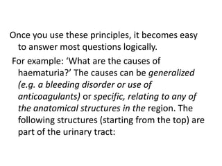
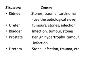
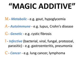
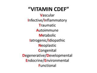
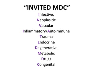
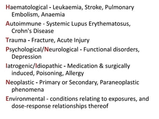
A surgical sieve is useful when trying to answer questions on the cause of a sign or symptom.
It helps to bring to mind lots of different conditions which could be potential causes.
It can also be helpful in formulating a differential diagnosis for patients where the diagnosis is not clear.
This document discusses neoplasms (tumors) of the salivary glands. It begins by describing the major and minor salivary glands. The most common benign tumors are pleomorphic adenoma, Warthin's tumor, and oncocytoma. The most common malignant tumors are mucoepidermoid carcinoma and adenoid cystic carcinoma. Factors like size and location of the gland affect likelihood of malignancy. Surgery is the main treatment and complications can include facial nerve paralysis, fluid collections, and Frey's syndrome.
This document discusses dentigerous cysts, including their definition, pathogenesis, clinical features, radiological features, histological features, investigation, treatment, and potential complications. A dentigerous cyst is an odontogenic cyst that forms around the crown of an unerupted tooth due to fluid accumulation between the reduced enamel epithelium and enamel surface. Clinically, they usually appear as asymptomatic swellings but can cause expansion of the bone. Radiographically, they appear as well-defined radiolucencies that surround the crown of an unerupted tooth. Treatment involves surgical removal of the cyst lining either through enucleation or marsupialization along with removal of the associated tooth. Complications can include
This document provides information on tumors of the salivary glands. It discusses the anatomy and histology of salivary glands, classification of salivary gland tumors, and specifics on certain tumor types including pleomorphic adenoma and Warthin's tumor. Pleomorphic adenoma is the most common benign salivary gland tumor, characterized by epithelial and mesenchymal differentiation. Warthin's tumor commonly occurs bilaterally in the parotid glands of older smoking males. The document covers epidemiology, etiology, histogenesis, clinical features, investigation, pathology and treatment of various salivary gland tumors.
This document provides information on salivary gland diseases presented over multiple sessions. It begins with the objectives and overview of topics to be covered, including applied anatomy of the salivary glands, autonomic innervation and effects on function, inflammatory and obstructive disorders, neoplasms, and investigations. It then describes in detail the anatomy, physiology and investigations of the major salivary glands. Salivary gland diseases are classified as functional, obstructive, non-neoplastic and neoplastic. Specific conditions like sialadenitis, sialolithiasis, mucocele and ranula are explained. Imaging modalities like ultrasound, sialography, CT, MRI and sialendoscopy
This document summarizes different types of jaw swellings and their characteristics. It begins by classifying jaw swellings based on their origin, such as from tooth germs, mucoperiosteum, or being osseous, inflammatory, or malignant. Specific types of swellings arising from these origins are then described in more detail such as epulis, odontogenic tumors, fibrous dysplasia, Paget's disease, and osteomyelitis. Clinical features, radiographic findings, histological characteristics, and treatment approaches are provided for many of these conditions. In particular, ameloblastoma, a benign odontogenic tumor, is discussed in depth covering its pathogenesis, clinical presentation, classification, management,
This document discusses various causes of midline neck swellings, including congenital, infectious, inflammatory, and neoplastic etiologies. It provides details on specific conditions such as thyroglossal duct cyst, laryngocele, Ludwig's angina, sublingual dermoid cyst, reactive lymphadenopathy, thyroid disorders, and laryngeal malignancy. Treatment options vary depending on the underlying cause but may include antibiotics, incision and drainage, surgery, radiotherapy, or a combination of therapies.
Pleomorphic adenoma is the most common salivary gland tumor, accounting for 60% of cases. It occurs most often in the parotid glands and presents as a painless, firm mass. Histologically, it contains both epithelial and mesenchymal elements arranged in a trabecular pattern within a fibrous stroma. Treatment involves complete surgical removal of the tumor with adequate margins to prevent recurrence due to microscopic projections outside the capsule. Imaging such as CT or MRI is used to identify the location and characteristics of the tumor prior to surgery.
Based on the information provided, the key differentials would be:
- Radicular cyst: Most common cyst in jaws, associated with non-vital tooth. Location and association with tooth fits.
- Dentigerous cyst: Second most common, associated with crown of unerupted tooth. Location fits.
- Odontogenic keratocyst: Aggressive cyst, often multilocular radiolucency. Less likely based on description.
- Aneurysmal bone cyst: Often multilocular "soap bubble" appearance. Less likely based on description.
- Traumatic bone cyst: Often interradicular in location. Possible based on location described.
Further investigation with tooth
This document discusses the evaluation and treatment of solitary thyroid nodules (STN). STNs occur in 1-5% of the population and require evaluation to detect disorders of thyroid function or malignancy. Evaluation includes history, exam, thyroid stimulating hormone levels, ultrasound of the neck, and fine needle aspiration biopsy. Fine needle aspiration biopsy has a 70-97% accuracy rate and can determine if a nodule is benign, malignant, or requires further testing. Treatment options include surgery such as hemithyroidectomy or total thyroidectomy, cyst aspiration, or in rare cases, thyroid hormone suppression.
Salivary glands Disorders and management.Manish Shetty
1. The parotid gland is the largest salivary gland located below and in front of the ear. The submandibular gland is located beneath the jawbone. Saliva produced by these glands contains enzymes and antibodies that support oral health.
2. Sialography uses dye and x-rays to visualize the salivary ducts and identify any obstructions like stones. Benign tumors and infections are common salivary gland conditions. Surgery may be used to treat tumors or remove obstructions in the ducts.
3. The most common salivary gland tumor is the pleomorphic adenoma, which is generally benign. Sjögren's syndrome is an autoimmune condition that
A cyst is an epithelium-lined sac containing fluid or semisolid material. In the formation of a cyst, the epithelial cells first proliferate and later undergo degeneration and liquefaction. The liquefied material exerts equal pressure on the walls of the cyst from within. Cysts grow by expansion and thus displace the adjacent teeth by pressure. May can produce expansion of the cortical bone. On a radiograph, the radiolucency of a cyst is usually bordered by a radiopaque periphery of dense sclerotic bone. The radiolucency may be unilocular or multilocular. Odontogenic cysts are those which arise from the epithelium associated with the development of teeth. The source of epithelium is from the enamel organ, the reduced enamel epithelium, the cell rests of Malassez or the remnants of the dental lamina.
Neurofibroma is a benign tumor that arises from the connective tissue of nerves. It consists of neural and fibrous elements. Neurofibromas can be single or multiple, and form fusiform swellings along nerves. They can occur in cranial, spinal, or peripheral nerves. The main types are nodular, plexiform, generalized (von Recklinghausen's disease), elephantiatic, and pachydermatocoele. Nodular neurofibromas commonly affect peripheral nerves in adults as single, firm nodules. Plexiform neurofibromas often involve the face and can cause vision problems or bone erosion. Generalized neurofibromatosis is an inherited condition with
This document provides information on various types of salivary gland tumors. It begins with an overview of the major and minor salivary gland tumors that are most commonly seen in adults and children. Examples include Warthin tumor, acinic cell carcinoma, and polymorphous low-grade adenocarcinoma. The document then describes the histological features of many benign and malignant epithelial and nonepithelial salivary gland tumors at both the macroscopic and microscopic levels. Key diagnostic characteristics of tumors like pleomorphic adenoma, myoepithelioma, oncocytoma, and adenoid cystic carcinoma are highlighted.
The document discusses different types of cysts that can occur in the oral region, dividing them into odontogenic cysts and non-odontogenic cysts. Odontogenic cysts include radicular, dentigerous, primordial, odontogenic keratocyst, and lateral periodontal cysts. Non-odontogenic cysts include globulomaxillary, nasolabial, median palatal, and nasopalatine canal cysts. Each cyst type is described in terms of etiology, clinical features, radiographic appearance, histology, and treatment.
The document discusses the anatomy and triangles of the neck, describing boundaries, contents, and clinical significance. It also covers common neck masses including cysts, sinuses, fistulas, ulcers, tumors, and infections. Lymphatic drainage is described for deep cervical nodes along vertical and circular chains.
LYMPH NODES OF HEAD AND NECK AND DIFFERENTIAL DIAGNOSIS OF CERVICAL LYMPHA...Dr. Monali Prajapati
1. Introduction
a. Anatomy
b. Structure
c. Function
2. Lymph nodes of head and neck
3. Drainage
4. Lymph node levels and sublevels
5. Clinical examination of nodes
6. Diagnosis
7. Causes of cervical lymphadenopathy
8. Differential diagnosis of cervical lymphadenopathy
9. References
The document discusses several types of non-odontogenic cysts that develop in the oral cavity. It describes nasopalatine duct cysts, which originate from remnants of the nasopalatine duct in the maxilla. These cysts typically appear as well-defined radiolucencies between the central incisors. Median palatal cysts and globulomaxillary cysts are also discussed, which develop from epithelial remnants during fusion of facial processes. Palatal cysts of newborns are extraosseous cysts that commonly appear on the hard palate of infants.
Solitary oral ulcers and systemic diseasesDr. Harsh Shah
A brief overview of different ulcerative lesions seen in the oral cavity linked to the dangerous systemic diseases and preventive measures for the disease before it turns lerhal
SDDCH, Parbhani
This document discusses various types of pseudocysts and true cysts found in the body. It begins by defining the key differences between a true cyst and a pseudocyst. Pseudocysts are lined by granulation and/or fibrous tissue rather than an epithelial cell layer. The document then classifies and describes different types of pseudocysts including traumatic bone cysts, aneurysmal bone cysts, and developmental cysts. It provides details on the etiology, location, patient demographics, clinical presentation, radiographic features, histopathology and treatment for each type.
This document discusses second branchial cleft anomalies, which present as a sinus, cyst, or fistula in the neck anterior to the sternocleidomastoid muscle. Symptoms include a skin pit or palpable cord that can express milky discharge. Treatment involves complete surgical excision to avoid infection. Remnants of the thyroglossal duct are also discussed, presenting as midline neck swellings that move with swallowing and can become infected. Ultrasound and nuclear scans help differentiate these from other neck masses. Surgical excision is treatment.
This document discusses diseases of the salivary glands. It outlines benign and malignant lesions, as well as non-neoplastic disorders like sialadenitis and sialectasis. Common benign tumors described include ranula, mucocele, and dermoid cysts. Premalignant lesions include leukoplakia and erythroplakia. Common sites of oral cancer are the lip, buccal mucosa, tongue, hard palate and floor of mouth. Non-squamous cancers and lymphomas are also discussed. The diagnosis and treatment of various salivary gland disorders are described.
This document discusses various types of odontogenic cysts. It begins with introducing cysts in general and then classifies odontogenic cysts based on etiology and tissue of origin. Several specific types of odontogenic cysts are then described in more detail, including their clinical features, radiographic features, and differential diagnosis. These include dentigerous cysts, eruption cysts, odontogenic keratocysts, gingival cysts of newborn and adult, lateral periodontal cysts, calcifying odontogenic cysts, periapical cysts, residual cysts, and paradental cysts.
Branchial anomalies result from improper development of the branchial apparatus during embryogenesis. They present as cysts, sinuses, or fistulas in the neck region due to failure of branchial clefts or pouches to regress normally. The definitive treatment is complete surgical excision to prevent recurrent infections while protecting important nerves like the spinal accessory and recurrent laryngeal. Second branchial cleft cysts are the most common type and manifest as neck masses anterior to the sternocleidomastoid muscle.
The document discusses various cysts and masses that can occur in the neck region. It covers thyroglossal duct cysts, branchial cleft cysts, cystic hygroma, dermoid cysts, ranula, tuberculosis lymphadenitis, cervical ribs, carotid body tumors, and lymphomas among other conditions. For each, it discusses etiology, clinical features, diagnosis and treatment.
Common Benign Oral cavity disorders by. Dr.vijay kumarvijaymgims
The document discusses various types of oral lesions and conditions. It begins by describing the anatomy of the oral cavity and defines a lesion. It then classifies lesions based on their depth and texture. Specific lesion types are defined such as ulcers, erosions, abscesses, cysts, blisters, pustules, hematomas and plaques. Causes of oral lesions including congenital conditions, inflammatory/traumatic conditions, autoimmune diseases and precancerous lesions are listed. Finally, examples of benign tumors such as fibromas and pyogenic granulomas are provided along with more detailed descriptions of torus, lingual thyroid and inflammatory diseases like candidiasis and Vincent's angina.
The document discusses salivary gland disorders. It begins with definitions and classifications of salivary glands. It then discusses the anatomy, functions, and disorders of the parotid, submandibular, and sublingual salivary glands. Diagnostic aids are outlined including clinical history, physical examination, imaging such as CT, MRI, ultrasound and sialography. Cystic conditions of the minor salivary glands such as mucoceles are also summarized. Disorders are classified and inflammatory, obstructive, neoplastic and other conditions are described.
This document provides an overview of pulp and periapical pathologies. It begins by defining the dental pulp and pulpitis. It then covers causes of pulp disease including physical, chemical, and bacterial factors. It classifies pulp disease and discusses factors affecting the pulp response. It details the pathways of bacterial invasion of the pulp and describes different types of reversible and irreversible pulpitis. It also discusses chronic hyperplastic pulpitis, gangrenous necrosis of the pulp, and diseases of the periapical tissues like acute and chronic apical periodontitis and periapical abscess.
Emergency management of oral and maxillofacial trauma including_100844.pptxNdayishimiyeSamuel1
Oral and Maxillofacial area is a crucial area for respiration, digestive, and esthetic functions. When traumatized, a backup of knowledge and skills is required to restore pleasing look and function. This ppt details how to optimize the emergency and late better outcomes of patients with oral and maxillofacial trauma.
The document provides an introduction to general surgery. It outlines the overall and specific objectives of the general surgery course, which are to develop students' skills in evaluating and diagnosing surgical problems through history taking, physical examination, and ordering appropriate investigations. It also describes the different types of assessments and references used in the course. The document covers various topics related to general surgery including the history and classifications of surgery, as well as approaches to the surgical history and physical examination of patients.
Dr. Sharmin Nahar and Dr. Olivia Akhter are presenting a seminar on pediatric hematology and oncology at BSMMU. They discuss the case of a 2-year-old girl admitted with headaches, vomiting, and seizures for the past month as well as left-sided weakness. Imaging shows a mass in the 4th ventricle causing hydrocephalus, possibly an ependymoma or medulloblastoma. They then provide an introduction to brain tumors in children, discussing types, incidence, risk factors, classification, and clinical features. Treatment options for brain tumors including surgery, radiation, and chemotherapy are also summarized.
This document discusses maxillofacial oncology and tumor diagnosis. It defines key terms like oncology, tumor, neoplasm, benign and malignant tumors. It describes the multi-step process of metastasis. The importance of a thorough health history, clinical examination including lymph nodes, radiographic examination and laboratory tests in tumor diagnosis is highlighted. Different types of biopsies including exfoliative cytology, oral brush cytology and incisional/excisional biopsies are also discussed.
This document provides tips and instructions for using a PowerPoint presentation on Hydatid disease:
1. The PPT can be freely downloaded, edited, and modified. It includes blank slides to engage students by asking questions before providing information.
2. The presentation is to be used for active learning sessions where blank slides are first shown to elicit student responses, followed by the content slides.
3. This format allows for repetition and revision of content through multiple iterations of questioning and answering.
This document discusses brain abscess, cranial subdural empyema, and epidural abscess. It covers the epidemiology, etiology, pathogenesis, clinical findings, diagnosis, and management of these conditions. Brain abscesses are typically caused by bacteria spreading from contiguous sites of infection or through the bloodstream. Clinical findings depend on the location and size of the abscess. Diagnosis involves neuroimaging and culture of aspirated contents. Treatment involves antibiotics and sometimes surgery. Outcomes depend on early diagnosis and treatment.
The document discusses the local and systemic effects of tumors on the host. Locally, both benign and malignant tumors can cause compression, mechanical obstruction, and tissue destruction. Malignant tumors tend to have more serious local effects. Systemically, cancers can cause cachexia, fever, tumor lysis syndrome, and paraneoplastic syndromes. Grading describes a tumor's differentiation, while staging evaluates its extent based on primary tumor size, lymph node involvement, and metastasis.
gyanaecology.endometriosis and adenomyosis.(dr.salama)student
The document summarizes endometriosis and adenomyosis. Endometriosis occurs when endometrial tissue grows outside the uterus, most commonly on the ovaries, uterine ligaments and pelvis. It causes pain and infertility. Adenomyosis involves endometrial tissue in the uterine wall. Both can be diagnosed by laparoscopy and treated through drugs or surgery, with hysterectomy providing definitive treatment for severe adenomyosis.
MBBS 2nd Year Pathology - Neoplasia : IntroductionNida Us Sahr
Chapter 7 (Neoplasia) from Robbins and Cotran Pathologic Basis of Disease (9th Edition) for MBBS 2nd Year.
After going through this presentation, it will be easy to understand Neoplasia from Robbins.
This document discusses the importance of conducting a thorough clinical examination for medically compromised patients. It involves taking a detailed medical history, performing a physical exam, and considering any relevant lab tests or medical consultations needed to assess a patient's health risks. The medical history covers symptoms, medical conditions, medications, and social factors. The physical exam checks vital signs and examines major organ systems. Laboratory tests and medical consultations can aid in risk assessment. Proper risk assessment is key to determining if dental treatment can be conducted safely and what modifications may be needed based on the patient's health status and procedures planned. The overall goal is to minimize health risks through modified treatment approaches.
Neuroblastoma is the most common cancer in babies and the third-most common cancer in children after leukemia and brain cancer, proper diagnosis, treatment must be done in appropriate time. As it a fatal condition psychosocial support is most important for patient and family.
Sézary syndrome is an aggressive form of cutaneous T-cell lymphoma characterized by erythroderma (red, scaly skin over large areas of the body), enlarged lymph nodes, and the presence of malignant T-cells known as Sézary cells in the blood, skin, and lymph nodes. If left untreated, Sézary syndrome can be fatal within 3 years. Treatment options include chemotherapy, immunotherapy, targeted therapy, extracorporeal photopheresis, radiation therapy, retinoids, and phototherapy, with the choice of treatment depending on disease stage, symptoms, and tumor burden.
The document discusses tumor lysis syndrome, which occurs when large numbers of tumor cells are destroyed rapidly, releasing their contents into the bloodstream. This can cause hyperuricemia, hyperkalemia, hyperphosphatemia, and hypocalcemia, potentially leading to acidosis and renal failure. It also summarizes paraneoplastic syndromes, which are symptom complexes not directly caused by the tumor's location or metastasis. Common types include endocrine, neurological, mucocutaneous, and hematological syndromes. The grading and staging of cancers is also summarized, with grading based on differentiation and growth rate, and staging assessing tumor size, lymph node involvement, and metastasis.
Importance of investigations in HOMOEOPATHYRashmiVerma72
Investigations play an important role in homeopathy by helping to ascertain the accurate diagnosis, pathology, prognosis, and line of treatment. Physical examinations and laboratory/radiological tests can detect underlying causes, disease processes, and structural changes, which aids in differentiating common and uncommon symptoms. Knowledge of pathology and diagnosis allows homeopaths to understand the functional disturbances that precede structural changes in chronic diseases. A variety of blood, imaging, endoscopy, and other investigations are commonly used in homeopathic case analysis and management.
Oncology is that department of medicine that deals with tumors. A medical professional who practices oncology is an oncologist.This includes the detection of any cancer in person , therapies, post care of cancer patients after treatments in the superspeciality hospitals
This document presents a case of neurocysticercosis in a 38-year-old male. On examination, he had decreased consciousness, unequal pupils, spastic muscles, and decreased strength. Investigations showed cysts in the brain and spleen. Neurocysticercosis is caused by the pork tapeworm Taenia solium and can infect the brain, eye, muscle or subcutaneous tissue. Symptoms depend on the location of cysts and include seizures, headaches, strokes and hydrocephalus. Diagnosis involves blood tests, imaging and biopsy. Treatment involves anti-parasitic drugs and corticosteroids.
What is Informed Consent?
Informed consent is the process by which a patient voluntarily confirms his/her willing participation in an operation after having been informed about all the aspects of the operation that is its benefits, its prognosis, and complications
Informed consent must be in written form and documented with the patients signature and date of consent
This document summarizes the key anatomical and histological features of the normal heart and mechanisms of various cardiac diseases. It describes that the normal heart weighs 250-350g and has thicker left ventricular walls. The myocytes that make up the heart have specific organelles and structures. Various valves and blood vessels supply the heart. Cardiac dysfunction can arise from failures of contraction, flow obstruction, regurgitation, conduction disorders, or discontinuity of circulation. Heart failure results from the heart's inability to pump sufficient blood. Hypertrophy is an adaptive response to pressure/volume overload but causes abnormal tissue deposition over time. Congenital heart diseases include defects causing left-right or right-left shunts and obstructions. I
Histololgy of Female Reproductive System.pptxAyeshaZaid1
Dive into an in-depth exploration of the histological structure of female reproductive system with this comprehensive lecture. Presented by Dr. Ayesha Irfan, Assistant Professor of Anatomy, this presentation covers the Gross anatomy and functional histology of the female reproductive organs. Ideal for students, educators, and anyone interested in medical science, this lecture provides clear explanations, detailed diagrams, and valuable insights into female reproductive system. Enhance your knowledge and understanding of this essential aspect of human biology.
8 Surprising Reasons To Meditate 40 Minutes A Day That Can Change Your Life.pptxHolistified Wellness
We’re talking about Vedic Meditation, a form of meditation that has been around for at least 5,000 years. Back then, the people who lived in the Indus Valley, now known as India and Pakistan, practised meditation as a fundamental part of daily life. This knowledge that has given us yoga and Ayurveda, was known as Veda, hence the name Vedic. And though there are some written records, the practice has been passed down verbally from generation to generation.
share - Lions, tigers, AI and health misinformation, oh my!.pptxTina Purnat
• Pitfalls and pivots needed to use AI effectively in public health
• Evidence-based strategies to address health misinformation effectively
• Building trust with communities online and offline
• Equipping health professionals to address questions, concerns and health misinformation
• Assessing risk and mitigating harm from adverse health narratives in communities, health workforce and health system
Cell Therapy Expansion and Challenges in Autoimmune DiseaseHealth Advances
There is increasing confidence that cell therapies will soon play a role in the treatment of autoimmune disorders, but the extent of this impact remains to be seen. Early readouts on autologous CAR-Ts in lupus are encouraging, but manufacturing and cost limitations are likely to restrict access to highly refractory patients. Allogeneic CAR-Ts have the potential to broaden access to earlier lines of treatment due to their inherent cost benefits, however they will need to demonstrate comparable or improved efficacy to established modalities.
In addition to infrastructure and capacity constraints, CAR-Ts face a very different risk-benefit dynamic in autoimmune compared to oncology, highlighting the need for tolerable therapies with low adverse event risk. CAR-NK and Treg-based therapies are also being developed in certain autoimmune disorders and may demonstrate favorable safety profiles. Several novel non-cell therapies such as bispecific antibodies, nanobodies, and RNAi drugs, may also offer future alternative competitive solutions with variable value propositions.
Widespread adoption of cell therapies will not only require strong efficacy and safety data, but also adapted pricing and access strategies. At oncology-based price points, CAR-Ts are unlikely to achieve broad market access in autoimmune disorders, with eligible patient populations that are potentially orders of magnitude greater than the number of currently addressable cancer patients. Developers have made strides towards reducing cell therapy COGS while improving manufacturing efficiency, but payors will inevitably restrict access until more sustainable pricing is achieved.
Despite these headwinds, industry leaders and investors remain confident that cell therapies are poised to address significant unmet need in patients suffering from autoimmune disorders. However, the extent of this impact on the treatment landscape remains to be seen, as the industry rapidly approaches an inflection point.
These lecture slides, by Dr Sidra Arshad, offer a simplified look into the mechanisms involved in the regulation of respiration:
Learning objectives:
1. Describe the organisation of respiratory center
2. Describe the nervous control of inspiration and respiratory rhythm
3. Describe the functions of the dorsal and respiratory groups of neurons
4. Describe the influences of the Pneumotaxic and Apneustic centers
5. Explain the role of Hering-Breur inflation reflex in regulation of inspiration
6. Explain the role of central chemoreceptors in regulation of respiration
7. Explain the role of peripheral chemoreceptors in regulation of respiration
8. Explain the regulation of respiration during exercise
9. Integrate the respiratory regulatory mechanisms
10. Describe the Cheyne-Stokes breathing
Study Resources:
1. Chapter 42, Guyton and Hall Textbook of Medical Physiology, 14th edition
2. Chapter 36, Ganong’s Review of Medical Physiology, 26th edition
3. Chapter 13, Human Physiology by Lauralee Sherwood, 9th edition
Local Advanced Lung Cancer: Artificial Intelligence, Synergetics, Complex Sys...Oleg Kshivets
Overall life span (LS) was 1671.7±1721.6 days and cumulative 5YS reached 62.4%, 10 years – 50.4%, 20 years – 44.6%. 94 LCP lived more than 5 years without cancer (LS=2958.6±1723.6 days), 22 – more than 10 years (LS=5571±1841.8 days). 67 LCP died because of LC (LS=471.9±344 days). AT significantly improved 5YS (68% vs. 53.7%) (P=0.028 by log-rank test). Cox modeling displayed that 5YS of LCP significantly depended on: N0-N12, T3-4, blood cell circuit, cell ratio factors (ratio between cancer cells-CC and blood cells subpopulations), LC cell dynamics, recalcification time, heparin tolerance, prothrombin index, protein, AT, procedure type (P=0.000-0.031). Neural networks, genetic algorithm selection and bootstrap simulation revealed relationships between 5YS and N0-12 (rank=1), thrombocytes/CC (rank=2), segmented neutrophils/CC (3), eosinophils/CC (4), erythrocytes/CC (5), healthy cells/CC (6), lymphocytes/CC (7), stick neutrophils/CC (8), leucocytes/CC (9), monocytes/CC (10). Correct prediction of 5YS was 100% by neural networks computing (error=0.000; area under ROC curve=1.0).
Rasamanikya is a excellent preparation in the field of Rasashastra, it is used in various Kushtha Roga, Shwasa, Vicharchika, Bhagandara, Vatarakta, and Phiranga Roga. In this article Preparation& Comparative analytical profile for both Formulationon i.e Rasamanikya prepared by Kushmanda swarasa & Churnodhaka Shodita Haratala. The study aims to provide insights into the comparative efficacy and analytical aspects of these formulations for enhanced therapeutic outcomes.
Adhd Medication Shortage Uk - trinexpharmacy.comreignlana06
The UK is currently facing a Adhd Medication Shortage Uk, which has left many patients and their families grappling with uncertainty and frustration. ADHD, or Attention Deficit Hyperactivity Disorder, is a chronic condition that requires consistent medication to manage effectively. This shortage has highlighted the critical role these medications play in the daily lives of those affected by ADHD. Contact : +1 (747) 209 – 3649 E-mail : sales@trinexpharmacy.com
Clinic ^%[+27633867063*Abortion Pills For Sale In Tembisa Central19various
Clinic ^%[+27633867063*Abortion Pills For Sale In Tembisa Central Clinic ^%[+27633867063*Abortion Pills For Sale In Tembisa CentralClinic ^%[+27633867063*Abortion Pills For Sale In Tembisa CentralClinic ^%[+27633867063*Abortion Pills For Sale In Tembisa CentralClinic ^%[+27633867063*Abortion Pills For Sale In Tembisa Central
One health condition that is becoming more common day by day is diabetes.
According to research conducted by the National Family Health Survey of India, diabetic cases show a projection which might increase to 10.4% by 2030.
2. WHAT IT IS?
• A surgical sieve is useful when trying to
answer questions on the cause of a sign or
symptom.
• It helps to bring to mind lots of different
conditions which could be potential causes.
• It can also be helpful in formulating a
differential diagnosis for patients where the
diagnosis is not clear.
3. • A sieve allows you to gather your thoughts,
working from first principles, and come up
with at least some sensible statements. When
you answer a question, you should really talk
about the most common things first and the
rarities at the end.
• One disadvantage of using a sieve is that you
may not be able to rapidly reorganise your
thoughts in this way, but still it is useful when
all else fails and is invaluable in essay writing.
4. THE AETIOLOGICAL SIEVE
“ CAT IN CAN MED DIP”Congenital
Acquired
Traumatic
Inflammatory (physical, chemical, infective)
Neoplastic (benign or malignant, primary or secondary)
Circulatory
Autoimmune
Nutritional
Metabolic
Endocrine
Drugs
Degenerative
Iatrogenic
Psychosomatic
5. THE ANATOMICAL SIEVE
• This can apply to anatomical sites, structures or tissue
types.
• If asked ‘What are the causes of mechanical bowel
obstruction?’, you could say ‘Adhesions.’ This is a
correct answer but an incorrect way of saying it.
• Start by saying that the bowel is a structure consisting
of several anatomical regions and hence obstruction
can occur anywhere along its length, for example,
stomach outflow obstruction, small bowel obstruction
and large bowel obstruction.
6. • The examiner will then usually pick one route
and lead you along it.
• If writing an essay, you obviously need to
discuss all three.
• Then, add that the bowel is a hollow tube, and
like any hollow tube (cf. ureters) it can become
blocked at three sites: from outside the tube
pressing in (extramural), within the wall of the
tube (intramural) and within the lumen of the
tube (luminal). Where appropriate, answers
should be structured in this way.
8. GENERAL AND SPECIFIC
• Tell me about postoperative complications.
• Here, we can use two types of classification: one
applies to the type of complication, and the other
gives a time scale.
• Postoperative complications can be generalized,
i.e. applying to any operation (such as the effects
of anaesthesia), or specific, i.e. applying to a
particular operation (such as damage to the
recurrent laryngeal nerve in thyroidectomy).
• Once classified into general and specific the
complications can be broken down further into
time scales. These complications can be
immediate, early or late.
9. Once you use these principles, it becomes easy
to answer most questions logically.
For example: ‘What are the causes of
haematuria?’ The causes can be generalized
(e.g. a bleeding disorder or use of
anticoagulants) or specific, relating to any of
the anatomical structures in the region. The
following structures (starting from the top) are
part of the urinary tract:
11. TISSUE TYPES
• Try to list the causes of a lump in the groin
A good method is to use tissue types, i.e. say that this lump
can arise from any of the tissue types in this region
Tissue type Example
Skin Sebaceous cyst
Adipose tissue Lipoma
Connective tissue Fibroma
Lymphatics Enlarged lymph node
Blood vessels Saphena varix, femoral artery aneurysm
Inguinal canal Inguinal hernia, hydrocoele of the cord
Femoral canal Femoral hernia
Testes Undescended testes
12.
13. “MAGIC ADDITIVE”
M - Metabolic - e.g. gout, hypoglycemia
A- Autoimmune - e.g. lupus, Crohn's disease
G- Genetic - e.g. cystic fibrosis
I - Infective (bacterial, viral, fungal, protozoal,
parasitic) - e.g. gastroenteritis, pneumonia
C- Cancer - e.g. lung cancer, lymphoma
14. A- Acquired (environmental factors) - e.g. COPD,
asbestosis
D - Degenerative - e.g. osteoarthritis, dementia
D- Drugs - e.g. penicillin allergy, Clostridium
difficile infection after antibiotics
I - Insanity - psychiatric conditions e.g. depression,
factitious diarrhea
T- Trauma - gun shot wound, surgical trauma
I - Idiopathic - conditions with unknown cause e.g.
idiopathic intracranial hypertension
V- Vascular - e.g. stroke, angina
E- Endocrine - e.g. diabetes, acromegally
25. INVESTIGATIONS
Always break down investigations in the following
manner:
1. Simple urine and faecal tests (e.g. urine dipstix,
microscopy and culture, pregnancy tests, faecal
occult blood)
2. Haematological tests (routine, e.g. FBC, or
special, e.g. tumour markers)
3. Radiological tests (e.g. CXR, ultrasound or CT)
4. Special investigations (e.g. gastroscopy, V/Q
scans)
26. MANAGEMENT
• ‘Discuss the treatment of benign prostatic
hypertrophy’ is a different question from ‘Discuss
the management of benign prostatic
hypertrophy.’
• Management involves discussing all of the steps
that deal with a clinical problem, including the
history, examination, investigations, formation of
a diagnosis and treatment.
• When discussing treatment you can again break
down your answer into subheadings.
27. • For example: treatment can be conservative, medical
or surgical.
• For example, in this case:
Conservative.
This usually means ruling out cancer. A prostate specific
antigen (PSA) 4 and a normal examination would help
the doctor reassure the patient and a policy of watchful
waiting may be adopted until the symptoms get worse.
Medical. For example, drugs such as 1-adreno-
receptor blockers or 5--reductase inhibitors.
Surgical. For example, trans-urethral resection of the
prostate (TURP).
28. Mind Bender
It is very important for medical students to be able
to “think outside the box”.
Learn to think outside the box with the help of a
Mind Bender Book.
Mind Bender books are designed to help you think.
Are you smart enough to solve a Mind Bender?
Get a Mind Bender Here: Mind Bender – Payhip
Link: payhip.com/mindbender
ADHESION- an abnormal union of membranous surfaces due to inflammation or injury
Answering an Essay
Essay questions nowadays are not common, but if they do feature tend to be quite generalized; for example, ‘Minimal access surgery — discuss.’ There will, however, always be the odd question based on a detailed knowledge of one condition
The following is a guide for the headings you can use in writing such an essay; some surgeons refer to this as the pathological sieve.
Definition
Aetiology (incidence, age, sex, geography)/risk factors
Histology (macro and micro)
Clinical features (signs and symptoms)
Diagnosis (and differential)
Clinical staging (if appropriate)
Investigations/treatment/management
Complications
Prognosis
A saphena varix, or a saphenous varix is a dilation of the saphenous vein at its junction with the femoral vein in the groin.
History of a Lump
Surgery is full of lumps. No matter where the lump is, there are only five questions you need to remember when taking the history of a lump:
When and how did you first notice the lump?
How has the lump changed since you first not
noticed it?
What symptoms does it cause you?
Have you got any more or have you had this
before?
What do you think it is?
SIADH- SYNDROME OF INAPPROPRIATE ANTI-DIURETIC HORMONE PRODUCTION
Management involves discussing all of the steps that deal with a clinical problem including the history, examination investigations, formation of a diagnosis and treatment
Remember that management depends on diagnosis and that diagnosis depends on history, examination and special investigations. Therefore ‘management’ refers to all of the steps of clinical assessment and investigation as well as treatment.
For example, if i asked how you would manage a case of acute cholecystitis, you need to say that you would give the patient adequate analgesia, arrange admission to a surgical bed, put up a drip, keep nil by mouth, etc., before talking about liver function tests or ultrasound scans (which would not normally be available immediately).
It is very important for medical students to be able to “think outside the box”.
Learn to think outside the box with the help of a Mind Bender Book.
Mind Bender books are designed to help you think.
Are you smart enough to solve a Mind Bender?
Get a Mind Bender Here: Mind Bender – Payhip
Alternative: payhip.com/mindbender