Surgical Anatomy of Salivary Glands and its Applied aspects
•Download as PPTX, PDF•
16 likes•1,320 views

Detailed discussion on surgical anatomy of salivary glands with special focus on major glands. Relationship of facial nerve and its branhes to parotid gland is also discussed. Complications are also discussed. Surgical approaches are also discussed.

Recommended
More Related Content
What's hot
What's hot (20)
Similar to Surgical Anatomy of Salivary Glands and its Applied aspects
Similar to Surgical Anatomy of Salivary Glands and its Applied aspects (20)
More from Dibya Falgoon Sarkar
More from Dibya Falgoon Sarkar (18)
Recently uploaded
Recently uploaded (20)
Surgical Anatomy of Salivary Glands and its Applied aspects
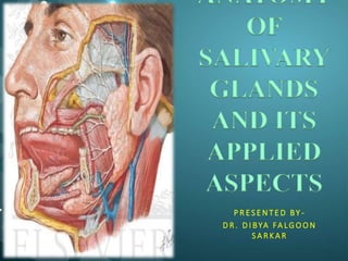
- 1. ANATOMY OF SALIVARY GLANDS AND ITS APPLIED ASPECTS P R ES E N T E D BY - D R . D I BYA FA LG O O N S A R K A R
- 2. INTRODUCTION • The salivary glands are exocrine glands, glands with ducts, that produce saliva and pour their secretion in the oral cavity • The major function of the salivary glands is to secrete saliva, which plays a significant role in lubrication, digestion, immunity, and the overall maintenanceof homeostasis within the human body . Major (Paired) • Parotid • Submandibular • Sublingual Minor - Those in the Tongue, Palatine Tonsil, Palate,Lips and Cheeks
- 3. PAROTID GLAND • Largest major salivary gland (15-30g weight) • Pyramidal shaped irregular lobulated mass lying mainly below the external acoustic meatus between mandible and sternomastoid. • They are compound tubuloalveolar glands with acini lined by seromucous cells
- 4. PAROTID CAPSULE • Derived from investing layer of deep cervical fascia which splits into superficial and deep layers to enclose the parotid gland. • Superficial lamina-Extends superiorly from the masseter to zygomatic arch. Thick and closely adherent-sends fibrous septa into the gland. • Deep lamina-thin- attached to styloid process, mandible and tympanic plate. Extends upto the stylomandibular ligament.
- 5. • Stylomandibular ligament- Separates the superficial and deep lobes of the parotid gland and also the submandibular gland from the parotid. The stylomandibular ligamentis an important surgical landmark when considering the resection of deep lobe tumors . In fact,stylomandibular tenotomy can be a crucial maneuver in providing exposure for en bloc resections of deep-lobe parotid or other parapharyngeal space tumors . ( Holsinger & Bui - Springer Publication)
- 6. ANATOMY OF PAROTID GLANDS
- 7. ANATOMY OF PAROTID GLAND • Resembles an inverted 3 sided pyramid • Four surfaces 1. Superior(Base of the Pyramid) 2.Superficial 3. Anteromedial 4. Posteromedial • Superior Surface is Concave : Related to 1. Cartilaginous part of ext acoustic meatus 2. Post. Aspect of temperomandibular joint 3. Auriculotemporal Nerve 4. Superficial temporal vessels • Apex : Overlaps posterior belly of digastric and adjoining part of carotid triangle • Superficial Surface : Covered by 1. Skin 2. Superficial fascia containing facial branches of great auricular Nerve 3. Superficial parotid lymph nodes and post fibers of platysma • Anteromedial Surface : Grooved by posterior border of ramus of mandible Related to • Masseter • Lateral Surface of temperomandibular joint • Medial pterygoid muscles • Emerging branches of Facial N • Posteromedial Surface: Related • to mastoid process with sternomastoid and posterior belly of digastric. •Styloid process with structures attached to it. • External Carotid A. which enters the gland through the surface • Internal Carotid A. which lies deep
- 8. BORDERS OF PAROTID GLAND
- 9. ACCESSORY PAROTID GLAND • Accessory parotid gland mayalso be present lying anteriorly over the masseter muscle between the parotid duct and zygoma .Its ducts empty directly into the parotid duct through one tributary . • Accessory glandular tissue is histologicallydistinct from parotid tissue in thatit maycontain mucinous acinar cellsin addition to the serous acinar cells
- 10. STRUCTURES WITHIN PAROTID GLAND • Facial Nerve • Retromandibular vein • External carotid artery
- 11. FACIAL NERVE • FACIAL NERVE Is intimately aasociated with the parotid gland, and understanding its anatomy is important for parotid surgeries. • It divides the parotid into superficial and deep lobes • Exits from the stylomastoid foramen and gives the posterior auricular and to posterior belly of digastric, before entering into the parotid • Within the parotid gland facial nerve forms the pes anserinus pattern of branching
- 12. • Davis et al described six different facial nerve branching patterns with no predominant pattern. • The major variations are in the origin of the buccal branch and the degree of cross innervations between adjacent terminal branches
- 13. VEINS PASSING THROUGH PAROTID GLAND
- 14. ARTERIES PASSING THROUGH PAROTID GLAND
- 15. PAROTID DUCT Ductus parotideus; Stensen’s duct • 5 cm in length • Appears in the anterior border of the gland • Runs anteriorly and downwards on the masseter b/w the upper and lower buccal branches of facial N. •At the anterior border of masseter it pierces • Buccal pad of fat • Buccopharyngeal fascia • Buccinator Muscle •It opens into the vestibule of mouth opposite to the 2nd upper molar
- 17. NERVE SUPPLY OF PAROTID GLAND
- 18. INCISIONS USED IN PAROTID SURGERY
- 19. OPERATIVE TECHNIQUES FOR PAROTIDECTOMY • A modified Blair incision is given in the natural crease just anterior to the helix and extends underneath the earlobe and superiorly over mastoid process • Then the incision curves inferiorly along anterior border of SCM • Incision continues below angle of mandible at distance safe from marginal mandibular nerve • Dissection is carried to the depth of parotid fascia and the platysma • Tail of parotid is separated from the SCM • Greater auricular nerve is to be preserved to maintain sensation to earlobe ( Lore et al 2004)
- 20. SURGICAL LANDMARKS FOR IDENTIFICATION OF FACIAL NERVE • Tragal pointer - The main trunk of the facial nerve is located 1 cm anteroinferior and 1 cm deep to the tip of the tragal cartilage. • Digastric ridge- The main trunk is just superior to the attachment of the posterior belly of the digastric muscle to the digastric groove. This landmark also marks the approximate depth of the facial nerve. • Stylomastoid foramen - The base of the styloid process is 5 to 8 mm deep to the tympanomastoid suture line. The facial nerve can be identified as it exits the stylomastoid foramen and passes over the posterolateral aspect of the styloid process. • Tympanomastoid suture line - The nerve lies 6 to 8 mm deep to the inferior end of the tympanomastoid suture line. • Mastoid - For revision cases, extensive tumors or, as a last resort, a mastoidectomy can be performed to locate the vertical segment of the facial nerve, which can then be followed as it exits the mastoid.
- 21. Identifying facial nerve intra operatively
- 22. APPLIED ASPECTS • Parotid swellings are very painful due to the unyeilding nature of the parotid fascia. • Parotid abscess is best drained by horizontal incision according to Hiltons method of incision and drainage. Vertical incision on skin but transverse incision on the parotid fascia to safeguard facial nerve and branches • Gustatory sweating, also known as Frey’s syndrome, presents as redness and perspiration over the cheek and parotid region in anticipation of eating. This complication may develop several years after parotidectomy and occurs when severed parasympathetic nerves to the parotid gland regenerate in an aberrant fashion and innervate sweat glands in the dermis.
- 24. ANATOMY OF SUBMANDIBULAR GLAND • The submandibular gland (“the submaxillary gland”) is the second largest major salivary gland and weighs 7–16g. • The gland is located in the submandibular triangle. • Large superficial and small deeper part continous with each other around the posterior border of mylohyoid. • During neckdissection or subman- dibular gland excision, this mylohyoid muscle must be gently retracted anteriorlyto expose the lingual nerve and submandibular ganglion .
- 25. FASCIA ENCLOSING THE SUBMANDIBULAR GLAND • The middle layer of the deep cervical fascia encloses the submandibular gland . • This fascia is clinically relevant because the marginal mandibular branch of the facial nerve is superficial to it, and care must be taken to preserve the nerve during surgery in the submandibular region . Thus, division of the submandibular gland fascia, when oncologically appropriate, is a reliable method of preserving and protecting the marginal mandibular branch of the facial nerve during neckdissection and/or submandibular gland resection .
- 26. RELATIONS OF SUBMANDIBULAR GLAND • Inferiorly- covered by • Skin • Superficial fascia containing platysma and cervical branches of facial N • Deep Fascia • Facial Vein • Submandibular Nodes • Lateral surface • Related to submandibular fossa on the mandible • Mandibular attachment of Medial pterygoid • Facial Artery • Medial surface • Anterior part- is related to mylohyoid muscle, nerve and vessels • Middle part - Hyoglossus, styloglossus, lingual nerve, submandibular ganglion, hypoglossal nerve and deep lingual vein. • Posterior Part - Styloglossus, stylohyoid ligament,9th nerve and wall of pharynx • Deep part • Small in size •Lies deep to mylohyoid and superficial to hyoglossus and styloglossus • Posteriorly continuous with superficial part around the posterior border mylohyoid
- 27. SUBMANDIBULAR DUCT • Wharton’s duct (5 cm long) • Emerges at the anterior end of deep part of the gland • Runs forwards on hyoglossus b/w lingual and hypoglossal N • At the ant. Border of hyoglossus it is crossed by lingual nerve • Opens in the floor of mouth at the side of frenulum of tongue
- 28. BLOOD SUPPLY AND LYMPHATIC DRAINAGE OF SUBMANDIBULAR GLANDS • Arteries - Branches of facial and lingual arteries • Veins - Drains to the corresponding veins • Lymphatics - Deep Cervical Nodes via submandibular nodes
- 30. APPLIED ASPECTS • The formation of calculus is more common in the submandibular gland than in the parotid. • For excision of the submandibular salivary gland( for calculus or tumour), a skin crease incision is as a rule, given more than 1inch( 2.5cm) below the angle of the jaw . • A stone in the submandibular duct(wharton’s duct) can be palpated bimanually in the floor of the mouth and can even be seen if sufficiently large.
- 31. EXTRAORAL INCISIONS USED IN SUBMANDIBULAR GLAND RESECTION • Placed 2-4 cm below the mandible, parallel to it • Preserve : • Marginal mandibular nerve • Lingual nerve • Hypoglossal nerve
- 33. KEY POINTS DURING SUBMANDIBULAR GLAND EXCISION • A safe way to preserve the marginal nerve is to ligate the facial vein and incise the submandibular gland fascia at the inferior border of the gland. These tissues can then be lifted from the gland along with the marginal nerve. • The hypoglossal nerve should be identified adjacent to the intermediate tendon of the digastric muscle. • The facial artery is encountered on the posterior aspect of the gland as it passes deep to the posterior belly of the digastric and enters the submandibular gland. The facial artery is then ligated, taking care to preserve the hypoglossal nerve. • The lingual nerve and efferent fibers of the chorda tympani can be identified deep to the mylohyoid muscle along with the submandibular ganglion. Just medial to the lingual nerve is Wharton’s duct. • Wharton’s duct should be ligated to prevent retrograde spread of infection from mouth. (Hsu et al 2009)
- 34. INTRAORAL APPROACH TO SUBMANDIBULAR GLANDS • This procedure is anatomically safe and can be performed with minimal morbidity • Infiltration with Xylocaine plus epinephrine with an adequate waiting period for hemostasis; The intraoral approach (IOA) consisted of an incision on the floor of mouth from the caruncle of Wharton's duct to the retromolar trigone • careful identification of the submandibular duct/lingual nerve relationship; • Anterior retraction of the mylohyoid muscle to expose the superficial lobe; • superiorly directed, extraoral, manipulation of the submandibular gland; • close and blunt dissection to the gland laterally to avoid injury to the facial artery and vein. (PMID: 10839409 [PubMed - indexed for MEDLINE] Division of Plastic and Reconstructive Surgery at the University of California, Los Angeles 90095-1665, USA.)
- 35. COMPLICATIONS • Transient weakness can also occur from stretch injury to the marginal nerve during surgery • Haematoma - The management of postoperative hematoma is wound exploration, clot evacuation, and hemostasis with bipolar cautery to avoid thermal injury to the marginal mandibular nerve.
- 36. SUBLINGUAL GLAND
- 37. ANATOMY OF SUBLINGUAL GLAND • Smallest of the three glands • Weighs nearly 3-4 gm • Lies beneath the oral mucosa in contact with the sublingual fossa on lingual aspect of mandible Duct (Ducts of Rivinus ) • 8-20 ducts • Most of them open directly into the floor of mouth • Few join the submandibular duct
- 38. RELATIONS OF SUBLINGUAL GLAND •Above - Mucosa of oral floor, raised as sublingual fold •Below - Mylohyoid Infront , Anterior end of its fellow •Behind -Deep part of Submandibular gland •Lateral -Mandible above the anterior part of mylohyoid line •Medial -Genioglossus and separated from it by lingual nerve and submandibular duct
- 39. •Blood supply • Arterial from sublingual and submental arteries • Venous drainage corresponds to the arteries •Nerve Supply Similar to that of submandibular glands(via lingual nerve , chorda tympani and sympathetic fibers)
- 40. SURGICAL APPROACH TO SUBLINGUAL GLAND - linear incision is made parellel and lateral to submandibular duct - incision shouldn’t extend more posteriorly to 1st molar tooth to avoid damage to lingual nerve - the submandibular duct is carefully identified and retracted medially - stay sutures-passing through margins of mucosa to aid in retention
- 41. • Using blunt dissection(scissors) lingual nerve is identified -the sublingual gland lying adjacent to inner cortex of mandible is mobilized and its multiple ducts are divided carefully to avoid damage to it. • The anterolateral part of sublingual gland may be attached to periosteum of mandible by fibrous tissue which must be divided carefully. followed by removal of gland as and when necessary
- 42. APPLIED ASPECTS • The structures at risk during dissection of the gland are the submandibular duct and the lingual nerve. • The duct lies superficially in the floor of the mouth medial to the sublingual fold, and is crossed inferiorly by the nerve which then enters the tongue • The sublingual artery and vein also lie on the medial aspect of the gland close to the submandibular duct and lingual nerve.
- 43. CONCLUSION • Surgical techniques for salivary gland surgery will continue to evolve as technologies develop. • A thorough understanding of the disease processes and anatomy will remain of paramount importance in the successful surgical management of salivary gland diseases.
Editor's Notes
- Composition of saliva
- Approximately 80% of the gland overlies the masseter muscle anteriorly and 20% extends medially through the stylomandibular tunnel, which is bordered by the posterior edge of the mandibular ramus, the posterior belly of digastric , the upper portion of sternocleidomastoid and the stylomandibular ligament
- Parotidomasseteric fascia- as it also covers the masseter deeply
- Tumors of superficial lobe – Superficial parotidectomy Tumors involving both lobes – Total Parotidectomy
- Submand glands are also resected in oral carcinoma cases with metastasising submand lymph nodes
- If malignancy is suspected, a submandibular lymph node dissection should be performed, including removal of perivascular nodes near the facial artery. High grade malignancies or malignancies with lymph node metastases may also require a complete neck dissection to remove metastatic disease.