
Recommended
More Related Content
Similar to Submandibular region and suprahyoid muscles.pptx
Similar to Submandibular region and suprahyoid muscles.pptx (20)
Recently uploaded
Recently uploaded (20)
Submandibular region and suprahyoid muscles.pptx
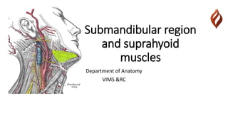
- 1. Submandibular region and suprahyoid muscles Department of Anatomy VIMS &RC
- 2. Submandibular region • The region extends from the under cover of the body of the mandible to hyoid bone • Contents are 1. Submandibular gland 2. Sublingual gland 3. Suprahyoid muscles 4. Extrinsic muscles of tongue 5. Lingual nerve 6. Submandibular ganglion 7. Lingual artery and facial artery
- 4. Submandibular gland • One among the paired salivary glands • Walnut in appearance • It’s a mixed salivary gland • Placed in digastric triangle • Partially occupying the submandibular fossa of mandible • It has a large superficial part and a small deep part • Continues around the posterior border of mylohyoid muscle
- 6. Parts • Superficial part has • Anterior end –extends up to the anterior belly of digastric • Posterior end –stylomandibular ligament • Inferior surface –investing layer of deep cervical fascia • Medial surface - investing layer of deep cervical fascia • Lateral surface –submandibular fossa of mandible
- 7. Relations • Relations of inferior surface • Skin • Superficial fascia • Platysma • Deep cervical fascia • Common facial vein • Cervical part of the facial nerve • Submandibular lymph nodes
- 9. Relations • Relations of lateral surface • Submandibular fossa • Medial pterygoid muscle • Facial artery –loops downwards and forwards between the bone and the gland ,winds around the anterio-inferior angle of the masseter before appearing on the face
- 10. Relations • Medial surface • Anterior pat -Rests on mylohyoid muscle • Separated by mylohyoid muscles and nerves • Posterior part-Styloid apparatus • Posterior belly of digastric muscle • Glossopharyngeal nerve • Middle constrictor of pharynx • Intermediate area – • hyoglossus muscle • Lingual nerve • Submandibular ganglion
- 11. Relations • Deep part • Extends from the interval between the mylohyoid and hyoglossus muscle to the sublingual salivary glands • Related to • Medially -hyoglossus muscle • Laterally-mylohyoid muscle • Above –lingual nerve and submandibular ganglion • Below –hypoglossal nerve
- 12. • Blood supply – • Branches of facial artery and lingual artery • Corresponding veins drain to Internal jugular vein • Lymphatic drainage • Submandibular lymph nodes –jugulo-digastric lymph nodes
- 13. Submandibular duct • Wharton’s duct • 5 cm long • Starts -Middle of the deep surface of the superficial part of the gland • Direction-Upward and backward ,forward and upward • Opens -On the floor of the mouth at the sublingual papillae on each side of the frenulum linguae
- 15. Nerve supply-preganglionic fibres Superior salivatory nucleus Facial nerve Chorda typmani nerve Lingual nerve(post root ) Submandibular ganglion
- 16. Nerve supply –post ganglionic fibres Submandibular ganglion Lingual nerve (ant root) Submandibular Sublingual glands
- 17. Sublingual gland • Smallest • Located in the floor of the mouth • Lodges the sublingual fossa • Between the mucous membrane and mylohyoid muscle • Ducts -8-20 small ducts • Most of them opens separately on the floor of the mouth on the sublingual fold (Ducts of Rivinus ) • Some of them unite to form sublingual duct and joins the submandibular duct (Ducts of Bartholin )
- 19. Sublingual gland • Blood supply –sublingual and submental arteries -facial artery • Lymphatics- submental group of lymph nodes • Nerve supply –same as submandibular gland • Relations – • Infront -Meets the opposite side gland at symphysis menti • Behind –deep part of submandibular gland • Above –covered by mucous membrane of mouth forming the sublingual fold • Below –mylohyoid muscle • Laterally-sublingual fossa • Medially –rests on genioglossus
- 20. Applied anatomy • The submandibular gland may enlarge because of the obstruction of its duct due to a calculus or by tumour • Sialography – visualization of the duct system of the major salivary gland dusts in x ray after injecting radio opaque dye • Ranula –cystic swelling of the floor of the mouth due to obstruction of the ducts of sublingual gland
- 22. Supra hyoid muscles • Geniohyoid –Genial tubercle –c1 via hypoglossal nerve • Anterior belly of digastric-digastric fossa –mylohyoid nerve • Posterior belly of digastric –mastoid notch –facial nerve • Mylohyoid -mylohyoid line –mylohyoid nerve • Stylohyoid-styloid process -facial nerve
- 25. Extrinsic tongue muscles • Genioglossus • Hyoglossus • Styloglossus
- 26. Lingual nerve • Branch of posterior division of mandibular nerve • Lies in infratemporal fossa • Where chorda tympani joins the nerve • Comes in direct contact with mandible covered by mucous membrane ,medial to the third molar tooth • Rests on hyoglossus • Submandibular ganglion suspends from the lower border with two roots • Winds around the lower border of the submandibular duct • Sensory nerve supply to the pre-sulcal part of the tongue
- 28. Submandibular ganglion • Small fusiform ganglion • Part of Parasympathetic system • Structurally related to lingual • Functionally connected to facial nerve and Chordatympani • Consists of parasympathetic and sympathetic roots • Parasympathetic root – secretomotor pathway to submandibular and sublingual glands • Sympathetic root –primary vasomotor supply to the blood vessels around the glands
- 30. Thank you