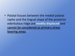
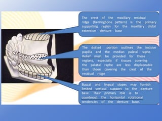
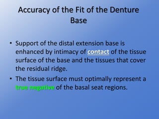
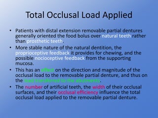
- Distal extension removable partial dentures depend on the residual ridge for support since they do not have full tooth support.
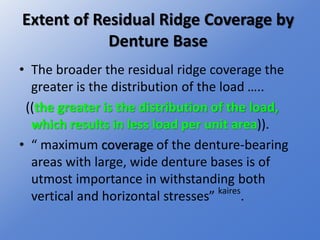
- Several factors influence the support provided by the residual ridge, including its contour, the extent of coverage by the denture base, impression accuracy, fit of the denture base, framework design, and occlusal load.
- The ideal residual ridge for support would have dense cortical bone covering cancellous bone with a broad, rounded crest and sloping sides covered by dense tissue.