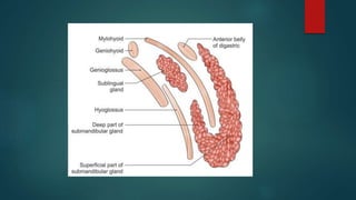
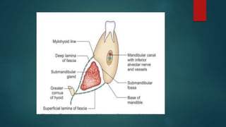
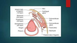
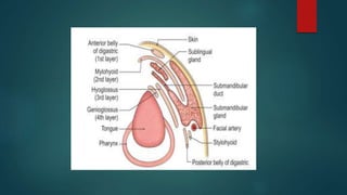
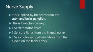
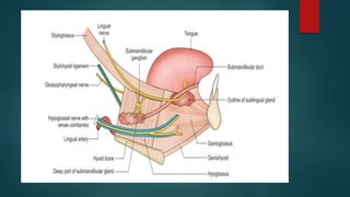
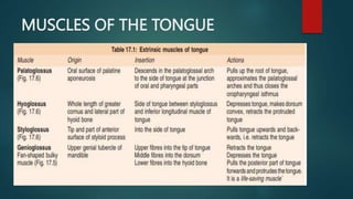
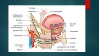
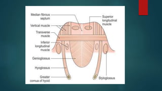
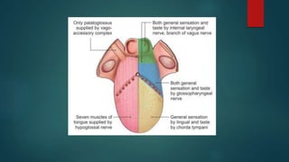
ANATOMY OF
SUBMANDIBULAR GLAND
AND TONGUE
DR MESHWA P. OZA
ENT RESIDENT
MK SHAH MEDICAL COLLEGE AHMEDABAD
SUBMANDIBULAR GLAND
 It is situated in the anterior part of the
digastric triangle.
 The gland is about the size of a walnut
weighing about 15 to 20 g.
 It is roughly J-shaped, being indented by the
posterior border of the mylohyoid
 It divides the gland into a larger part
superficial to the muscle, and a small part
lying deep to the muscle
COVERINGS
 The gland is partially enclosed between two layers of
deep cervical fascia.
 The superficial layer of fascia covers the inferior
surface of the gland and is attached to the base of
the mandible.
 The deep layer covers the medial surface of the
gland and is superiorly to the mylohyoid line of the
mandible
Superficial Part
 This part of the gland fills the digastric triangle.
 It extends superiorly deep to the mandible up to the
mylohyoid line.
 Inferiorly, It overlaps stylohyoid and the posterior
belly of digastric.
 It has three surfaces:
 a. Inferior
 b. Lateral
 c. Medial
Relations
 The inferior surface is covered by:
 a. Skin
 b. Platysma
 c. Cervical branch of the facial nerve
 d. Deep fascia
 e. Facial vein
 f. Submandibular lymph nodes
 The lateral surface is related to:
 a. The submandibular fossa on the mandible
 b. Insertion of the medial pterygoid
 c. The facial artery
 The medial surface is related to:
 • Anterior part: Mylohyoid, submental branch of facial
artery, mylohyoid nerve and vessels
 • Middle part: Hyoglossus, styloglossus, lingual artery,
artery, XII nerve
 • Posterior part: Stylohyoid, styloglossus, IX nerve
Deep Part
 This part is small in size.
 It lies deep to the mylohyoid, and superficial to the
hyoglossus and the styloglossus .
 Posteriorly, it is continuous with the superficial part
around the posterior border of the mylohyoid.
 Anteriorly, it extends up to the posterior end of the
sublingual gland.
Relations
 Present in between mylohyoid and hyoglossus.
 Laterally – Mylohyoid
 Medially – Hyoglossus
 Above – Lingual nerve with submandibular
ganglion
 Below – Hypoglossal nerve
Blood Supply and Lymphatic Drainage
 Arterial supply- Facial artery.
 The facial artery arises from the external
carotid just above the tip of the greater cornua
of the hyoid bone.
 The veins drain into the common facial or
lingual vein.
 Lymph passes to submandibular lymph nodes
Nerve Supply
 It is supplied by branches from the
submandibular ganglion.
 These branches convey:
 1 Secretomotor fibres
 2 Sensory fibres from the lingual nerve
 3 Vasomotor sympathetic fibres from the
plexus on the facial artery
SUBMANDIBULAR DUCT/
WHARTON’S DUCT
 It is thin walled, and is about 5 cm long.
 It emerges at the anterior end of the deep part of the gland and
runs upwards and forwards on the hyoglossus, between the
lingual and hypoglossal nerves.
 At the anterior border of the hyoglossus, the duct is crossed by
the lingual nerve .

 It opens on the floor of the mouth, on the summit of the
sublingual papilla, at the side of the frenulum of the tongue
Tongue
 The tongue is a muscular organ situated in the floor
of the mouth.
 It is associated with the functions of
 (i) taste
 (ii) speech
 (iii) chewing
 (iv) deglutition
 (v) cleansing of mouth.
PARTS OF TONGUE
 1 A root
 2 A tip
 3 A body, which has:
 a. A curved upper surface or dorsum
 b. An inferior surface confined to the oral part only.
MUSCLES OF THE TONGUE
Arterial Supply
 It is derived from the tortuous lingual artery, a branch
of the external carotid artery.
 The root of the tongue is also supplied by the
tonsillar artery, a branch of facial artery, and
ascending pharyngeal branch of external carotid
artery
Venous Drainage
 1 Deep lingual vein: The chief vein of tongue, seen on
the inferior surface of tongue near median plane.
 2 Venae comitantes, accompany lingual artery. They
are joined by dorsal lingual veins.
 3 Venae comitantes accompanying the hypoglossal
nerve.
Lymphatic Drainage
 1 The tip of the tongue drains bilaterally to the
submental nodes.
 2 The right and left halves of the remaining part of
the anterior two-thirds of the tongue drain
unilaterally to the submandibular nodes.
 A few central lymphatics drain bilaterally to the deep
cervical nodes.
 3 The posteriormost part and posterior one-
third of the tongue drain bilaterally into the
upper deep cervical lymph nodes including
jugulodigastric nodes.
 4 The whole lymph finally drains to the
jugulo-omohyoid nodes.
 These are known as the lymph nodes of the
tongue.
Nerve Supply
Motor Nerves
All the intrinsic and extrinsic muscles, except the
palatoglossus, are supplied by the hypoglossal nerve.
The palatoglossus is supplied by the cranial root of the
accessory nerve through the pharyngeal plexus.
So seven out of eight muscles are supplied by XII nerve
Sensory Nerves
 The lingual nerve is the nerve of general sensation for anterior
two-thirds of the tongue
 The chorda tympani is the nerve of taste for the anterior two-
thirds of the tongue except vallate papillae.
 The glossopharyngeal nerve is the nerve for both general
sensation and taste for the posterior one-third of the tongue
including the circumvallate papillae.
 The posteriormost part of the tongue is supplied by the vagus
nerve through the internal laryngeal branch
Taste pathway
THANK YOU

meshwa ppt tongue final.pptx

  • 1.
    ANATOMY OF SUBMANDIBULAR GLAND ANDTONGUE DR MESHWA P. OZA ENT RESIDENT MK SHAH MEDICAL COLLEGE AHMEDABAD
  • 2.
    SUBMANDIBULAR GLAND  Itis situated in the anterior part of the digastric triangle.  The gland is about the size of a walnut weighing about 15 to 20 g.
  • 3.
     It isroughly J-shaped, being indented by the posterior border of the mylohyoid  It divides the gland into a larger part superficial to the muscle, and a small part lying deep to the muscle
  • 5.
    COVERINGS  The glandis partially enclosed between two layers of deep cervical fascia.  The superficial layer of fascia covers the inferior surface of the gland and is attached to the base of the mandible.  The deep layer covers the medial surface of the gland and is superiorly to the mylohyoid line of the mandible
  • 7.
    Superficial Part  Thispart of the gland fills the digastric triangle.  It extends superiorly deep to the mandible up to the mylohyoid line.  Inferiorly, It overlaps stylohyoid and the posterior belly of digastric.  It has three surfaces:  a. Inferior  b. Lateral  c. Medial
  • 8.
    Relations  The inferiorsurface is covered by:  a. Skin  b. Platysma  c. Cervical branch of the facial nerve  d. Deep fascia  e. Facial vein  f. Submandibular lymph nodes
  • 9.
     The lateralsurface is related to:  a. The submandibular fossa on the mandible  b. Insertion of the medial pterygoid  c. The facial artery
  • 11.
     The medialsurface is related to:  • Anterior part: Mylohyoid, submental branch of facial artery, mylohyoid nerve and vessels  • Middle part: Hyoglossus, styloglossus, lingual artery, artery, XII nerve  • Posterior part: Stylohyoid, styloglossus, IX nerve
  • 13.
    Deep Part  Thispart is small in size.  It lies deep to the mylohyoid, and superficial to the hyoglossus and the styloglossus .  Posteriorly, it is continuous with the superficial part around the posterior border of the mylohyoid.  Anteriorly, it extends up to the posterior end of the sublingual gland.
  • 14.
    Relations  Present inbetween mylohyoid and hyoglossus.  Laterally – Mylohyoid  Medially – Hyoglossus  Above – Lingual nerve with submandibular ganglion  Below – Hypoglossal nerve
  • 16.
    Blood Supply andLymphatic Drainage  Arterial supply- Facial artery.  The facial artery arises from the external carotid just above the tip of the greater cornua of the hyoid bone.  The veins drain into the common facial or lingual vein.  Lymph passes to submandibular lymph nodes
  • 17.
    Nerve Supply  Itis supplied by branches from the submandibular ganglion.  These branches convey:  1 Secretomotor fibres  2 Sensory fibres from the lingual nerve  3 Vasomotor sympathetic fibres from the plexus on the facial artery
  • 19.
    SUBMANDIBULAR DUCT/ WHARTON’S DUCT It is thin walled, and is about 5 cm long.  It emerges at the anterior end of the deep part of the gland and runs upwards and forwards on the hyoglossus, between the lingual and hypoglossal nerves.  At the anterior border of the hyoglossus, the duct is crossed by the lingual nerve .   It opens on the floor of the mouth, on the summit of the sublingual papilla, at the side of the frenulum of the tongue
  • 21.
  • 22.
     The tongueis a muscular organ situated in the floor of the mouth.  It is associated with the functions of  (i) taste  (ii) speech  (iii) chewing  (iv) deglutition  (v) cleansing of mouth.
  • 23.
    PARTS OF TONGUE 1 A root  2 A tip  3 A body, which has:  a. A curved upper surface or dorsum  b. An inferior surface confined to the oral part only.
  • 25.
  • 30.
    Arterial Supply  Itis derived from the tortuous lingual artery, a branch of the external carotid artery.  The root of the tongue is also supplied by the tonsillar artery, a branch of facial artery, and ascending pharyngeal branch of external carotid artery
  • 31.
    Venous Drainage  1Deep lingual vein: The chief vein of tongue, seen on the inferior surface of tongue near median plane.  2 Venae comitantes, accompany lingual artery. They are joined by dorsal lingual veins.  3 Venae comitantes accompanying the hypoglossal nerve.
  • 32.
    Lymphatic Drainage  1The tip of the tongue drains bilaterally to the submental nodes.  2 The right and left halves of the remaining part of the anterior two-thirds of the tongue drain unilaterally to the submandibular nodes.  A few central lymphatics drain bilaterally to the deep cervical nodes.
  • 33.
     3 Theposteriormost part and posterior one- third of the tongue drain bilaterally into the upper deep cervical lymph nodes including jugulodigastric nodes.  4 The whole lymph finally drains to the jugulo-omohyoid nodes.  These are known as the lymph nodes of the tongue.
  • 35.
    Nerve Supply Motor Nerves Allthe intrinsic and extrinsic muscles, except the palatoglossus, are supplied by the hypoglossal nerve. The palatoglossus is supplied by the cranial root of the accessory nerve through the pharyngeal plexus. So seven out of eight muscles are supplied by XII nerve
  • 36.
    Sensory Nerves  Thelingual nerve is the nerve of general sensation for anterior two-thirds of the tongue  The chorda tympani is the nerve of taste for the anterior two- thirds of the tongue except vallate papillae.  The glossopharyngeal nerve is the nerve for both general sensation and taste for the posterior one-third of the tongue including the circumvallate papillae.  The posteriormost part of the tongue is supplied by the vagus nerve through the internal laryngeal branch
  • 38.
  • 39.