The document discusses the anatomy and physiology of the heart and pericardium. It describes the fibrous skeleton that forms a structural foundation around the atrioventricular and arterial orifices. It then details the specialized conduction system of the heart, including the sinoatrial node, atrioventricular node, bundle of His, and Purkinje fibers. Finally, it explains the coronary circulation, noting the right and left coronary arteries and their branches that supply the heart muscle.
Join live classes, download study aids, sell your documents, join or host your own classes online, get tutoring, tutor students, take practices tests and more at Examville.com
The heart has four chambers. The two superior receiving chambers are the atria (= entry halls or chambers), and the two inferior pumping chambers are the ventricles (= little bellies).
On the anterior surface of each atrium is a wrinkled pouchlike structure called an auricle
USMLE CVS 005 Blood vessels – Arteries and veins.pdfAHMED ASHOUR
The major blood vessels in the human body form an extensive network that facilitates the transportation of blood, oxygen, and nutrients to various tissues and organs.
Understanding the anatomy and function of major blood vessels is essential for comprehending the circulatory system and diagnosing and treating cardiovascular conditions.
Synthetic Fiber Construction in lab .pptxPavel ( NSTU)
Synthetic fiber production is a fascinating and complex field that blends chemistry, engineering, and environmental science. By understanding these aspects, students can gain a comprehensive view of synthetic fiber production, its impact on society and the environment, and the potential for future innovations. Synthetic fibers play a crucial role in modern society, impacting various aspects of daily life, industry, and the environment. ynthetic fibers are integral to modern life, offering a range of benefits from cost-effectiveness and versatility to innovative applications and performance characteristics. While they pose environmental challenges, ongoing research and development aim to create more sustainable and eco-friendly alternatives. Understanding the importance of synthetic fibers helps in appreciating their role in the economy, industry, and daily life, while also emphasizing the need for sustainable practices and innovation.
Introduction to AI for Nonprofits with Tapp NetworkTechSoup
Dive into the world of AI! Experts Jon Hill and Tareq Monaur will guide you through AI's role in enhancing nonprofit websites and basic marketing strategies, making it easy to understand and apply.
Unit 8 - Information and Communication Technology (Paper I).pdfThiyagu K
This slides describes the basic concepts of ICT, basics of Email, Emerging Technology and Digital Initiatives in Education. This presentations aligns with the UGC Paper I syllabus.
June 3, 2024 Anti-Semitism Letter Sent to MIT President Kornbluth and MIT Cor...Levi Shapiro
Letter from the Congress of the United States regarding Anti-Semitism sent June 3rd to MIT President Sally Kornbluth, MIT Corp Chair, Mark Gorenberg
Dear Dr. Kornbluth and Mr. Gorenberg,
The US House of Representatives is deeply concerned by ongoing and pervasive acts of antisemitic
harassment and intimidation at the Massachusetts Institute of Technology (MIT). Failing to act decisively to ensure a safe learning environment for all students would be a grave dereliction of your responsibilities as President of MIT and Chair of the MIT Corporation.
This Congress will not stand idly by and allow an environment hostile to Jewish students to persist. The House believes that your institution is in violation of Title VI of the Civil Rights Act, and the inability or
unwillingness to rectify this violation through action requires accountability.
Postsecondary education is a unique opportunity for students to learn and have their ideas and beliefs challenged. However, universities receiving hundreds of millions of federal funds annually have denied
students that opportunity and have been hijacked to become venues for the promotion of terrorism, antisemitic harassment and intimidation, unlawful encampments, and in some cases, assaults and riots.
The House of Representatives will not countenance the use of federal funds to indoctrinate students into hateful, antisemitic, anti-American supporters of terrorism. Investigations into campus antisemitism by the Committee on Education and the Workforce and the Committee on Ways and Means have been expanded into a Congress-wide probe across all relevant jurisdictions to address this national crisis. The undersigned Committees will conduct oversight into the use of federal funds at MIT and its learning environment under authorities granted to each Committee.
• The Committee on Education and the Workforce has been investigating your institution since December 7, 2023. The Committee has broad jurisdiction over postsecondary education, including its compliance with Title VI of the Civil Rights Act, campus safety concerns over disruptions to the learning environment, and the awarding of federal student aid under the Higher Education Act.
• The Committee on Oversight and Accountability is investigating the sources of funding and other support flowing to groups espousing pro-Hamas propaganda and engaged in antisemitic harassment and intimidation of students. The Committee on Oversight and Accountability is the principal oversight committee of the US House of Representatives and has broad authority to investigate “any matter” at “any time” under House Rule X.
• The Committee on Ways and Means has been investigating several universities since November 15, 2023, when the Committee held a hearing entitled From Ivory Towers to Dark Corners: Investigating the Nexus Between Antisemitism, Tax-Exempt Universities, and Terror Financing. The Committee followed the hearing with letters to those institutions on January 10, 202
Palestine last event orientationfvgnh .pptxRaedMohamed3
An EFL lesson about the current events in Palestine. It is intended to be for intermediate students who wish to increase their listening skills through a short lesson in power point.
Instructions for Submissions thorugh G- Classroom.pptxJheel Barad
This presentation provides a briefing on how to upload submissions and documents in Google Classroom. It was prepared as part of an orientation for new Sainik School in-service teacher trainees. As a training officer, my goal is to ensure that you are comfortable and proficient with this essential tool for managing assignments and fostering student engagement.
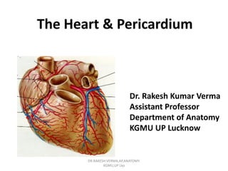
1. The Heart & Pericardium
Dr. Rakesh Kumar Verma
Assistant Professor
Department of Anatomy
KGMU UP Lucknow
DR.RAKESH VERMA,AP,ANATOMY.
KGMU,UP Lko
2. Fibrous skeleton
• Dense fibrous connective
tissue forms a structural
foundation around AV &
arterial orifices.
• It is site of attachment to atria
& ventricles.
• Musculature of atria &
ventricles does not cross this
fibrous ring.
• It is also an electrical insulator
between atria and ventricles
except via AV Bundle/Bundle
of His.
DR.RAKESH VERMA,AP,ANATOMY.
KGMU,UP Lko
A
P
L R
3. Conduction system
– Specialized cardiac muscle fibers
– Self-excitable
– Repeatedly generate action potentials that
trigger heart contractions
– Act as pacemaker & Form conduction
system
– Consist of SA , AV Node,Bundle of His &
Purkinje fibers
DR.RAKESH VERMA,AP,ANATOMY.
KGMU,UP Lko
4. Conduction System
• SA node acts as natural pacemaker
– Faster than other auto rhythmic fibers
– Initiates 100 times per second
• Nerve impulses from autonomic nervous
system (ANS) and hormones modify timing
and strength of each heartbeat
DR.RAKESH VERMA,AP,ANATOMY.
KGMU,UP Lko
5. Conduction system
1. Begins in sinoatrial (SA) node in right atrial wall
• Propagates through atria via gap junctions
• Atria contact
2. Reaches atrioventricular (AV) node in interatrial
septum
3. Enters atrioventricular (AV) bundle (Bundle of His)
• Only site where action potentials can conduct from
atria to ventricles due to fibrous skeleton
4. Enters right and left bundle branches which extends
through interventricular septum toward apex
5. Finally, large diameter Purkinje fibers conduct action
potential to remainder of ventricular myocardium
DR.RAKESH VERMA,AP,ANATOMY.
KGMU,UP Lko
6. Frontal plane
Right atrium
Right ventricle
Left atrium
Left ventricle
Anterior view of frontal section
Frontal plane
Left atrium
Left ventricle
Anterior view of frontal section
SINOATRIAL (SA) NODE
1
Right atrium
Right ventricle
Frontal plane
Left atrium
Left ventricle
Anterior view of frontal section
SINOATRIAL (SA) NODE
ATRIOVENTRICULAR
(AV) NODE
1
2
Right atrium
Right ventricle
Frontal plane
Left atrium
Left ventricle
Anterior view of frontal section
SINOATRIAL (SA) NODE
ATRIOVENTRICULAR
(AV) NODE
ATRIOVENTRICULAR (AV)
BUNDLE (BUNDLE OF HIS)
1
2
3
Right atrium
Right ventricle
Frontal plane
Left atrium
Left ventricle
Anterior view of frontal section
SINOATRIAL (SA) NODE
ATRIOVENTRICULAR
(AV) NODE
ATRIOVENTRICULAR (AV)
BUNDLE (BUNDLE OF HIS)
RIGHT AND LEFT
BUNDLE BRANCHES
1
2
3
4
Right atrium
Right ventricle
Frontal plane
SINOATRIAL (SA) NODE
ATRIOVENTRICULAR
(AV) NODE
Left atrium
Left ventricle
Anterior view of frontal section
ATRIOVENTRICULAR (AV)
BUNDLE (BUNDLE OF HIS)
RIGHT AND LEFT
BUNDLE BRANCHES
PURKINJE FIBERS
1
2
3
4
5
Right atrium
Right ventricle
DR.RAKESH VERMA,AP,ANATOMY.
KGMU,UP Lko
8. Coronary Circulation
• Coronary circulation is the functional blood
supply to the heart muscle itself
• Collateral routes ensure blood delivery to
heart even if major vessels are occluded
DR.RAKESH VERMA,AP,ANATOMY.
KGMU,UP Lko
9. Coronary Circulation: Arterial Supply
• Heart is supplied by
TWO CORONARY
arteries:
1- Right coronary
artery(RCA)
2- Left coronary
artery(LCA)
• These coronary arteries
arise at the root of the
aorta.
DR.RAKESH VERMA,AP,ANATOMY.
KGMU,UP Lko
11. Coronary artery & their branches
• LCA supplies ant.
and apical parts of
heart ,and
ant.2/3rd of
interventricular
septum via LAD .
• Circumflex branch-
- supplies the
lateral and
post.surface of
heart.
DR.RAKESH VERMA,AP,ANATOMY.
KGMU,UP Lko
12. Coronary artery & their branches
RCA supplies:
• Right ventricle
• Interventricular
septum (posterior
1/3rd part)
• Inferior part of left
ventricle
• Conducting system
except Left branch
of bundle of His
supplied by LCA
DR.RAKESH VERMA,AP,ANATOMY.
KGMU,UP Lko
13. Cardiac Dominance
• The right coronary gives
posterior interventricular
artery &supply
conduction system, such
hearts are Right
Dominance
• In 10% cases posterior
interventricular artery
come from circumflex
artery which supply
posterior interventricular
septum as well as AV
node, such hearts are Left
Dominance
DR.RAKESH VERMA,AP,ANATOMY.
KGMU,UP Lko
14. Venous return of Heart
• Most of the venous
blood return to heart
occurs through the
coronary sinus
• Venae cardis
minimi/Thebesian vein
& Anterior cardiac veins
directerly drain into the
right atrium
DR.RAKESH VERMA,AP,ANATOMY.
KGMU,UP Lko
15. Venous return of Heart
• Great cardiac vein
• Middle cardiac vein
• Small cardiac vein
• Posterior vein of left
ventricle
• Right marginal vein
• Oblique vein of left
atrium of Marshall
DR.RAKESH VERMA,AP,ANATOMY.
KGMU,UP Lko
16. Blood flow to Heart during Systole & Diastole
• During systole when heart muscle contracts it
compresses the coronary arteries therefore
blood flow is less.
• As we know blood flow to subendocardial
surface of left ventricle during systole is not
there, therefore, this region is prone to
ischemic damage and most common site of
Myocardial infarction.
DR.RAKESH VERMA,AP,ANATOMY.
KGMU,UP Lko
17. CORONARY BLOOD FLOW
• Coronary blood flow to the right side is not
much affected during systole.
Reason---Pressure difference between
aorta and right ventricle is greater during
systole than during diastole, therefore more
blood flow to right ventricle occurs during
systole.
DR.RAKESH VERMA,AP,ANATOMY.
KGMU,UP Lko
18. CORONARY BLOOD FLOW
• Coronary blood flow in Humans at rest is
about 225-250 ml/minute, about 5% of
cardiac output.
• At rest, the heart extracts 60-70% of oxygen
from each unit of blood delivered to heart
[other tissue extract only 25% of O2.
DR.RAKESH VERMA,AP,ANATOMY.
KGMU,UP Lko
20. Moral of the Story
Well-deserved Royalty:
Not just to have a
crown….but to have a well-
fitting ONE!
DR.RAKESH VERMA,AP,ANATOMY.
KGMU,UP Lko
21. Lymphatic Drainage
• Right trunk- to brachiocephalic node
• Left trunk-to tracheobronchial node
DR.RAKESH VERMA,AP,ANATOMY.
KGMU,UP Lko
22. Innervation of Heart
• Parasympathetic-
Vagus &
recurrent
laryngeal
• Sympathetic-
Upper 4 to 5
thoracic segment
of spinal cord
• Both forming
superficial &
deep cardiac
plexus
DR.RAKESH VERMA,AP,ANATOMY.
KGMU,UP Lko
23. Innervation of Heart
• The superficial
plexus lies in the
concavity of
the aortic arch, and
a deep plexus
between the aortic
arch and
the trachea.
• Both are closely
connected.
DR.RAKESH VERMA,AP,ANATOMY.
KGMU,UP Lko
24. Innervation of Heart
superficial plexus
• It is formed by the
superior cardiac branch
of the left sympathetic
trunk and the lower
superior cervical cardiac
branch of the left vagus
nerve.
• Its branches are-
• (a) to the deep part of
the plexus;
• (b) to the anterior
coronary plexus; and
• (c) to the left
anterior pulmonary
plexus. DR.RAKESH VERMA,AP,ANATOMY.
KGMU,UP Lko
25. Innervation of Heart
• The deep cardiac plexus
is formed by the cardiac
nerves derived from the
cervical ganglia of the
sympathetic trunk, and
the cardiac branches of
the vagus and recurrent
laryngeal nerves except
superior cardiac branch
of the left sympathetic
trunk and the lower
superior cervical cardiac
branch of the left vagus
nerve which form
superficial plexus .
DR.RAKESH VERMA,AP,ANATOMY.
KGMU,UP Lko
26. Innervation of Heart
• The branches from the right half of the deep part
of the cardiac plexus form the anterior
pulmonary plexus, and the anterior coronary
plexus.
• Those behind the pulmonary artery to form part
of the posterior coronary plexus.
• The left half of the deep part of the plexus is
connected with the superficial cardiac plexus,
and to the anterior pulmonary plexus, and is
then continued to form the greater part of the
posterior coronary plexus.
DR.RAKESH VERMA,AP,ANATOMY.
KGMU,UP Lko
27. Anatomical basis of referred pain from heart
• The pain sensation is carried by middle and
inferior cervical cardiac branches and thoracic
cardiac branches of sympathetic chain to
thoracic ganglion of sympathetic chain.
• From here , impulses passes via the white rami
to thoracic spinal nerve(T-1 to T-5) .
• The central process of the neurons in dorsal
root ganglion carry impulses to posterior horn
of T-1 to T-5 spinal segment mainly of left side.
DR.RAKESH VERMA,AP,ANATOMY.
KGMU,UP Lko
28. Anatomical basis of referred pain from heart
• The pain sensation of ischemic myocardium
reach the spinal cord, they stimulate the
somatic sensory neurons of corresponding
spinal segment. Therefore pain is felt in
medial side of left arm, forearm &
anterosuperior part of chest & back as well .
DR.RAKESH VERMA,AP,ANATOMY.
KGMU,UP Lko
29. Applied Anatomy
• Pericarditis & pericadial effusion
• Cardiac tamponade
• Ligation of ascending of aorta & pulmonary
trunk through transverse sinus during cardiac
surgery
• Dextrocardia (situs inversus)
• Apex shift in Ventricular hypertrophy
• Arrythymia,tachycardia,bradycardia
DR.RAKESH VERMA,AP,ANATOMY.
KGMU,UP Lko
30. Applied Anatomy
Rheumatic heart ds.
• Pericarditis, Myocarditis & endocarditis
• Mitral valve stenosis and regurgitation
• Tricuspid valve stenosis and regurgitation
• Aortic valve stenosis and regurgitation
• Pulmonary valve stenosis
IHD-
• Angina pectoris
• MI
• Angiography & Angioplasty
• Bypass surgery
DR.RAKESH VERMA,AP,ANATOMY.
KGMU,UP Lko
31. Question-1
Crista terminalis is a feature of
a. Right ventricle
b. Left ventricle
c. Right atrium
d. Left atrium
DR.RAKESH VERMA,AP,ANATOMY.
KGMU,UP Lko
32. Question-2
Right bundle branch (RBB) travels in
a. Triangle of Koch
b. Moderator band
c. Membranous interventricular septum
d. Supraventricular crest
DR.RAKESH VERMA,AP,ANATOMY.
KGMU,UP Lko
33. Question-3
The most anterior valve of heart is
a. Tricuspid
b. Mitral
c. Pulmonary
d. Aortic
DR.RAKESH VERMA,AP,ANATOMY.
KGMU,UP Lko
34. Question-4
Conducting tissue of the heart is a modification
of
a. Epicardium
b. Myocardium
c. Endocardium
d. Nerve fibers
DR.RAKESH VERMA,AP,ANATOMY.
KGMU,UP Lko
35. Question-5
The right margin of cardiac shadow in a
radiograph is formed by all of the following
except
a. Superior vena cava
b. Right atrium
c. Right ventricle
d. Right brachiocephalic vein
DR.RAKESH VERMA,AP,ANATOMY.
KGMU,UP Lko
36. Question-6
An unconscious patient involved in a major car
accident was brought to the casualty with an
injury of sharp object on his chest in the middle
of the sternum at the level of 4th costal cartilage.
Which chamber of heart is likely to be
punctured?
a. Left atrium
b. Left ventricle
c. Right atrium
d. Right ventricle
DR.RAKESH VERMA,AP,ANATOMY.
KGMU,UP Lko
37. Question-7
The acute margin of heart is
a. Inferior
b. Superior
c. Left
d. Right
DR.RAKESH VERMA,AP,ANATOMY.
KGMU,UP Lko