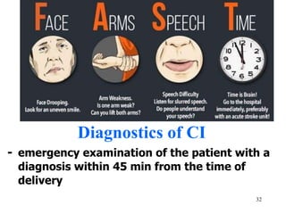
1
VASCULAR DISORDERS
2
Сerebrovascular diseases
- 3 place on death rate
- leading cause of disability
- 30% of stroke sufferers die in
the course of the first month from
the moment of stroke
- 45-48% of patients die by the
end of the year
3
Cerebrovascular diseases
Clinical classification
I. Acute
1.Transient strokes
- Transient ischemic attack (TIA)
- Cerebral hypertensive crises
- Acute hypertensive encephalopathy
2. Permanent strokes
- Cerebral infarction (85%)
- Subarachnoid haemorrhage (1-5%)
- Intracerebral haemorrhage (10-12%) II.
Chronic diseases
4
Cerebrovascular diseases
Classification (ICD-10):
I60 Subarachnoid haemorrhage
I61 Intracerebral haemorrhage
I62 Other nontraumatic intracranial
haemorrhage
I63 Cerebral infarction
I64 Stroke, not specified as
haemorrhage or infarction G45
Transient cerebral ischemic
attacks and related syndromes
5
Transient strokes
Transient ischemic attack (TIA) ―
is an acute episode of temporary
neurologic dysfunction resulting from
focal cerebral ischemia not associated
with permanent cerebral infarction
and lasting less than 24h.
• TIA is caused by a temporary
blockage of one of the arteries that
supply the brain.
6
Symptoms of TIA in carotid
arteries
• hemiparesis
• hemihypesthesia
• aphasia and apraxia (in damage of
the dominant hemisphere)
• anosognosia
• blindness or decreased vision in one
eye
• central paresis of the facial nerve
7
Symptoms of TIA in in vertebral
and basilar arteries
- dizziness
- nausea and vomiting
- double vision
- dysarthria and dysphagia
- ataxia
- amnesia
- cortical blindness
- tetraparesis
- hearing loss
- peripheral paresis of the facial nerve
8
Cerebral hypertensive crises
are associated with hypertensive
diseases and accompanied by
symptoms of increased intracranial
pressure.
Symptoms: occipital headache and
visual disorders (photopsy);
hyperhydrosis; hyperemia of face;
a sharp headache; a nausea;
vomiting; nonsystemic vertigo;
epileptic
attacks.
9
Acute hypertensive
encephalopathy is typically associated with
hypertensive renal disease
Symptoms:
-elevated blood pressure,
-psychomotor agitation,
-nausea, vomiting,
-visual and sensitive disorders,
-impairment of consciousness,
-symptoms of increased intracranial
pressure,
-meningeal symptoms,
10
-local symptoms and epileptic attacks are
probable.
Permanent strokes
Ischemic Stroke is a syndrome
characterized by a sudden loss of
brain function caused by the
interruption of cerebral blood flow
and lasting more than 24 hours.
Etiology / risk factors
1. Atherosclerosis
2. Arterial hypertension
11
4. Heart diseases (atrial fibrillation,
myocardial infarction, left ventricular
aneurysm,artificial heart valve, rheumatic
disease of the heart valves,
myocardiopathy, bacterial endocarditis)).
5. Vasculitis
6. Diabetes
Etiology / risk factors
6. Hematological diseases (erythremia, sickle
cell anemia, thrombocytosis, leukemia)
7. Immunological disorders (antiphospholipid
syndrome)
12
8. Venous thrombosis
9. Dissection of the precerebral or cerebral
arteries
10. Migraine
11. In women - taking oral contraceptives
12. Smoking
Pathogenesis of the Ischemic Stroke
- thrombosis
- Embolism
- decreased perfusion
Pathogenetic types:
- aterotrombotic (15-20%)
13
- cardioembolic (15-20%)
- hemodynamic (5-10%)
- lacunar (infarct size ≤1.5 cm) (15-20%)
- hemorheologic
- cryptogenic (20-30%)
14
Pathogenesis of the Ischemic Stroke
15
Metabolic changes in reply to the acute
focal ischemia of the brain:
1.Decrease of cerebral blood flow below
55 ml (100 g/min) causes
deceleration of proteins’ synthesis in
neurons.
2. Decrease of the cerebral blood flow
below 35 ml (100 g/min) stimulates
anaerobic glycolysis.
3. Decrease of the cerebral blood flow
below 20 ml leads to redundant
16
release of the excitatory
neurotransmitters and to the energy
metabolism disorders.
4. Anoxic cell membrane depolarization
― apoptosis (death of neuron cells)
occurs due to reduction of the
cerebral blood flow below 10 ml.
Сentral zone of the Cerebral infarction
(«nucleus») is formed in 6-8 minutes after
acute focal ischemia development. In the
course of several hours cerebral infarction
17
«nucleus» is surrounded by a zone of
«ischemic penumbra».
Penumbra is an area of dynamic
metabolic changes, in which brain
bloodflow is reduced to 20-40 ml (100
g/min) and only functional changes take
place.
Duration of penumbra existence defines
time period called «a therapeutic
window»
18
― the period when medical actions
are most perspective and can limit
cerebral infarction area.
The great mass of cerebral infarction
is formed in 3-6 h after the first
clinical symptoms occurrence, and its
formation comes to the end in 48-56
h.
Periods of the stroke:
• up to 6 hours;
• acute (from 1 day to 1 month);
19
• early rehabilitation period (up to 3
months);
• late rehabilitation period (from 3
months to 1 year);
• sequelas of stroke (from a year and
more).
Symptoms of Cerebral infarction
in MCA:
• contralateral hemiparesis,
hemihypesthesia, homonymous
hemianopsia
20
• paresis of gaze
• motor and/or sensory aphasia
• autotopagnosia, anosognosia
(neglect)
21
• MCA Superior Division Infarction
22
• MCA Inferior Division Infarction
23
• Bilateral MCA proximal main stem terriotry
infarct.
24
• MCA cortical branch Infarction
25
Symptoms of Cerebral infarction
in ACA:
• contralateral paresis of the legs, less
significant paresis of the arm
• contralateral hemianesthesia, mainly in the
leg
• urinary incontinence
• contralateral grasping reflex, sucking reflex
• abulic-akinetic syndrome, akinetic mutism
• apraxia of walking; apraxia in the
extremities
• emotional lability, euphoria
26
• Anterior Cerebral Artery Infarction
27
Symptoms of Cerebral infarction
in PCA:
• contralateral homonymous hemianopsia;
bilateral lesions can lead to cortical blindness
• Occlusion of the PCA in the dominant
hemisphere can cause amnestic aphasia,
alexia, agraphia, or amnesia
• Blockage of the thalamoperforating arteries
can cause depression of consciousness,
paresis of the gaze up; contralateral
hemianesthesia, ataxia, transient
hemiparesis, choreoathetosis, pain and
paresthesia.
28
• Posterior Cerebral Artery Infarction
29
Symptoms of Cerebral infarction
in vertebral, basilar arteries:
• bilateral hemiparesis, hemihypesthesia
• cerebellar ataxia and nystagmus
• classical alternating syndromes
• impaired consciousness and vital functions
• cortical blindness, oculomotor disorders,
hyperkinesis, sleep disorders, hallucinations,
amnesia
• Bulbar syndrome
30
• Obstruction of the artery of the labyrinth
may manifest as dizziness and sudden
unilateral deafness.
• Multiple Lacunar State
Lateral Medullary Infarction
31
Inferior Cerebellar Infarction
32
Diagnostics of CI
- emergency examination of the patient with a
diagnosis within 45 min from the time of
delivery
33
-assessment of neurological deficit according to
NIHSS
- general analysis of blood and urine; blood
chemistry (+lipid profile); coagulogram
- blood pressure monitoring
• ECG; chest x-ray
• CT or MRI of the brain (+with angioprogram)
Diagnostics of CI
• Ultrasound of the heart
• Ultrasound of the precebral arteries
• lumbar puncture in the absence of
contraindications
34
• EEG, consultation of a cardiologist,
angiosurgeon according to indications
• Occluded middle cerebral artery on CT
angiography
35
Occluded middle cerebral artery on CT angiography
Carotid ultrasound
36
Carotid ultrasound
37
Treatment of CI
• treatment in intensive care unit
• ALV, installation of a nasogastric tube,
urinary catheter according to indications
• raising the head end of the bed by 30⁰
• blood pressure correction is performed on
the first day if systolic blood pressure is
˃220 mm Hg, diastolic blood pressure is
˃120 mm Hg
• If necessary, blood pressure is reduced by no
more than 15% from the initial level within
38
24 hours (urapidil iv, ACE inhibitors,
angiotensin receptor antagonists)
Treatment
• maintaining normovolemia
• neuroprotective therapy: glycine, magnesium
sulfate
• parasympathomimetic drugs: ipidacrine,
choline alfoscerate
• In presence of cerebral edema: osmotic
diuretics + furosemide
• In big cerebellar infarction- consultation of a
neurosurgeon
39
• Antiplatelet agents (acetylsalicylic acid) and
anticoagulants if indicated
• In hyperlipidemia: atorvastatin, rosuvastatin
Treatment of CI
• in vomiting: domperidone, metoclopramide
• in epileptic syndrome: phenytoin,
carbomazepine, valproic acid
• in spastic syndrome: baclofen, tolperisone
• in dizziness: betahistine
• in extrapyramidal syndrome: amantadine
• in psychomotor agitation: tofisopam,
diazepam, chlorprotixen
40
• Early rehabilitation of patients in a stable
state, physiotherapy, exercise therapy,
speech therapist
Systemic thrombolysis
ALTEPLAZA 0.9 mg / kg (maximum dose 90
mg) -10% of the entire dose i/v in a bolus
for 1 min, the remaining dose is
administered in i/v infusion for 1 hour
Conditions:
1. absence of hemorrhagic changes on brain
CT
2. BP less than 180/105 mm Hg
41
3. age 18-80 years
4. the onset of symptoms less than 4.5
hours before treatment
Systemic thrombolysis
Contraindications:
- Hemorrhage on CT or signs of cerebral infarction with
size more than 1/3 of the cerebral hemisphere;
- minimal symptoms of CI
- suspicion of SAH
- internal bleeding in the last 3 weeks
- hemorrhagic diseases
- intracranial surgery or head injury last 3 months
- major surgical interventions last 14 days
42
- lumbar puncture last 7 days
- intracranial hemorrhage, aneurysm, AVM in anamnesis
- CI + epileptic seizures
- myocardial infarction last 3 months
Emergency endovascular procedures
• Intra-arterial thrombolysis: medications
delivered directly to the brain.
• Removing the clot with a stent retriever
43
(thrombectomy, thrombaspiration) Carotid
endarterectomy Angioplasty and stents
44
Intracerebral hemorrhage
is a clinical form of stroke,
45
caused by blood vessel rupture
causing blood penetration into
cerebral parenchyma.
Сauses
- arterial hypertension and rupture of
microaneurysms
- vascular malformations
- saccular cerebral aneurysms
- coagulopathies (hemorrhagic diathesis)
- use of anticoagulants
- vasculitis
-brain tumors
46
-amyloid angiopathy
-alcohol, cocaine usage
Symptoms of intracranial
hemorrhage :
• abrupt onset during physical or emotional
stress;
• sharp headache, nausea, vomiting
• epileptic seizures are possible
• impaired consciousness (from stunning to
coma)
• autonomic disorders: the skin is
hyperemic, hyperhidrosis, tense pulse,
increased blood pressure, hyperthermia
47
• meningeal symptoms;
• retinal edema and hemorrhages on the
eye-ground;
• Severe focal neurological signs.
48
Intracerebral hemorrhage
49
The subarachnoid hemorrhage
in most cases (to 80%) occurs
owing to:
• ruptured cerebral aneurysm;
• arteriovenous malformation;
• systemic connective tissue
disorders;
• blood disorders;
• hypertensive diseases and
cerebral atherosclerosis.
50
51
Symptoms:
• sudden sharp headache, nausea,
vomiting
• epileptic seizures are possible
• impaired consciousness (from stunning
to coma)
• psychomotor agitation
• main syndrome is meningeal
• autonomic disorders: hyperemia of the
skin, change in heart rate, increased
blood pressure, hyperthermia, changes
in the ECG
52
• focal neurological symptoms are usually
absent, but possible
53
54
Diagnostics of SAH
- general analysis of blood and urine; blood
chemistry; coagulogram
• ECG; chest x-ray X-ray of the skull (if head
injury is suspected)
• CT or MRI of the brain with angioprogram
• digital subtraction angiography
• transcranial dopplerography
• lumbar puncture in the absence of
contraindications
• neurosurgeon consultation
55
• consultation of an ophthalmologist and
therapist
subarachnoid hemorrhage
56
A: Digital subtraction angiography of the cerebral vessels,
demonstrating an aneurysm (black arrow) at the junction of the A1
57
and A2 segments of the right
ACA. B: Complete
embolization of the aneurysm
sac after coiling.
Magnetic
Resonance
Angiography
(MRA)
Treatment of
ICH and SAH
• treatment in
intensive care unit
58
• ALV, installation of a nasogastric tube,
urinary catheter according to indications
• raising the head end of the bed by 30⁰
• maintaining blood pressure not more than
150 mm Hg (to reduce BP - urapidil)
• For the prevention and treatment of
vasospasm: nimodipine orally or i/v
• maintaining normovolemia
• analgesia and sedation during all
manipulations
Treatment
• neuroprotective therapy: emoxipin, mexibel,
magnesium sulfate
• in hypertensive hemorrhages by the type of
hemorrhagic impregnation: vitamin K, and
etamzilat
• In presence of cerebral edema: osmotic
diuretics + furosemide
• in psychomotor agitation: tofisopam,
diazepam, chlorprotixen
60
• in pain and hyperthermia: analgin,
paracetamol, diclofenac, ketoprofen,
ketorolac, meloxicam, ibuprofen, tramadol59
Treatment
• in vomiting: domperidone, metoclopramide
• in epileptic syndrome: phenytoin,
carbomazepine, valproic acid
• in spastic syndrome: baclofen, tolperisone
• in dizziness: betahistine
• in extrapyramidal syndrome: amantadine
61
• IN ICH early rehabilitation of patients in a
stable state, physiotherapy, exercise therapy
• Surgical treatment according to
indications
Indications for Surgical treatment in ICH
1.Increase of intracranial pressure in patients
with putamenny and subcortical hematomas
with a volume of more than 40 cm³ and
cerebellar hematomas more than 15 cm³
62
2.Hemispheric hematoma with a volume of 30-
40 cm³ and the absence of the effect of
treatment for 3-5 days
3.Thalamus hemorrhage + ventricular
tamponade and / or occlusive hydrocephalus
Contraindications for Surgical treatment
in ICH
Relative contraindication: age older than 70
years; somatic pathology in the stage of
subcompensation or decompensation;
hemorrhage in brainstem and thalamus
63
Absolute contraindications: coma (GCS ≤7).
In acute occlusive hydrocephalus -
installation of external ventricular drainage
Chronic cerebral ischemia ―
discirculatory encephalopathy
3 stages:
First stage.
Subjective patient complains of
headaches, weakness, fatigability,
emotional lability, memory and
64
attention decrease, dizziness,
instability at walking, sleep
disturbance.
Second stage.
Patient’ s complaints are similar
to the first stage, but they
degrade.
The focal signs progress:
65
- coordination and oculomotor
disorders;
- pyramidal insufficiency;
- amyostatic syndrome; -
- memory and emotional disorders.
Third stage.
The quantity of complaints
decreases.
66
Neurological symptoms :
coordination disorders, pyramidal,
bulbar, pseudobulbar, amyostatic,
psychoorganic syndromes.
Patients are invalid; social and
communal maladjustment is
observed.

Vascular Disorders.docx

  • 1.
  • 2.
    2 Сerebrovascular diseases - 3place on death rate - leading cause of disability - 30% of stroke sufferers die in the course of the first month from the moment of stroke - 45-48% of patients die by the end of the year
  • 3.
    3 Cerebrovascular diseases Clinical classification I.Acute 1.Transient strokes - Transient ischemic attack (TIA) - Cerebral hypertensive crises - Acute hypertensive encephalopathy 2. Permanent strokes - Cerebral infarction (85%) - Subarachnoid haemorrhage (1-5%) - Intracerebral haemorrhage (10-12%) II. Chronic diseases
  • 4.
    4 Cerebrovascular diseases Classification (ICD-10): I60Subarachnoid haemorrhage I61 Intracerebral haemorrhage I62 Other nontraumatic intracranial haemorrhage I63 Cerebral infarction I64 Stroke, not specified as haemorrhage or infarction G45 Transient cerebral ischemic attacks and related syndromes
  • 5.
    5 Transient strokes Transient ischemicattack (TIA) ― is an acute episode of temporary neurologic dysfunction resulting from focal cerebral ischemia not associated with permanent cerebral infarction and lasting less than 24h. • TIA is caused by a temporary blockage of one of the arteries that supply the brain.
  • 6.
    6 Symptoms of TIAin carotid arteries • hemiparesis • hemihypesthesia • aphasia and apraxia (in damage of the dominant hemisphere) • anosognosia • blindness or decreased vision in one eye • central paresis of the facial nerve
  • 7.
    7 Symptoms of TIAin in vertebral and basilar arteries - dizziness - nausea and vomiting - double vision - dysarthria and dysphagia - ataxia - amnesia - cortical blindness - tetraparesis - hearing loss - peripheral paresis of the facial nerve
  • 8.
    8 Cerebral hypertensive crises areassociated with hypertensive diseases and accompanied by symptoms of increased intracranial pressure. Symptoms: occipital headache and visual disorders (photopsy); hyperhydrosis; hyperemia of face; a sharp headache; a nausea; vomiting; nonsystemic vertigo; epileptic attacks.
  • 9.
    9 Acute hypertensive encephalopathy istypically associated with hypertensive renal disease Symptoms: -elevated blood pressure, -psychomotor agitation, -nausea, vomiting, -visual and sensitive disorders, -impairment of consciousness, -symptoms of increased intracranial pressure, -meningeal symptoms,
  • 10.
    10 -local symptoms andepileptic attacks are probable. Permanent strokes Ischemic Stroke is a syndrome characterized by a sudden loss of brain function caused by the interruption of cerebral blood flow and lasting more than 24 hours. Etiology / risk factors 1. Atherosclerosis 2. Arterial hypertension
  • 11.
    11 4. Heart diseases(atrial fibrillation, myocardial infarction, left ventricular aneurysm,artificial heart valve, rheumatic disease of the heart valves, myocardiopathy, bacterial endocarditis)). 5. Vasculitis 6. Diabetes Etiology / risk factors 6. Hematological diseases (erythremia, sickle cell anemia, thrombocytosis, leukemia) 7. Immunological disorders (antiphospholipid syndrome)
  • 12.
    12 8. Venous thrombosis 9.Dissection of the precerebral or cerebral arteries 10. Migraine 11. In women - taking oral contraceptives 12. Smoking Pathogenesis of the Ischemic Stroke - thrombosis - Embolism - decreased perfusion Pathogenetic types: - aterotrombotic (15-20%)
  • 13.
    13 - cardioembolic (15-20%) -hemodynamic (5-10%) - lacunar (infarct size ≤1.5 cm) (15-20%) - hemorheologic - cryptogenic (20-30%)
  • 14.
    14 Pathogenesis of theIschemic Stroke
  • 15.
    15 Metabolic changes inreply to the acute focal ischemia of the brain: 1.Decrease of cerebral blood flow below 55 ml (100 g/min) causes deceleration of proteins’ synthesis in neurons. 2. Decrease of the cerebral blood flow below 35 ml (100 g/min) stimulates anaerobic glycolysis. 3. Decrease of the cerebral blood flow below 20 ml leads to redundant
  • 16.
    16 release of theexcitatory neurotransmitters and to the energy metabolism disorders. 4. Anoxic cell membrane depolarization ― apoptosis (death of neuron cells) occurs due to reduction of the cerebral blood flow below 10 ml. Сentral zone of the Cerebral infarction («nucleus») is formed in 6-8 minutes after acute focal ischemia development. In the course of several hours cerebral infarction
  • 17.
    17 «nucleus» is surroundedby a zone of «ischemic penumbra». Penumbra is an area of dynamic metabolic changes, in which brain bloodflow is reduced to 20-40 ml (100 g/min) and only functional changes take place. Duration of penumbra existence defines time period called «a therapeutic window»
  • 18.
    18 ― the periodwhen medical actions are most perspective and can limit cerebral infarction area. The great mass of cerebral infarction is formed in 3-6 h after the first clinical symptoms occurrence, and its formation comes to the end in 48-56 h. Periods of the stroke: • up to 6 hours; • acute (from 1 day to 1 month);
  • 19.
    19 • early rehabilitationperiod (up to 3 months); • late rehabilitation period (from 3 months to 1 year); • sequelas of stroke (from a year and more). Symptoms of Cerebral infarction in MCA: • contralateral hemiparesis, hemihypesthesia, homonymous hemianopsia
  • 20.
    20 • paresis ofgaze • motor and/or sensory aphasia • autotopagnosia, anosognosia (neglect)
  • 21.
    21 • MCA SuperiorDivision Infarction
  • 22.
    22 • MCA InferiorDivision Infarction
  • 23.
    23 • Bilateral MCAproximal main stem terriotry infarct.
  • 24.
    24 • MCA corticalbranch Infarction
  • 25.
    25 Symptoms of Cerebralinfarction in ACA: • contralateral paresis of the legs, less significant paresis of the arm • contralateral hemianesthesia, mainly in the leg • urinary incontinence • contralateral grasping reflex, sucking reflex • abulic-akinetic syndrome, akinetic mutism • apraxia of walking; apraxia in the extremities • emotional lability, euphoria
  • 26.
    26 • Anterior CerebralArtery Infarction
  • 27.
    27 Symptoms of Cerebralinfarction in PCA: • contralateral homonymous hemianopsia; bilateral lesions can lead to cortical blindness • Occlusion of the PCA in the dominant hemisphere can cause amnestic aphasia, alexia, agraphia, or amnesia • Blockage of the thalamoperforating arteries can cause depression of consciousness, paresis of the gaze up; contralateral hemianesthesia, ataxia, transient hemiparesis, choreoathetosis, pain and paresthesia.
  • 28.
    28 • Posterior CerebralArtery Infarction
  • 29.
    29 Symptoms of Cerebralinfarction in vertebral, basilar arteries: • bilateral hemiparesis, hemihypesthesia • cerebellar ataxia and nystagmus • classical alternating syndromes • impaired consciousness and vital functions • cortical blindness, oculomotor disorders, hyperkinesis, sleep disorders, hallucinations, amnesia • Bulbar syndrome
  • 30.
    30 • Obstruction ofthe artery of the labyrinth may manifest as dizziness and sudden unilateral deafness. • Multiple Lacunar State Lateral Medullary Infarction
  • 31.
  • 32.
    32 Diagnostics of CI -emergency examination of the patient with a diagnosis within 45 min from the time of delivery
  • 33.
    33 -assessment of neurologicaldeficit according to NIHSS - general analysis of blood and urine; blood chemistry (+lipid profile); coagulogram - blood pressure monitoring • ECG; chest x-ray • CT or MRI of the brain (+with angioprogram) Diagnostics of CI • Ultrasound of the heart • Ultrasound of the precebral arteries • lumbar puncture in the absence of contraindications
  • 34.
    34 • EEG, consultationof a cardiologist, angiosurgeon according to indications • Occluded middle cerebral artery on CT angiography
  • 35.
    35 Occluded middle cerebralartery on CT angiography Carotid ultrasound
  • 36.
  • 37.
    37 Treatment of CI •treatment in intensive care unit • ALV, installation of a nasogastric tube, urinary catheter according to indications • raising the head end of the bed by 30⁰ • blood pressure correction is performed on the first day if systolic blood pressure is ˃220 mm Hg, diastolic blood pressure is ˃120 mm Hg • If necessary, blood pressure is reduced by no more than 15% from the initial level within
  • 38.
    38 24 hours (urapidiliv, ACE inhibitors, angiotensin receptor antagonists) Treatment • maintaining normovolemia • neuroprotective therapy: glycine, magnesium sulfate • parasympathomimetic drugs: ipidacrine, choline alfoscerate • In presence of cerebral edema: osmotic diuretics + furosemide • In big cerebellar infarction- consultation of a neurosurgeon
  • 39.
    39 • Antiplatelet agents(acetylsalicylic acid) and anticoagulants if indicated • In hyperlipidemia: atorvastatin, rosuvastatin Treatment of CI • in vomiting: domperidone, metoclopramide • in epileptic syndrome: phenytoin, carbomazepine, valproic acid • in spastic syndrome: baclofen, tolperisone • in dizziness: betahistine • in extrapyramidal syndrome: amantadine • in psychomotor agitation: tofisopam, diazepam, chlorprotixen
  • 40.
    40 • Early rehabilitationof patients in a stable state, physiotherapy, exercise therapy, speech therapist Systemic thrombolysis ALTEPLAZA 0.9 mg / kg (maximum dose 90 mg) -10% of the entire dose i/v in a bolus for 1 min, the remaining dose is administered in i/v infusion for 1 hour Conditions: 1. absence of hemorrhagic changes on brain CT 2. BP less than 180/105 mm Hg
  • 41.
    41 3. age 18-80years 4. the onset of symptoms less than 4.5 hours before treatment Systemic thrombolysis Contraindications: - Hemorrhage on CT or signs of cerebral infarction with size more than 1/3 of the cerebral hemisphere; - minimal symptoms of CI - suspicion of SAH - internal bleeding in the last 3 weeks - hemorrhagic diseases - intracranial surgery or head injury last 3 months - major surgical interventions last 14 days
  • 42.
    42 - lumbar puncturelast 7 days - intracranial hemorrhage, aneurysm, AVM in anamnesis - CI + epileptic seizures - myocardial infarction last 3 months Emergency endovascular procedures • Intra-arterial thrombolysis: medications delivered directly to the brain. • Removing the clot with a stent retriever
  • 43.
  • 44.
    44 Intracerebral hemorrhage is aclinical form of stroke,
  • 45.
    45 caused by bloodvessel rupture causing blood penetration into cerebral parenchyma. Сauses - arterial hypertension and rupture of microaneurysms - vascular malformations - saccular cerebral aneurysms - coagulopathies (hemorrhagic diathesis) - use of anticoagulants - vasculitis -brain tumors
  • 46.
    46 -amyloid angiopathy -alcohol, cocaineusage Symptoms of intracranial hemorrhage : • abrupt onset during physical or emotional stress; • sharp headache, nausea, vomiting • epileptic seizures are possible • impaired consciousness (from stunning to coma) • autonomic disorders: the skin is hyperemic, hyperhidrosis, tense pulse, increased blood pressure, hyperthermia
  • 47.
    47 • meningeal symptoms; •retinal edema and hemorrhages on the eye-ground; • Severe focal neurological signs.
  • 48.
  • 49.
    49 The subarachnoid hemorrhage inmost cases (to 80%) occurs owing to: • ruptured cerebral aneurysm; • arteriovenous malformation; • systemic connective tissue disorders; • blood disorders; • hypertensive diseases and cerebral atherosclerosis.
  • 50.
  • 51.
    51 Symptoms: • sudden sharpheadache, nausea, vomiting • epileptic seizures are possible • impaired consciousness (from stunning to coma) • psychomotor agitation • main syndrome is meningeal • autonomic disorders: hyperemia of the skin, change in heart rate, increased blood pressure, hyperthermia, changes in the ECG
  • 52.
    52 • focal neurologicalsymptoms are usually absent, but possible
  • 53.
  • 54.
    54 Diagnostics of SAH -general analysis of blood and urine; blood chemistry; coagulogram • ECG; chest x-ray X-ray of the skull (if head injury is suspected) • CT or MRI of the brain with angioprogram • digital subtraction angiography • transcranial dopplerography • lumbar puncture in the absence of contraindications • neurosurgeon consultation
  • 55.
    55 • consultation ofan ophthalmologist and therapist subarachnoid hemorrhage
  • 56.
    56 A: Digital subtractionangiography of the cerebral vessels, demonstrating an aneurysm (black arrow) at the junction of the A1
  • 57.
    57 and A2 segmentsof the right ACA. B: Complete embolization of the aneurysm sac after coiling. Magnetic Resonance Angiography (MRA) Treatment of ICH and SAH • treatment in intensive care unit
  • 58.
    58 • ALV, installationof a nasogastric tube, urinary catheter according to indications • raising the head end of the bed by 30⁰ • maintaining blood pressure not more than 150 mm Hg (to reduce BP - urapidil) • For the prevention and treatment of vasospasm: nimodipine orally or i/v • maintaining normovolemia • analgesia and sedation during all manipulations
  • 59.
    Treatment • neuroprotective therapy:emoxipin, mexibel, magnesium sulfate • in hypertensive hemorrhages by the type of hemorrhagic impregnation: vitamin K, and etamzilat • In presence of cerebral edema: osmotic diuretics + furosemide • in psychomotor agitation: tofisopam, diazepam, chlorprotixen
  • 60.
    60 • in painand hyperthermia: analgin, paracetamol, diclofenac, ketoprofen, ketorolac, meloxicam, ibuprofen, tramadol59 Treatment • in vomiting: domperidone, metoclopramide • in epileptic syndrome: phenytoin, carbomazepine, valproic acid • in spastic syndrome: baclofen, tolperisone • in dizziness: betahistine • in extrapyramidal syndrome: amantadine
  • 61.
    61 • IN ICHearly rehabilitation of patients in a stable state, physiotherapy, exercise therapy • Surgical treatment according to indications Indications for Surgical treatment in ICH 1.Increase of intracranial pressure in patients with putamenny and subcortical hematomas with a volume of more than 40 cm³ and cerebellar hematomas more than 15 cm³
  • 62.
    62 2.Hemispheric hematoma witha volume of 30- 40 cm³ and the absence of the effect of treatment for 3-5 days 3.Thalamus hemorrhage + ventricular tamponade and / or occlusive hydrocephalus Contraindications for Surgical treatment in ICH Relative contraindication: age older than 70 years; somatic pathology in the stage of subcompensation or decompensation; hemorrhage in brainstem and thalamus
  • 63.
    63 Absolute contraindications: coma(GCS ≤7). In acute occlusive hydrocephalus - installation of external ventricular drainage Chronic cerebral ischemia ― discirculatory encephalopathy 3 stages: First stage. Subjective patient complains of headaches, weakness, fatigability, emotional lability, memory and
  • 64.
    64 attention decrease, dizziness, instabilityat walking, sleep disturbance. Second stage. Patient’ s complaints are similar to the first stage, but they degrade. The focal signs progress:
  • 65.
    65 - coordination andoculomotor disorders; - pyramidal insufficiency; - amyostatic syndrome; - - memory and emotional disorders. Third stage. The quantity of complaints decreases.
  • 66.
    66 Neurological symptoms : coordinationdisorders, pyramidal, bulbar, pseudobulbar, amyostatic, psychoorganic syndromes. Patients are invalid; social and communal maladjustment is observed.