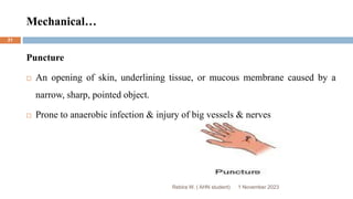
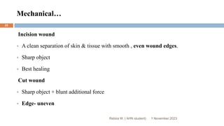
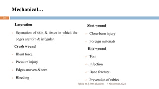
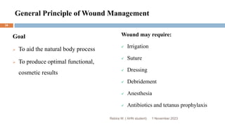
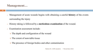
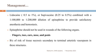
1. Institute of Health Science
Department of Nursing
Postgraduate Program of Adult Health Nursing
Wound Management Presentation
Set By: Rebira Workineh (AHN Student)
Rebira W. ( AHN student)
1 November 2023
1
2. Objectives
Rebira W. ( AHN student)
At the end of this lesson, the participants will be able to:
Describe the structures and functions of the skin
Understand the adverse impact of wound on quality of life
List types of wounds
Express wound healing
Identify factors affecting wound healing
Know how to manage wounds
1 November 2023
2
3. Objectives
Rebira W. ( AHN student)
Describe complications of wound healing
Explain how to manage chronic wounds
Differentiate pressure sores
Describe diabetic foot ulcer
Differentiate hypertrophic scars from keloid scars
Know how to develop nursing care plan for a patient with wounds
1 November 2023
3
4. Outlines
Rebira W. ( AHN student)
Overview of the integument
Introduction to wound
Types of wounds
Wound healing
Factors affecting wound healing
General principle of wound management
Complications of wound healing
Managing chronic wound
1 November 2023
4
5. Outlines
Rebira W. ( AHN student)
Pressure sores
Diabetic foot ulcers
Hypertrophic scars
Keloid scars
Develop nursing care plan
References
1 November 2023
5
6. Anatomy of the Integument
The skin is the largest organ of the body and provides the interface between
the body and the rest of the world
The skin provides the first line of host defense mechanisms and protects the
integrity and functioning of internal organ systems
The psychosocial aspect of skin appearance is extremely important to a
person’s well-being.
Rebira W. ( AHN student) 1 November 2023
6
7. Skin Layers
Epidermis
Outermost layer of skin, which is thin & avascular
Further divided into five structurally and functionally distinct layers
Corneum
Lucidum
Granulosum
Spinosum
Germinativum (basal layer)
Stratum
Rebira W. ( AHN student) 1 November 2023
7
8. Skin Layers…
These several thin layers of the epidermis contain the following:
Melanocytes
Produce melanin, a pigment that gives skin its color and protects it from the
damaging effects of ultraviolet radiation.
Keratinocytes
Produce keratin, a water repellent protein that gives the epidermis its tough,
protective quality.
Rebira W. ( AHN student) 1 November 2023
8
9. Skin Layers…
Rebira W. ( AHN student)
Dermis
The layer of skin lying beneath the epidermis
Is highly vascular and tough connective tissue
Contains collagen and elastic fibers, nerve fibers, lymphatics, sweat and
sebaceous glands, and hair follicles.
1 November 2023
9
10. Skin Layers…
Rebira W. ( AHN student)
SubcutaneousTissue
Made up of dense connective & adipose tissue
Houses major blood vessels, lymphatics, and nerves
Acts as a heat insulator
Provides a nutritional depot that is used during illness or starvation
Also acts as a mechanical shock absorber and helps the skin move easily over the
underlying structures
1 November 2023
10
11. Skin Layers
Fascia
Superficial fascia is below the subcutaneous tissue of the skin
Is a type of dense, firm, membranous connective tissue
Connects the skin to subjacent parts and facilitates movement
Rebira W. ( AHN student) 1 November 2023
11
13. Age-Related Changes
Rebira W. ( AHN student)
Sweat glands diminish in number
Epithelial and fatty layers of tissue atrophy and become thin
Thickness of subcutaneous fat on the legs or forearms diminishes, even if
abdominal or hip fat remains abundant
Collagen and elastin shrink and degenerate
1 November 2023
13
14. Age…
Rebira W. ( AHN student)
Collagen content of the skin decreases by approximately 1% per year
throughout adult life.
The net effect of all these changes is thin, dry, and inelastic skin that is
increasingly susceptible to separation of dermis and epidermis as minor
friction or shearing forces cause an injury known as skin tear.
1 November 2023
14
15. Physiology of the integument
Protection: intact skin prevents invasion of the body by bacteria
Thermoregulation: intact skin facilitates heat loss and cools the body when
necessary
Fluid and electrolyte balance: intact skin prevents the escape of water and
electrolytes from the body
Vitamin D Synthesis
Sensation
Psychosocial
Rebira W. ( AHN student) 1 November 2023
15
16. Introduction to Wound
Rebira W. ( AHN student)
Wounds have substantial adverse impact on client’s quality of life and have
a predictive risk with mortality.
Better understanding of the etiology of the wound & following the evidenced
based management protocols can:
o Extremely improve client’s health outcomes
o Lower the cost of the medical care
1 November 2023
16
17. Definition
Rebira W. ( AHN student)
Wound is defined as a break in the normal continuity of a tissue.
Is a disruption in the integrity of body tissue which may be intentional or
unintentional.
Caused by a transfer of any form of energy into the body w/c can be either to:
An externally visible structure like the skin
Deeper structures like muscles, tendons or internal organs
1 November 2023
17
18. Rebira W. ( AHN student)
CLASSIFICATION OF WOUNDS
1 November 2023
18
19. Based on the Origin
1. Mechanical
Incisions or incised wounds
Lacerations
Abrasions
Avulsions
Ulceration
Puncture
Crush wound
Shot wound
2. Chemical
Acid
Base
3. Wound caused by radiation
4. Wound caused by thermal forces
Burning
Freezing
5. Special
Exotic, poisonous animals
Rebira W. ( AHN student) 1 November 2023
19
20. Mechanical wounds
Abrasions
A wound in which the surface layers of skin are scraped away.
Superficial part of the epidermal layer.
Good wound healing
Rebira W. ( AHN student) 1 November 2023
20
21. Mechanical…
Puncture
An opening of skin, underlining tissue, or mucous membrane caused by a
narrow, sharp, pointed object.
Prone to anaerobic infection & injury of big vessels & nerves
Rebira W. ( AHN student) 1 November 2023
21
22. Mechanical…
Incision wound
A clean separation of skin & tissue with smooth , even wound edges.
Sharp object
Best healing
Cut wound
Sharp object + blunt additional force
Edge- uneven
Rebira W. ( AHN student) 1 November 2023
22
23. Mechanical…
Laceration
Separation of skin & tissue in which the
edges are torn & irregular.
Crush wound
Blunt force
Pressure injury
Edges-uneven & torn
Bleeding
Shot wound
Close-burn injury
Foreign materials
Bite wound
Torn
Infection
Bone fracture
Prevention of rabies
Rebira W. ( AHN student) 1 November 2023
23
24. Wound Caused by Radiation
Rebira W. ( AHN student)
Symptoms and severity depends on:
Amount of radiation
Length of exposure
Body part that was exposed
Symptoms may occur immediately, after a few days, or even as long as months.
Body parts mostly sensitive to radiation sickness:
Bone marrow
Gastrointestinal tract
1 November 2023
24
25. Wounds caused by thermal forces
Rebira W. ( AHN student)
Burning
1st degree – superficial injury (epidermis)
2nd degree –partial or deep partial thickness (epidermis+superficial or deep dermis)
3rd degree – full thickness (epidermis + entire dermis)
4th degree – (skin + subcutaneous tissue + muscle and bone)
Freezing
Mild, moderate, severe (redness, bullas, necrosis)
Rewarm – not only the frozen area but the whole body
1 November 2023
25
26. Surgical Wound Infection
Traditional surgical wound classification scheme introduced in 1964 classifies the wounds
based on the rate of infection
Clean
Non-traumatic , non-infected
No break in sterility technique
Respiratory tract , GI, & Gut not entered
Clean-contaminated
Minor break in sterility technique
Rebira W. ( AHN student) 1 November 2023
26
27. Surgical…
Rebira W. ( AHN student)
GI , Resp tracts entered, no sign.
Spillage GUT ,biliary tracts entered , no infected urine or bile
Contaminated
Fresh traumatic wounds
Major break in sterility
Gross spillage from GI tract
Entrance of Gut or biliary tract in the presence of infected urine or bile
1 November 2023
27
28. Surgical…
Rebira W. ( AHN student)
Dirty & infected
Acute bacterial inflammation without pus
Wound with heavy contamination
Transection of clean wounds for the purpose of surgical access to collect pus,
traumatic wound with retained devitalized tissue
1 November 2023
28
29. SWI Risk Factors
Rebira W. ( AHN student)
Age > 60 years
Female
Obesity
Infections
Underlying disease
Diabetes, Congestive heart failure
Liver disease, renal failure
Duration of preoperative stay hospitalization > 72 hours, ICU stay
Immunosuppression
1 November 2023
29
30. Wound Healing
Rebira W. ( AHN student)
Wound healing is a mechanism whereby the body attempts to restore the
integrity of the injured part.
The disruption in the integrity of tissue, whether surgical or traumatic,
stimulates a series of events that attempts to restore the injured tissue to a
normal state.
1 November 2023
30
32. Clinical Types of Healing
1 November 2023
Rebira W. ( AHN student)
32
Healing by first intention
Clean, straight line, edges well approximated with sutures, rapid healing
Healing occurs by epithelialization, minimal scar formation
Healing by second intention
Larger wounds with tissue loss, edges not approximated, heals from the inside out, granulation
tissue fills in the wound, longer healing time, larger scars
Healing by third intention (delayed primary closure)
Delay 3-5 days before injury is sutured, greater access for pathogens to invade, greater inflammation,
more granulation, larger scars.
33. Factors Affecting Wound Healing
Ischemia
Infection
Foreign body
Edema, elevated tissue pressure
Age and gender
Sex hormones
Stress
Ischemia
Disease
Obesity
Medication
Alcoholism and smoking
Immunocompromised condition
Nutrition
Rebira W. ( AHN student)
Local Factors Systemic Factors
1 November 2023
33
34. General Principle of Wound Management
Goal
To aid the natural body process
To produce optimal functional,
cosmetic results
Wound may require:
Irrigation
Suture
Dressing
Debridement
Anesthesia
Antibiotics and tetanus prophylaxis
1 November 2023
34
Rebira W. ( AHN student)
35. 1 November 2023
Rebira W. ( AHN student)
35
Reading assignment, read the following two points in details!
Suture
Dressing
36. Management…
1 November 2023
Rebira W. ( AHN student)
36
Management of acute wounds begins with obtaining a careful history of the events
surrounding the injury
History taking is followed by a meticulous examination of the wound.
Examination assessment include:
The depth and configuration of the wound
The extent of nonviable tissue
The presence of foreign bodies and other contamination
37. Management…
1 November 2023
Rebira W. ( AHN student)
37
Lidocaine ( 0.5 to 1%), or bupivacaine (0.25 to 0.5%) combined with a
1:100,000 to 1:200,000 dilution of epinephrine to provide satisfactory
anesthesia and hemostasis.
Epinephrine should not be used in wounds of the following organs.
o Fingers, toes, ears, nose, and penis
B/c of risk of tissue necrosis secondary to terminal arteriole vasospasm in
these structures.
38. Antibiotics
1 November 2023
Rebira W. ( AHN student)
38
Antibiotics should be used when there is an obvious wound infection
Signs of infection
Erythema
Cellulitis
Swelling
Purulent discharge
39. Irrigation
1 November 2023
Rebira W. ( AHN student)
39
Irrigation to visualize all areas of the wound and remove foreign materials is best
accomplished with normal saline.
High-pressure wound irrigation is more effective in achieving complete debridement
of foreign material and nonviable tissues
Iodine, povidone-iodine and H2O3 are organically based antibacterial preparations.
They have been shown to impair wound healing b/c of injury to wound neutrophils
and macrophages, and thus should not be used.
40. Management…
1 November 2023
Rebira W. ( AHN student)
40
After the wound has been anesthetized, explored, irrigated, and debrided, the
area surrounding the wound should be cleaned, inspected, and the
surrounding hair clipped.
The area surrounding the wound should be prepared with povidone-iodine or
similar solution and draped with sterile towels.
41. Management…
1 November 2023
Rebira W. ( AHN student)
41
Having ensured hemostasis and adequate debridement of nonviable tissues
and removal of any remaining foreign bodies, irregular, macerated, or beveled
wound edges should be debrided to provide a fresh edge for re-
approximation.
Initial sutures the realign the edges of these tissue types will speed and greatly
enhance the anesthetic outcome of the wound repair.
42. Management…
1 November 2023
Rebira W. ( AHN student)
42
In general, the smallest suture required to hold the various layers of the
wound in approximation should be selected to minimize suture related
inflammation.
In areas with significant superficial tissue loss, split-thickness skin grafting
may be required.
After closing deep tissues and replacing significant tissue deficits, skin edges
should be re-approximated for cosmetic and rapid wound healing.
43. …
1 November 2023
Rebira W. ( AHN student)
43
Failure to remove the sutures by 7 to 10 days after repair will result in a
cosmetically inferior wound.
44. Complications of wound healing
1. Early complications
Seroma
Hematoma
Wound dehiscence & evisceration
Superficial wound infection
Deep wound infection
Mixed wound infection
Rebira W. ( AHN student) 1 November 2023
44
45. Complications…
2. Late complications
Hyperthrophic scar
Keloid formation
Necrosis
Inflammatory infiltration
Abscesses
Foreign body containing abscesses
Rebira W. ( AHN student) 1 November 2023
45
46. Managing Chronic Wounds
Rebira W. ( AHN student)
Pressure sore
Diabetic foot ulcer
Hypertrophic scars
Keloids
NB: A chronic ulcer, unresponsive to dressings and simple treatments should be
biopsied to rule out neoplastic change.
1 November 2023
46
47. Brainstorming???????
Rebira W. ( AHN student)
Which one is true or false? Why?
1. Low pressure endured for long periods of time is believed to be more
significant in producing pressure ulcers than higher pressure of short
duration.
2. Higher pressure endured for short periods of time is believed to be more
significant in producing pressure ulcers than low pressure of long
duration.
1 November 2023
47
48. Pressure Sores
Rebira W. ( AHN student)
These can be defined as tissue necrosis with ulceration due to prolonged
pressure.
Less preferable terms are bed sores, pressure ulcers and decubitus ulcers.
They should be regarded as preventable but occur in approximately 5% of all
hospitalized patients.
Ulcers can form when a person constantly maintains any position causing
pressure to a site.
1 November 2023
48
49. Etiology of Pressure Ulcers
Rebira W. ( AHN student)
Pressure ulcers usually occur in soft tissue over bony prominences that remain in
contact with compressing surfaces.
Pressure ulcers are the clinical manifestation of local tissue death.
Cellular metabolism depends on blood vessels to carry nutrients to the tissues and to
remove waste products.
When the soft tissue is subject to prolonged pressure & insufficient nutrients the
result is cell death.
1 November 2023
49
50. Pressure
Rebira W. ( AHN student)
Muscle is more sensitive to compression than skin
The deeper muscle tissue may be necrotic before damage to the overlying skin
is apparent
The force of pressure increases as the affected body surface area decreases
The normal response to prolonged pressure is a change in body position before
tissue ischemia occurs.
1 November 2023
50
51. Time-Pressure
Rebira W. ( AHN student)
Low pressure endured for long periods of time is believed to be more significant in
producing pressure ulcers than higher pressure of short duration.
If the time-pressure threshold is reached or exceeded, tissue damage continues even
after pressure is released.
Pressure ulceration can result from one period of sustained pressure
Most pressure ulcers occur secondary to repeated ischemic events without adequate
time for recovery
1 November 2023
51
54. Rebira W. ( AHN student)
Many other factors
Shear
Friction
Moisture
Infection
1 November 2023
54
55. Shear
Rebira W. ( AHN student)
Shear occurs when two surfaces move in the opposite direction.
For example, when a bed is elevated at the head, you can slide down in bed.
As the tailbone moves down, the skin over the bone might stay in place-essentially
pulling in the opposite direction
Shear is a mechanical force that is parallel rather than perpendicular to an area with
the main effect impacting deep tissues
Elevating the head of the bed increases shear and pressure in the sacral and coccygeal
areas
1 November 2023
55
56. Shear…
Rebira W. ( AHN student)
The mechanical forces can obstruct or tear and stretch blood vessels
Minimizing shearing forces involves raising the head of the bed to no more
than a 30 degree angle, except for short periods of time.
Shearing forces decreases the time tissue can remain under pressure.
If shear is present, vascular occlusion may occur at half the usual amount of
pressure.
1 November 2023
56
57. Shear & Friction
Rebira W. ( AHN student)
Shear injury will not be seen at the skin level because it happens beneath
the skin
Shear and friction go hand in hand—one rarely occurs without the other
Friction is the force of two surfaces moving across one another
Friction injury will be visible
1 November 2023
57
58. Friction
Rebira W. ( AHN student)
Erosion of surface tissue increases the potential for deeper tissue damage
because friction is the precursor of shear.
At risk for friction injuries:
o Individuals who have spastic conditions
o Patients who wear braces or appliances that rub against the skin
o The elderly
1 November 2023
58
59. Excessive Moisture
Rebira W. ( AHN student)
Moist skin is five times as likely to become ulcerated as dry skin
Constant exposure to wetness can waterlog or macerate the skin
Macerated epidermis is easily eroded
Wet skin surfaces increase the risk of friction as the patient is moved across
the surface of the bed linen.
1 November 2023
59
60. Causes of Moisture
Rebira W. ( AHN student)
Perspiration
Wound drainage
Soaking during bathing
Fecal and/or urinary incontinence
Maceration
1 November 2023
60
61. Stages of Pressure Sores
Rebira W. ( AHN student)
Stage 1: Non-blanchable erythema without a breach in the epidermis
Stage 2: Partial thickness skin loss involving the epidermis and dermis
Stage 3: Full-thickness skin loss extending into the subcutaneous tissue but not
through underlying fascia
Stage 4: Full-thickness skin loss through fascia with extensive tissue
destruction, may be involving muscle, tendon, joint or bone
1 November 2023
61
66. Unstageable Pressure Ulcer
Rebira W. ( AHN student)
Full thickness tissue loss in which the base of the ulcer is covered by slough
and/or eschar in the wound bed
Note: Until enough slough and/or eschar is removed to expose the base of the
wound, the true depth, and therefore stage, cannot be determined
1 November 2023
66
68. Evidence/Research
12 November 2023
Rebira W.( AHN student)
68
1. Study conducted at University of Turku in Finland (cross-sectional)
Among 270 studied populations, the prevalence of PU was 158 (5%)- November 2015 to January
2016.
The most prevalent stages of PU were stage II and I which was 39% and 37% respectively.
The most common sites identified were:
Heel=37%
Sacrum=26%
Buttocks=10.8%
Lateral malleolus=10%
Hip=8%
69. …
12 November 2023
Rebira W.( AHN student)
69
2. Study conducted at seven sites in Ethiopia (cross-sectional)
The prevalence of PU was 11.7% among 1881 studied participants (From January
1, 2000-June 1, 2019)
Stages of pressure ulcers:
Stage I=40.89%
Stage II=32.11%
Stage III=11.47%
Stage IV= 4.31%
70. …
12 November 2023
Rebira W.( AHN student)
70
3. Study conducted at Arba Minch, Jinka University in Southern Ethiopia (prospective
cohort study)
Among adult patients in ICU 216 followed prospectively, 25 (11.57%) developed
pressure ulcers after 6 days of admission-June 2021 to April 2022.
Predictors identified in this study were:
Age >= 40 years-three times risk for developing pressure ulcers
Friction/Shear-four times more risk for developing pressure ulcers than not
having friction/shear
71. …
12 November 2023
Rebira W.( AHN student)
71
Most common sites:
Sacrum =84%
Shoulder= 60%
Most prevalent stages:
Stage II=13(52%)
Stage I=12(48%)
72. Manage Pressure
Rebira W. ( AHN student)
Friction & shear management-use lift/turn sheets , and maintaining head of bed
at <30 degree
Turn & Reposition frequently
Pressure reduction & pressure redistribution utilizing low air loss mattress with
alternating pressure
Skin protection dressings over high risk areas
Pain management
1 November 2023
72
73. Manage Pressure Cont’d…
Rebira W. ( AHN student)
Optimal Nutrition & hydration management
Temperature - keeping skin cool, clean and dry
Off-loading of pressure areas e.g. heel protectors
Topical agents to relieve skin dryness
o Lotions
o Creams
o Ointments
1 November 2023
73
75. Causes
Peripheral neuropathy
Vascular disease
Trauma: acute-any injury to the foot or chronic – deformity
Decreased immune response
Delayed response to tissue injury
Rebira W. ( AHN student) 1 November 2023
76
76. Wagner Grading System
Grade 1: Superficial diabetic ulcer
Grade 2: Ulcer extension
o Involves ligament, tendon, joint capsule or fascia
o No abscess or osteomyelitis
Grade 3: Deep with abscess or osteomyelitis
Grade 4: Gangrene to portion of forefoot
Grade 5: Extensive gangrene of foot
Rebira W. ( AHN student) 1 November 2023
77
77. Treatment of Foot Ulcers
Wagner 0-2
Surgical if deformity present that will reulcerate
o Correct deformity
o Exostectomy
Wagner 3
Excision of infected bone
Wound allowed to granulate
Grafting (skin or bone) not generally effective
Wagner 4-5
Amputation
Rebira W. ( AHN student) 1 November 2023
78
78. Drug Treatment
First line
Ampicillin 1 g IV QID for 2-3 weeks
PLUS
Gentamicin 3-5 mg/kg IV QD in divided doses
PLUS
Metronidazole 500mg IV QID, for 10-14 days
Alternatives
Ceftazidime 1 gm IV TID
Plus
Gentamicin 3-5 mg/kg I.V. daily QD in divided doses
Plus
Metronidazole 500mg IV QID for 10-14 days
Rebira W. ( AHN student) 1 November 2023
79
79. Prevention of Foot Ulcer
Regular inspection & examination of foot & foot wear
Identification of high risk patient
Education of patient, family & health care providers
Appropriate foot wear
Treatment of non ulcerative pathology
Rebira W. ( AHN student) 1 November 2023
80
80. Hypertrophic Scars
Rebira W. ( AHN student)
These lesions are raised and thickened.
This process does not extend beyond the boundary of the scar.
Exacerbated by tension lines on the area of surgery: knee & elbow
Treatment
Nearly all hypertrophic scars undergo a degree of spontaneous resolvement
If present after six months, surgical excision is indicated
Pressure applied early to a lesion is also of benefit
Intractable lesions can be injected with triamcinolone
1 November 2023
81
81. Keloids Scars
Rebira W. ( AHN student)
Raised and thickened. This process extends beyond the boundary of the incision.
Keloids scars will keep on evolving for two years, these scars are not contagious.
Site: Chest, shoulder, upper back, back of neck and earlobes.
Treatment
Applying early pressure
Injection of triamcinolone, or corticosteroids
Excision with above activities
1 November 2023
82
83. NOTE
Rebira W. ( AHN student)
Hypertrophy and keloid formations are an overactive response to the
natural process of wound healing.
Hypertrophy scars don’t extend beyond the boundary of the incision.
Keloid scars extend beyond the boundary of the incision.
1 November 2023
84
84. Develop Nursing Care Plan (ADOPIE)
1 November 2023
Rebira W. ( AHN student)
85
1. Assessment: Data collection
2. Diagnosis: Analysis of data
3. Outcome Identification: Setting measurable criteria
4. Planning: Goals prioritized
5. Implementation: Intervention, action
6. Evaluation: Goal met? Reassessment
85. References
Rebira W. ( AHN student)
1. Bailey and loves, short practice of surgery, 26th edition
2. Schwartz, principle of surgery, 8th edition
3. Surgery lecture note for health officers
4. Demarco, Sharon. “Wound and Pressure Ulcer Management.” Johns Hopkins
Medicine. 11 March 2014.
5. Wilhelmy, Jennifer, RN, CWCN, CNP. “Save our Skin Heal our Holes: The
basics of pressure ulcer prevention and wound care. DDNA National
Conference 2014.
1 November 2023
86
Many other factors—primarily shear, friction, excessive moisture, and possibly infection—interact to mechanically damage soft tissue
Fecal incontinence exposes the skin to bacteria in the stool and adds the risk of infection. When a patient is incontinent of both urine and stool, the urea from the urine reacts chemically with the stool and causes further damage.