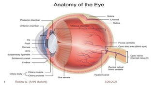
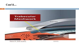
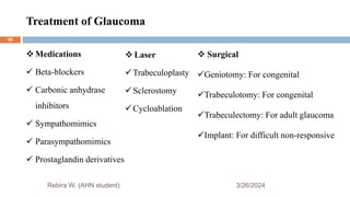
Institute of Health Science
Department of Nursing
Postgraduate Program of Adult Health Nursing
Glaucoma Presentation
Set By: Rebira Workineh (AHN Student)
Instructor: Mr. Adugna Oluma (BSc, MSc, & Assistant Professor)
3/26/2024
Rebira W. (AHN student) 1
Objectives
3/26/2024
At the end of this session, the participants will be able to:
 Understand the anatomy & physiology overview of eye & its structures
 Introduce, define, know analogy & distribution of glaucoma
 Explain risk factors, pathophysiology & clinical manifestations of glaucoma
 Identify diagnostic evaluation & management of glaucoma
 State how to develop nursing care plan for patient with glaucoma
 Describe complications of glaucoma
Rebira W. (AHN student)
2
Outlines
3/26/2024
 Overview of the eyes
 Introduction
 Epidemiology
 Risk factors
 Pathophysiology
 Classification of glaucoma
 Assessment & diagnostic evaluation
 Differential diagnosis
 Management of glaucoma
 Nursing process & health education
 Complications
 References
Rebira W. (AHN student)
3
3/26/2024
Rebira W. (AHN student)
4
Anatomy Cont’d…
3/26/2024
Rebira W. (AHN student)
 The optic nerve is a bundle of nerve
fibers
 It carries visual information from the
retina to the brain
5
Aqueous Humor Dynamics Include:
3/26/2024
Rebira W. (AHN student)
Anatomy of aqueous formation & drainage structures
 Ciliary body
 Posterior chamber
 Anterior chamber
 Angle of anterior chamber
 Aqueous outflow
6
Aqueous Humor Dynamics Cont’d…
3/26/2024
Rebira W. (AHN student)
Angle of anterior chamber formed by:
 Root of iris, anterior-most part of ciliary
body, scleral spur, trabecular meshwork
and Schwalbe’s line
7
Aqueous Humor Dynamics Cont’d…
3/26/2024
Rebira W. (AHN student)
Aqueous humor formation
 Is a complex pathway
 Ciliary process is a site of aqueous humor formation
 Mainly by three mechanisms:
1) Ultra filtration→ 20%
2) Active transfer → 70%
3) Diffusion→ 10%
8
Aqueous Humor Dynamics Cont’d…
Aqueous outflow system : Consists
of two pathways
1. Trabecular (conventional) outflow
2. Uveoscleral (unconventional)
outflow
3/26/2024
Rebira W. (AHN student)
9
Trabecular (Conventional) Outflow
3/26/2024
Rebira W. (AHN student)
10
Uveoscleral (Unconventional) Outflow
3/26/2024
Rebira W. (AHN student)
11
Fluid Circulation
3/26/2024
Rebira W. (AHN student)
 The eye has an internal fluid
circulation system
 Fluid is produced at the base of the iris
12
Fluid Cont’d…
3/26/2024
Rebira W. (AHN student)
The fluid flows through the pupil to the front of the iris
13
Fluid Cont’d…
3/26/2024
Rebira W. (AHN student)
 The fluid exits the eye at the angle between the iris
and the cornea where it drains through a spongy
meshwork
14
Brainstorming: What is Glaucoma?
3/26/2024
Rebira W. (AHN
student)
15
Introduction to Glaucoma
3/26/2024
 The term glaucoma is used to refer group of ocular conditions characterized by
the optic nerve damage.
 Glaucoma occurs as a result of increased intraocular pressure (IOP) caused by a
malformation or malfunction of the eyes drainage system.
 Normal range of IOP is 10 to 21 mm Hg (Brunner & Suddarth’s text book 12th
edition)
 The increased pressure causes compression of the retina & the optic nerve, & causes
progressive , permanent loss of eyesight if left untreated.
Rebira W. (AHN student)
16
Definition
3/26/2024
 Is a group of disorder characterized by an abnormally high IOP, optic nerve
dystrophy, & peripheral filed loss (Brunner & Suddarth’s text book 12th edition)
 A group of disorders characterized by a progressive optic neuropathy,
characteristic appearance of the optic disc, specific pattern of irreversible visual
field defects , associated frequently but not invariably with raised IOP.
 Is a symptomatic condition of the eye where the intraocular pressure is more than
normal (L.P Agarwal, 1922-2004)
Rebira W. (AHN student)
17
Cont’d…
3/26/2024
 Is an eye disorder that damages the optic nerves & certain
cells of the retina, resulting in gradual vision loss if
untreated.
 People with glaucoma often show no symptoms, although
one type can cause pain & is a medical emergency.
Rebira W. (AHN student)
18
IOP
3/26/2024
Rebira W. (AHN student)
 Depends on the balance between the production and removal of aqueous humor.
 Normal IOP is between 10 and 21mmHg
 IOP > 21.7 mmHg is abnormal
 IOP is determined by:
The rate of aqueous production
The resistance encountered by the aqueous humor as it flows out of the passage
The venous pressure of z episcleral veins that drain into z anterior ciliary vein
19
Dr. Joslin Glaucoma Analogy
3/26/2024
Think of the Eye as a Balloon
 To understand it better, think of the eye as a balloon
 When a balloon is inflated to the right level, all is well, but has the balloon ever over-
inflated to the point that it popped?
 While that’s not exactly what happens in glaucoma, one can think of the eye with
glaucoma like a balloon with too much air in it
 The eye tissues are too strong to pop, however when there is too much fluid in the
eye, it damages the weakest point of the eye
Rebira W. (AHN student)
20
Cont’d…
3/26/2024
 The optic nerve is the messenger that carries visual information from the eyes to the
brain so we can see
 When eye pressure increases due to too much fluid, the optic nerve gets squeezed &
pinched, causing damage
 The nerve fibers start to degenerate & can no longer carry visual information to brain
 Is a slow process, but eventually the tiny nerve axons & ganglions that are part of the
optic nerve become damaged & die
Rebira W. (AHN student)
21
Cont’d…
3/26/2024
 Once those nerve cells die, it is not possible to bring them back to life & the lost
vision with those cells won’t come back either
 That’s how glaucoma results in permanent vision loss
Rebira W. (AHN
student)
22
Think of the Eye as a Sink
3/26/2024
 IOP can be caused by too much fluid in the eye. It’s helpful to think of the eye as a
sink to understand this idea better.
 In the eye as a sink analogy, the eye’s faucet is always running, filling the eye
constantly with fluid, & the drain is always open to let that fluid drain out.
 The front part of the eye is filled with a clear fluid called the aqueous humor. It
nourishes the structures of the eye & is constantly produced.
 After it delivers nourishment to the cornea & lens, the aqueous humor flows out
through a tiny opening of spongy tissue called the trabecular meshwork.
Rebira W. (AHN student)
23
Cont’d…
3/26/2024
 It is meant tiny: the opening is only about 1/50 of an inch wide. The trabecular meshwork
is the open drain in the eye as a sink analogy.
 It is located in the front of the eye right at the angle made where the iris & cornea come
together.
 So, if the faucet is always running, making more & more aqueous humor fluid to nourish
the eye, & the drain is always open to let that fluid out, everything should be fine.
 However, in glaucoma, the drain can become clogged, & then the fluid can’t drain out of
the eye as fast as it is produced.
Rebira W. (AHN student)
24
Cont’d…
3/26/2024
 That causes the fluid to back up. That means there is more fluid than
necessary in the eye.
 Since the eye is a closed structure, the sink can’t overflow & instead gets
backed up with excess fluid.
 That built-up fluid is what causes pressure within the eye, & that’s when IOP
readings increase.
 High intraocular is one symptom of glaucoma.
Rebira W. (AHN student)
25
Epidemiology
3/26/2024
 Glaucoma is one of the leading causes of irreversible blindness in the world & is the
leading cause of blindness among adults in the United States
 It is estimated that at least 2 million Americans have glaucoma & that 5 to 10 million
more are at risk (Margolis et al., 2002)
 Glaucoma is more prevalent among people older than 40 years of age,& the incidence
increases with age
 Is also more prevalent among M than F & in the A-A & Asian populations
 There is no cure for glaucoma, but research continues
Rebira W. (AHN student)
26
Risk Factors for Glaucoma
3/26/2024
 Family history of glaucoma
 African American race
 Older age
 Diabetes
 Cardiovascular disease
 Migraine syndromes
 Nearsightedness (myopia)
 Eye trauma
 Prolonged use of topical or systemic corticosteroids
Rebira W. (AHN student)
27
Pathophysiology
3/26/2024
 There are two accepted theories regarding how increased IOP damages the optic
nerve in glaucoma
The direct mechanical theory
 Suggests that high IOP damages the retinal layer as it passes through the optic nerve
head
The indirect ischemic theory
 Suggests that high IOP compresses the microcirculation in the optic nerve head,
resulting in cell injury & death
 Some glaucomas appear as exclusively mechanical, & some are exclusively
ischemic types
Rebira W. (AHN student)
28
Cont’d…
3/26/2024
 Typically, most cases are a combination of both.
 Regardless of the cause of damage, glaucomatous changes typically evolve through
clearly discernible stages
1. Initiating events
 Precipitating factors include illness, emotional stress, congenital narrow angles,
long-term use of corticosteroids, and mydriatics (i.e., medications causing pupillary
dilation).
 These events lead to the 2nd stage
Rebira W. (AHN student)
29
Cont’d…
3/26/2024
2. Structural alterations in the aqueous outflow system
 Tissue & cellular changes caused by factors that affect aqueous humor dynamics lead to structural
alterations & to the third stage
3. Functional alterations
 Conditions such as ↑ IOP or impaired blood flow create functional changes that lead to the 4th stage
4. Optic nerve damage
 Atrophy of the optic nerve is characterized by loss of nerve fibers & blood supply, & this 4th stage
inevitably progresses to the 5th stage
5. Visual loss: Progressive loss of vision is characterized by visual field defects
(Brunner & Suddarth’s text book 10th edition)
Rebira W. (AHN
student)
30
Cont’d…
3/26/2024
Rebira W. (AHN student)
31
Cont’d…
3/26/2024
Rebira W. (AHN student)
32
Congenital Acquired
 True congenital
 Infantile
 Juvenile
 Primary Glaucoma
 Secondary Glaucoma
Classification of Glaucoma
3/26/2024
Rebira W. (AHN student)
Glaucoma
33
Classification Cont’d…
3/26/2024
Rebira W. (AHN student)
34
 Congenital glaucoma
 True congenital
 Infantile
 Juvenile
NB: Congenital glaucoma is not discussed in this presentation
Classification Cont’d…
3/26/2024
Rebira W. (AHN student)
 The two common clinical forms of glaucoma encountered under primary
glaucoma in adults are:
 Primary open-angle &
 Primary angle-closure glaucoma
(Brunner & Suddarth’s text book 10th edition)
35
Primary open-angle glaucoma
3/26/2024
Rebira W. (AHN student)
 Is the most common form of glaucoma
 Occurs when the fluid drainage is poor & fluid builds up in the eye & the
internal eye pressure goes up
 This increased pressure can cause damage to the optic nerve & vision loss
 Usually bilateral, but one eye may be more severely affected than the other.
 The exact mechanism of damage is still unknown
36
Cont’d…
3/26/2024
Rebira W. (AHN student)
 Vision is normal in the early stages
 POAG develops gradually and painlessly and has no initial symptoms
 If untreated, peripheral or side vision is slowly lost
 Tunnel vision:- Defective sight in which objects cannot be properly seen if not close
to the centre of the field of view
 Eventually,allvisionmaybelost
37
Chronic open-angle glaucoma
3/26/2024
Rebira W. (AHN student)
Clinical manifestation
 Optic nerve damage, visual field defects
 IOP >21 mm Hg
 May have fluctuating IOPs,
 Usually no symptoms but possible ocular pain, headache, & halos
38
Cont’d…
3/26/2024
Rebira W. (AHN student)
Treatment
 Decrease intraocular pressure 20% to 50%
 Additional topical & oral agents added as necessary
 Laser trabeculoplasty can provide a 20% drop in intraocular pressure.
 Glaucoma filtering surgery if continued optic nerve damage despite medication
therapy & trabeculoplasty
39
Normal Tension Glaucoma
3/26/2024
Rebira W. (AHN student)
Clinical manifestation
 IOP  21 mm Hg
 Optic nerve damage
 visual field defects
Treatment
 Treatment similar to COAG, however, the best management for normal tension
glaucoma management is yet to be established
 Goal is to lower the IOP by at least 30%
40
Ocular Hypertension
3/26/2024
Rebira W. (AHN student)
Clinical manifestation
 Elevated intraocular pressure
 Possible ocular pain or headache
Treatment
 Lower IOP by at least 20%
NB: In all three types of open-angle glaucoma, the anterior chamber angle is open &
appears normal.
41
Primary Angle-closure Glaucoma
3/26/2024
Rebira W. (AHN student)
 This type of glaucoma is an emergency situation, occurs suddenly & may
cause permanent vision loss in 48 to 72 hours
 Therefore, immediate treatment is required
 It occurs when the iris itself blocks the drainage angle and results in a sudden
increase in pressure
 Obstruction in aqueous humor outflow due to the complete or partial closure
of the angle from the forward shift of the peripheral iris to the trabecula
42
Pathogenesis
3/26/2024
Rebira W. (AHN student)
 Resistance of drainage of aqueous through the trabecular
meshwork, due to:
o Thickening of trabecular lamellae
o Reduction in number of lining trabecular cells
o Increased extracellular material in the trabecular meshwork
spaces
(AK Khurana, Textbook of Ophthalmic Nursing, 6th edition)
43
Acute angle-closure glaucoma
3/26/2024
Rebira W. (AHN student)
Clinical manifestation
 Rapidly progressive visual impairment, periocular pain, conjunctival hyperemia, &
congestion
 Pain may be associated with nausea, vomiting, bradycardia, & profuse sweating
 Reduced central visual acuity, severely elevated IOP, corneal edema
 Pupil is vertically oval, fixed in a semidilated position, & unreactive to light &
accommodation
44
Cont’d…
3/26/2024
Rebira W. (AHN student)
Treatment
 Ocular emergency: Administration of hyperosmotics, azetazolamide, &
topical ocular hypotensive agents, such as pilocarpine & beta-blockers
 Possible laser incision in the iris to release blocked aqueous & reduce IOP
 Other eye is also treated with pilocarpine eye drops &/or surgical
management to avoid a similar spontaneous attack
45
Subacute Angle-closure Glaucoma
3/26/2024
Rebira W. (AHN student)
Clinical manifestation
 Transient blurring of vision
 Halos around lights
 Temporal headaches and/or ocular pain
 Pupil may be semi-dilated
Treatment
 Prophylactic peripheral laser iridotomy
46
Chronic angle-closure glaucoma
3/26/2024
Rebira W. (AHN student)
Clinical manifestation
 Progression of glaucomatous cupping & significant visual field loss
 IOP may be normal or elevated
 Ocular pain
 Headache
Treatment
 Laser iridotomy & medications
47
Secondary Glaucoma
3/26/2024
Rebira W. (AHN
student)
 Glaucoma can develop as a complication from other
conditions:
 Eye injuries
 Uveitis
 Pigment dispersion
 Diabetes (Neovascular glaucoma
 Steroid use
48
Steroid Induced Glaucoma
3/26/2024
Rebira W. (AHN student)
Risk factors
 POAG
 Diabetes
 Myopia
 Stronger the steroid more the elevation
49
General C/F
3/26/2024
 Glaucoma is often called the silent thief of sight because most patients are unaware
that they have the disease until they have experienced visual changes and vision
loss.
 Blurred vision or halos around lights
 Difficulty focusing
 Difficulty adjusting eyes in low lighting
 Loss of peripheral vision
 Aching or discomfort around the eyes
 Headache
Rebira W. (AHN student)
50
“Halos”
3/26/2024
Rebira W. (AHN student)
51
Assessment & Diagnostic Findings
3/26/2024
Rebira W. (AHN
student)
Purpose
 To establish the diagnostic category
 To assess the optic nerve damage
 To formulate a treatment plan
 The patient’s ocular & medical history must be detailed to investigate the
history of predisposing factors
52
Assessment & Cont’d…
3/26/2024
Rebira W. (AHN student)
 There are four major types of examinations used in glaucoma evaluation, diagnosis,
& management.
 Tonometry: To measure the IOP
 Ophthalmoscopy : To inspect the optic nerve
 Gonioscopy: To examine the filtration angle of the anterior chamber
 Perimetry (Visual field test): To assess the visual fields
53
Assessment & Cont’d…
3/26/2024
Rebira W. (AHN student)
o Slit Lamp Examination: Used to examine z structure of the eye
o Fundus Photography: To monitor the disc for changes
o Pachymetry:
o Nerve fiber analysis: To asses the thickness of nerve fiber layer
54
Differential Diagnosis
3/26/2024
 Angle recession glaucoma
 Anterior ischemic optic neuropathy
 Optic atrophy
 Compressive optic neuropathy
 Drug-induced glaucoma
 Lens-particle glaucoma
 Pigmentary glaucoma
Rebira W. (AHN student)
55
Treatment of Glaucoma
3/26/2024
 Medications
 Beta-blockers
 Carbonic anhydrase
inhibitors
 Sympathomimics
 Parasympathomimics
 Prostaglandin derivatives
Rebira W. (AHN student)
 Laser
Trabeculoplasty
Sclerostomy
Cycloablation
 Surgical
Geniotomy: For congenital
Trabeculotomy: For congenital
Trabeculectomy: For adult glaucoma
Implant: For difficult non-responsive
56
3/26/2024
Nursing Process
and
Health Education
Rebira W. (AHN student)
57
Nursing Process
3/26/2024
 Nursing Diagnosis: Acute pain R/T increased IOP & surgical intervention AEB
facial expression of the patient
 Goal: Patient’s pain score will be reduced from 10 to 3 during hospitalization
 Nursing Intervention: Assess the IOP, elevate the head end of the bed at 30 degree
angle & administer analgesics for patient as prescribed
 Nursing Diagnosis: Self care deficit R/T loss of vision
 Goal: The client will verbalize understanding of the condition, prognosis & Rx.
Rebira W. (AHN student)
58
Nursing Process Cont’d…
3/26/2024
 Nursing Intervention: Demonstrating necessary procedures and
explaining the reasons for the action
 Nursing Diagnosis: Anxiety R/T potential loss of vision AEB halo
light & blurred vision
 Goal: The client will appear relaxed and reported anxiety will be
reduced to a manageable level
Rebira W. (AHN student)
59
Nursing Process Cont’d…
3/26/2024
 Nursing Intervention: Providing emotional support and counselling to reduce
anxiety, discuss the probability that careful monitoring & treatment can prevent
additional visual loss & identify helpful resources and support groups
 Nursing Diagnosis: Risk for infection
 Goal: Reduce the risk of infection during hospitalization
 Nursing Intervention: Encourage the patient to wear eye shield to prevent infection
Rebira W. (AHN student)
60
Health Education
3/26/2024
The medical & surgical management of glaucoma slows the progression of
the condition, but does not cure it
The lifelong therapeutic regimen mandates patient education
The nature of the disease and the importance of strict adherence to the
medication regimen must be explained to help ensure compliance
 Can’t be cured, but can be controlled, periodic checkup
 Avoid circumstances that may increase IOP, such as URTI, anxiety,
heavy lifting, etc.
Rebira W. (AHN student)
61
Complication
3/26/2024
 Complete loss of vision
 Choroidal detachment
 Retinal detachment
 Chronic hypotony
 Anterior segment necrosis
Rebira W. (AHN
student)
62
Research Studies
3/26/2024
Rebira W. (AHN student)
Safety & Efficiency of Biomatoprost Sustained Release in
Patients with Open Angle Glaucoma
63
Conclusion
3/26/2024
Rebira W. (AHN student)
Glaucoma is a condition that causes damage to the eyes optic nerve & gets
worse over the time
Without treatment the glaucoma can cause permanent blindness within few
years
So the treatment should be given at right time to prevent complications
64
References
3/26/2024
 K Khurana, (2008). Textbook of Ophthalmic Nursing, CBS Publishers, Banglore,6th
edition.
 Black.M.Joyce.Text book of Medical Surgical Nursing. Elsevier Publication.8th
edition.
 Chintamani, Lewis, (2011). Text book of Medical surgical Nursing, Elsevier
Publication 13th edition.
 Brunner & Suddarth’s, Textbook of Medical-Surgical Nursing, 10th edition.
Rebira W. (AHN student)
65
3/26/2024
Thanks!
Rebira W. (AHN student)
66

Glaucoma Seminar Presentation by Rebira.pptx

  • 1.
    Institute of HealthScience Department of Nursing Postgraduate Program of Adult Health Nursing Glaucoma Presentation Set By: Rebira Workineh (AHN Student) Instructor: Mr. Adugna Oluma (BSc, MSc, & Assistant Professor) 3/26/2024 Rebira W. (AHN student) 1
  • 2.
    Objectives 3/26/2024 At the endof this session, the participants will be able to:  Understand the anatomy & physiology overview of eye & its structures  Introduce, define, know analogy & distribution of glaucoma  Explain risk factors, pathophysiology & clinical manifestations of glaucoma  Identify diagnostic evaluation & management of glaucoma  State how to develop nursing care plan for patient with glaucoma  Describe complications of glaucoma Rebira W. (AHN student) 2
  • 3.
    Outlines 3/26/2024  Overview ofthe eyes  Introduction  Epidemiology  Risk factors  Pathophysiology  Classification of glaucoma  Assessment & diagnostic evaluation  Differential diagnosis  Management of glaucoma  Nursing process & health education  Complications  References Rebira W. (AHN student) 3
  • 4.
  • 5.
    Anatomy Cont’d… 3/26/2024 Rebira W.(AHN student)  The optic nerve is a bundle of nerve fibers  It carries visual information from the retina to the brain 5
  • 6.
    Aqueous Humor DynamicsInclude: 3/26/2024 Rebira W. (AHN student) Anatomy of aqueous formation & drainage structures  Ciliary body  Posterior chamber  Anterior chamber  Angle of anterior chamber  Aqueous outflow 6
  • 7.
    Aqueous Humor DynamicsCont’d… 3/26/2024 Rebira W. (AHN student) Angle of anterior chamber formed by:  Root of iris, anterior-most part of ciliary body, scleral spur, trabecular meshwork and Schwalbe’s line 7
  • 8.
    Aqueous Humor DynamicsCont’d… 3/26/2024 Rebira W. (AHN student) Aqueous humor formation  Is a complex pathway  Ciliary process is a site of aqueous humor formation  Mainly by three mechanisms: 1) Ultra filtration→ 20% 2) Active transfer → 70% 3) Diffusion→ 10% 8
  • 9.
    Aqueous Humor DynamicsCont’d… Aqueous outflow system : Consists of two pathways 1. Trabecular (conventional) outflow 2. Uveoscleral (unconventional) outflow 3/26/2024 Rebira W. (AHN student) 9
  • 10.
  • 11.
  • 12.
    Fluid Circulation 3/26/2024 Rebira W.(AHN student)  The eye has an internal fluid circulation system  Fluid is produced at the base of the iris 12
  • 13.
    Fluid Cont’d… 3/26/2024 Rebira W.(AHN student) The fluid flows through the pupil to the front of the iris 13
  • 14.
    Fluid Cont’d… 3/26/2024 Rebira W.(AHN student)  The fluid exits the eye at the angle between the iris and the cornea where it drains through a spongy meshwork 14
  • 15.
    Brainstorming: What isGlaucoma? 3/26/2024 Rebira W. (AHN student) 15
  • 16.
    Introduction to Glaucoma 3/26/2024 The term glaucoma is used to refer group of ocular conditions characterized by the optic nerve damage.  Glaucoma occurs as a result of increased intraocular pressure (IOP) caused by a malformation or malfunction of the eyes drainage system.  Normal range of IOP is 10 to 21 mm Hg (Brunner & Suddarth’s text book 12th edition)  The increased pressure causes compression of the retina & the optic nerve, & causes progressive , permanent loss of eyesight if left untreated. Rebira W. (AHN student) 16
  • 17.
    Definition 3/26/2024  Is agroup of disorder characterized by an abnormally high IOP, optic nerve dystrophy, & peripheral filed loss (Brunner & Suddarth’s text book 12th edition)  A group of disorders characterized by a progressive optic neuropathy, characteristic appearance of the optic disc, specific pattern of irreversible visual field defects , associated frequently but not invariably with raised IOP.  Is a symptomatic condition of the eye where the intraocular pressure is more than normal (L.P Agarwal, 1922-2004) Rebira W. (AHN student) 17
  • 18.
    Cont’d… 3/26/2024  Is aneye disorder that damages the optic nerves & certain cells of the retina, resulting in gradual vision loss if untreated.  People with glaucoma often show no symptoms, although one type can cause pain & is a medical emergency. Rebira W. (AHN student) 18
  • 19.
    IOP 3/26/2024 Rebira W. (AHNstudent)  Depends on the balance between the production and removal of aqueous humor.  Normal IOP is between 10 and 21mmHg  IOP > 21.7 mmHg is abnormal  IOP is determined by: The rate of aqueous production The resistance encountered by the aqueous humor as it flows out of the passage The venous pressure of z episcleral veins that drain into z anterior ciliary vein 19
  • 20.
    Dr. Joslin GlaucomaAnalogy 3/26/2024 Think of the Eye as a Balloon  To understand it better, think of the eye as a balloon  When a balloon is inflated to the right level, all is well, but has the balloon ever over- inflated to the point that it popped?  While that’s not exactly what happens in glaucoma, one can think of the eye with glaucoma like a balloon with too much air in it  The eye tissues are too strong to pop, however when there is too much fluid in the eye, it damages the weakest point of the eye Rebira W. (AHN student) 20
  • 21.
    Cont’d… 3/26/2024  The opticnerve is the messenger that carries visual information from the eyes to the brain so we can see  When eye pressure increases due to too much fluid, the optic nerve gets squeezed & pinched, causing damage  The nerve fibers start to degenerate & can no longer carry visual information to brain  Is a slow process, but eventually the tiny nerve axons & ganglions that are part of the optic nerve become damaged & die Rebira W. (AHN student) 21
  • 22.
    Cont’d… 3/26/2024  Once thosenerve cells die, it is not possible to bring them back to life & the lost vision with those cells won’t come back either  That’s how glaucoma results in permanent vision loss Rebira W. (AHN student) 22
  • 23.
    Think of theEye as a Sink 3/26/2024  IOP can be caused by too much fluid in the eye. It’s helpful to think of the eye as a sink to understand this idea better.  In the eye as a sink analogy, the eye’s faucet is always running, filling the eye constantly with fluid, & the drain is always open to let that fluid drain out.  The front part of the eye is filled with a clear fluid called the aqueous humor. It nourishes the structures of the eye & is constantly produced.  After it delivers nourishment to the cornea & lens, the aqueous humor flows out through a tiny opening of spongy tissue called the trabecular meshwork. Rebira W. (AHN student) 23
  • 24.
    Cont’d… 3/26/2024  It ismeant tiny: the opening is only about 1/50 of an inch wide. The trabecular meshwork is the open drain in the eye as a sink analogy.  It is located in the front of the eye right at the angle made where the iris & cornea come together.  So, if the faucet is always running, making more & more aqueous humor fluid to nourish the eye, & the drain is always open to let that fluid out, everything should be fine.  However, in glaucoma, the drain can become clogged, & then the fluid can’t drain out of the eye as fast as it is produced. Rebira W. (AHN student) 24
  • 25.
    Cont’d… 3/26/2024  That causesthe fluid to back up. That means there is more fluid than necessary in the eye.  Since the eye is a closed structure, the sink can’t overflow & instead gets backed up with excess fluid.  That built-up fluid is what causes pressure within the eye, & that’s when IOP readings increase.  High intraocular is one symptom of glaucoma. Rebira W. (AHN student) 25
  • 26.
    Epidemiology 3/26/2024  Glaucoma isone of the leading causes of irreversible blindness in the world & is the leading cause of blindness among adults in the United States  It is estimated that at least 2 million Americans have glaucoma & that 5 to 10 million more are at risk (Margolis et al., 2002)  Glaucoma is more prevalent among people older than 40 years of age,& the incidence increases with age  Is also more prevalent among M than F & in the A-A & Asian populations  There is no cure for glaucoma, but research continues Rebira W. (AHN student) 26
  • 27.
    Risk Factors forGlaucoma 3/26/2024  Family history of glaucoma  African American race  Older age  Diabetes  Cardiovascular disease  Migraine syndromes  Nearsightedness (myopia)  Eye trauma  Prolonged use of topical or systemic corticosteroids Rebira W. (AHN student) 27
  • 28.
    Pathophysiology 3/26/2024  There aretwo accepted theories regarding how increased IOP damages the optic nerve in glaucoma The direct mechanical theory  Suggests that high IOP damages the retinal layer as it passes through the optic nerve head The indirect ischemic theory  Suggests that high IOP compresses the microcirculation in the optic nerve head, resulting in cell injury & death  Some glaucomas appear as exclusively mechanical, & some are exclusively ischemic types Rebira W. (AHN student) 28
  • 29.
    Cont’d… 3/26/2024  Typically, mostcases are a combination of both.  Regardless of the cause of damage, glaucomatous changes typically evolve through clearly discernible stages 1. Initiating events  Precipitating factors include illness, emotional stress, congenital narrow angles, long-term use of corticosteroids, and mydriatics (i.e., medications causing pupillary dilation).  These events lead to the 2nd stage Rebira W. (AHN student) 29
  • 30.
    Cont’d… 3/26/2024 2. Structural alterationsin the aqueous outflow system  Tissue & cellular changes caused by factors that affect aqueous humor dynamics lead to structural alterations & to the third stage 3. Functional alterations  Conditions such as ↑ IOP or impaired blood flow create functional changes that lead to the 4th stage 4. Optic nerve damage  Atrophy of the optic nerve is characterized by loss of nerve fibers & blood supply, & this 4th stage inevitably progresses to the 5th stage 5. Visual loss: Progressive loss of vision is characterized by visual field defects (Brunner & Suddarth’s text book 10th edition) Rebira W. (AHN student) 30
  • 31.
  • 32.
  • 33.
    Congenital Acquired  Truecongenital  Infantile  Juvenile  Primary Glaucoma  Secondary Glaucoma Classification of Glaucoma 3/26/2024 Rebira W. (AHN student) Glaucoma 33
  • 34.
    Classification Cont’d… 3/26/2024 Rebira W.(AHN student) 34  Congenital glaucoma  True congenital  Infantile  Juvenile NB: Congenital glaucoma is not discussed in this presentation
  • 35.
    Classification Cont’d… 3/26/2024 Rebira W.(AHN student)  The two common clinical forms of glaucoma encountered under primary glaucoma in adults are:  Primary open-angle &  Primary angle-closure glaucoma (Brunner & Suddarth’s text book 10th edition) 35
  • 36.
    Primary open-angle glaucoma 3/26/2024 RebiraW. (AHN student)  Is the most common form of glaucoma  Occurs when the fluid drainage is poor & fluid builds up in the eye & the internal eye pressure goes up  This increased pressure can cause damage to the optic nerve & vision loss  Usually bilateral, but one eye may be more severely affected than the other.  The exact mechanism of damage is still unknown 36
  • 37.
    Cont’d… 3/26/2024 Rebira W. (AHNstudent)  Vision is normal in the early stages  POAG develops gradually and painlessly and has no initial symptoms  If untreated, peripheral or side vision is slowly lost  Tunnel vision:- Defective sight in which objects cannot be properly seen if not close to the centre of the field of view  Eventually,allvisionmaybelost 37
  • 38.
    Chronic open-angle glaucoma 3/26/2024 RebiraW. (AHN student) Clinical manifestation  Optic nerve damage, visual field defects  IOP >21 mm Hg  May have fluctuating IOPs,  Usually no symptoms but possible ocular pain, headache, & halos 38
  • 39.
    Cont’d… 3/26/2024 Rebira W. (AHNstudent) Treatment  Decrease intraocular pressure 20% to 50%  Additional topical & oral agents added as necessary  Laser trabeculoplasty can provide a 20% drop in intraocular pressure.  Glaucoma filtering surgery if continued optic nerve damage despite medication therapy & trabeculoplasty 39
  • 40.
    Normal Tension Glaucoma 3/26/2024 RebiraW. (AHN student) Clinical manifestation  IOP  21 mm Hg  Optic nerve damage  visual field defects Treatment  Treatment similar to COAG, however, the best management for normal tension glaucoma management is yet to be established  Goal is to lower the IOP by at least 30% 40
  • 41.
    Ocular Hypertension 3/26/2024 Rebira W.(AHN student) Clinical manifestation  Elevated intraocular pressure  Possible ocular pain or headache Treatment  Lower IOP by at least 20% NB: In all three types of open-angle glaucoma, the anterior chamber angle is open & appears normal. 41
  • 42.
    Primary Angle-closure Glaucoma 3/26/2024 RebiraW. (AHN student)  This type of glaucoma is an emergency situation, occurs suddenly & may cause permanent vision loss in 48 to 72 hours  Therefore, immediate treatment is required  It occurs when the iris itself blocks the drainage angle and results in a sudden increase in pressure  Obstruction in aqueous humor outflow due to the complete or partial closure of the angle from the forward shift of the peripheral iris to the trabecula 42
  • 43.
    Pathogenesis 3/26/2024 Rebira W. (AHNstudent)  Resistance of drainage of aqueous through the trabecular meshwork, due to: o Thickening of trabecular lamellae o Reduction in number of lining trabecular cells o Increased extracellular material in the trabecular meshwork spaces (AK Khurana, Textbook of Ophthalmic Nursing, 6th edition) 43
  • 44.
    Acute angle-closure glaucoma 3/26/2024 RebiraW. (AHN student) Clinical manifestation  Rapidly progressive visual impairment, periocular pain, conjunctival hyperemia, & congestion  Pain may be associated with nausea, vomiting, bradycardia, & profuse sweating  Reduced central visual acuity, severely elevated IOP, corneal edema  Pupil is vertically oval, fixed in a semidilated position, & unreactive to light & accommodation 44
  • 45.
    Cont’d… 3/26/2024 Rebira W. (AHNstudent) Treatment  Ocular emergency: Administration of hyperosmotics, azetazolamide, & topical ocular hypotensive agents, such as pilocarpine & beta-blockers  Possible laser incision in the iris to release blocked aqueous & reduce IOP  Other eye is also treated with pilocarpine eye drops &/or surgical management to avoid a similar spontaneous attack 45
  • 46.
    Subacute Angle-closure Glaucoma 3/26/2024 RebiraW. (AHN student) Clinical manifestation  Transient blurring of vision  Halos around lights  Temporal headaches and/or ocular pain  Pupil may be semi-dilated Treatment  Prophylactic peripheral laser iridotomy 46
  • 47.
    Chronic angle-closure glaucoma 3/26/2024 RebiraW. (AHN student) Clinical manifestation  Progression of glaucomatous cupping & significant visual field loss  IOP may be normal or elevated  Ocular pain  Headache Treatment  Laser iridotomy & medications 47
  • 48.
    Secondary Glaucoma 3/26/2024 Rebira W.(AHN student)  Glaucoma can develop as a complication from other conditions:  Eye injuries  Uveitis  Pigment dispersion  Diabetes (Neovascular glaucoma  Steroid use 48
  • 49.
    Steroid Induced Glaucoma 3/26/2024 RebiraW. (AHN student) Risk factors  POAG  Diabetes  Myopia  Stronger the steroid more the elevation 49
  • 50.
    General C/F 3/26/2024  Glaucomais often called the silent thief of sight because most patients are unaware that they have the disease until they have experienced visual changes and vision loss.  Blurred vision or halos around lights  Difficulty focusing  Difficulty adjusting eyes in low lighting  Loss of peripheral vision  Aching or discomfort around the eyes  Headache Rebira W. (AHN student) 50
  • 51.
  • 52.
    Assessment & DiagnosticFindings 3/26/2024 Rebira W. (AHN student) Purpose  To establish the diagnostic category  To assess the optic nerve damage  To formulate a treatment plan  The patient’s ocular & medical history must be detailed to investigate the history of predisposing factors 52
  • 53.
    Assessment & Cont’d… 3/26/2024 RebiraW. (AHN student)  There are four major types of examinations used in glaucoma evaluation, diagnosis, & management.  Tonometry: To measure the IOP  Ophthalmoscopy : To inspect the optic nerve  Gonioscopy: To examine the filtration angle of the anterior chamber  Perimetry (Visual field test): To assess the visual fields 53
  • 54.
    Assessment & Cont’d… 3/26/2024 RebiraW. (AHN student) o Slit Lamp Examination: Used to examine z structure of the eye o Fundus Photography: To monitor the disc for changes o Pachymetry: o Nerve fiber analysis: To asses the thickness of nerve fiber layer 54
  • 55.
    Differential Diagnosis 3/26/2024  Anglerecession glaucoma  Anterior ischemic optic neuropathy  Optic atrophy  Compressive optic neuropathy  Drug-induced glaucoma  Lens-particle glaucoma  Pigmentary glaucoma Rebira W. (AHN student) 55
  • 56.
    Treatment of Glaucoma 3/26/2024 Medications  Beta-blockers  Carbonic anhydrase inhibitors  Sympathomimics  Parasympathomimics  Prostaglandin derivatives Rebira W. (AHN student)  Laser Trabeculoplasty Sclerostomy Cycloablation  Surgical Geniotomy: For congenital Trabeculotomy: For congenital Trabeculectomy: For adult glaucoma Implant: For difficult non-responsive 56
  • 57.
  • 58.
    Nursing Process 3/26/2024  NursingDiagnosis: Acute pain R/T increased IOP & surgical intervention AEB facial expression of the patient  Goal: Patient’s pain score will be reduced from 10 to 3 during hospitalization  Nursing Intervention: Assess the IOP, elevate the head end of the bed at 30 degree angle & administer analgesics for patient as prescribed  Nursing Diagnosis: Self care deficit R/T loss of vision  Goal: The client will verbalize understanding of the condition, prognosis & Rx. Rebira W. (AHN student) 58
  • 59.
    Nursing Process Cont’d… 3/26/2024 Nursing Intervention: Demonstrating necessary procedures and explaining the reasons for the action  Nursing Diagnosis: Anxiety R/T potential loss of vision AEB halo light & blurred vision  Goal: The client will appear relaxed and reported anxiety will be reduced to a manageable level Rebira W. (AHN student) 59
  • 60.
    Nursing Process Cont’d… 3/26/2024 Nursing Intervention: Providing emotional support and counselling to reduce anxiety, discuss the probability that careful monitoring & treatment can prevent additional visual loss & identify helpful resources and support groups  Nursing Diagnosis: Risk for infection  Goal: Reduce the risk of infection during hospitalization  Nursing Intervention: Encourage the patient to wear eye shield to prevent infection Rebira W. (AHN student) 60
  • 61.
    Health Education 3/26/2024 The medical& surgical management of glaucoma slows the progression of the condition, but does not cure it The lifelong therapeutic regimen mandates patient education The nature of the disease and the importance of strict adherence to the medication regimen must be explained to help ensure compliance  Can’t be cured, but can be controlled, periodic checkup  Avoid circumstances that may increase IOP, such as URTI, anxiety, heavy lifting, etc. Rebira W. (AHN student) 61
  • 62.
    Complication 3/26/2024  Complete lossof vision  Choroidal detachment  Retinal detachment  Chronic hypotony  Anterior segment necrosis Rebira W. (AHN student) 62
  • 63.
    Research Studies 3/26/2024 Rebira W.(AHN student) Safety & Efficiency of Biomatoprost Sustained Release in Patients with Open Angle Glaucoma 63
  • 64.
    Conclusion 3/26/2024 Rebira W. (AHNstudent) Glaucoma is a condition that causes damage to the eyes optic nerve & gets worse over the time Without treatment the glaucoma can cause permanent blindness within few years So the treatment should be given at right time to prevent complications 64
  • 65.
    References 3/26/2024  K Khurana,(2008). Textbook of Ophthalmic Nursing, CBS Publishers, Banglore,6th edition.  Black.M.Joyce.Text book of Medical Surgical Nursing. Elsevier Publication.8th edition.  Chintamani, Lewis, (2011). Text book of Medical surgical Nursing, Elsevier Publication 13th edition.  Brunner & Suddarth’s, Textbook of Medical-Surgical Nursing, 10th edition. Rebira W. (AHN student) 65
  • 66.