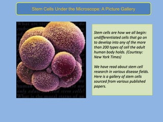
Stem Cells Under the Microscope: A Picture Gallery


                              Stem cells are how we all begin:
                              undifferentiated cells that go on
                              to develop into any of the more
                              than 200 types of cell the adult
                              human body holds. (Courtesy:
                              New York Times)

                              We have read about stem cell
                              research in various disease fields.
                              Here is a gallery of stem cells
                              sourced from various published
                              papers.
Rat brain slice showing neural stem cells (blue) that divide throughout life
to produce astrocytes (red) and mature neurons (green). Courtesy: Sarthak
Sinha
Human Embryonic Stem Cells forms spherical colonies
when cultured in-vitro. Courtesy: Sarthak Sinha
Human Embryonic Stem Cells used for human transplantation
PLoS Biology. Courtesy: Sarthak Sinha
International Journal of Stem Cell Research and
Transplantation
The germ line stem cell niche contains germ stem cells
(blue), somatic support cells (green), and spectrosomes (red).
IMAGE COURTESY OF ANDREA ZAMPARINI
Human hematopoietic stem cells. Image courtesy:
Lancet,Oncology
Image courtesy:
http://www.embryonicstemcellresearch.org/
Image courtesy: http://www.embryonicstemcellresearch.org/
Spontaneous embryonic stem cell differentiation in mouse
cells. The bright green cells show that some embryonic
stem cells differentiate into cardiac myocytes.
Important Embryonic Stem Cell
           Research Milestones

          Stem cells were discovered in human cord blood
•   1981- First in vitro stem cell line developed from mice
•   1988- Embryonic stem cell lines were created from
    hamsters
•   1995- First embryonic stem cell line derived from a primate
•   1997- Cloned lamb from stem cell and leukemia origin
    found as haematopoietic stem cell, which could indicate
    potential proof of cancer stem cells

Stem cells under the microscope a picture gallery

  • 1.
    Stem Cells Underthe Microscope: A Picture Gallery Stem cells are how we all begin: undifferentiated cells that go on to develop into any of the more than 200 types of cell the adult human body holds. (Courtesy: New York Times) We have read about stem cell research in various disease fields. Here is a gallery of stem cells sourced from various published papers.
  • 2.
    Rat brain sliceshowing neural stem cells (blue) that divide throughout life to produce astrocytes (red) and mature neurons (green). Courtesy: Sarthak Sinha
  • 3.
    Human Embryonic StemCells forms spherical colonies when cultured in-vitro. Courtesy: Sarthak Sinha
  • 4.
    Human Embryonic StemCells used for human transplantation PLoS Biology. Courtesy: Sarthak Sinha
  • 5.
    International Journal ofStem Cell Research and Transplantation
  • 6.
    The germ linestem cell niche contains germ stem cells (blue), somatic support cells (green), and spectrosomes (red). IMAGE COURTESY OF ANDREA ZAMPARINI
  • 7.
    Human hematopoietic stemcells. Image courtesy: Lancet,Oncology
  • 8.
  • 9.
  • 10.
    Spontaneous embryonic stemcell differentiation in mouse cells. The bright green cells show that some embryonic stem cells differentiate into cardiac myocytes.
  • 11.
    Important Embryonic StemCell Research Milestones Stem cells were discovered in human cord blood • 1981- First in vitro stem cell line developed from mice • 1988- Embryonic stem cell lines were created from hamsters • 1995- First embryonic stem cell line derived from a primate • 1997- Cloned lamb from stem cell and leukemia origin found as haematopoietic stem cell, which could indicate potential proof of cancer stem cells