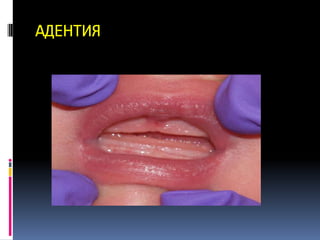
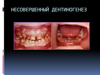
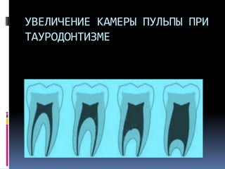
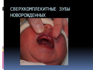
This document provides information about the dento-gingival unit, which refers to the junctional epithelium and gingival fibers that maintain the attachment between the gingiva and tooth. It discusses the development, structure, and functions of the junctional epithelium. In particular, it describes how the junctional epithelium forms a specialized attachment to the tooth through hemidesmosomes and an internal basal lamina. It also discusses the dynamic cellular processes and molecular components that allow the junctional epithelium to regenerate and maintain a strong yet permeable attachment between the oral cavity and periodontium.
The document discusses mid facial fractures, which involve the bones of the central face between the forehead and upper jaw. It describes the classification systems for mid facial fractures proposed by Le Fort and others. Le Fort I fractures involve the upper jaw, Le Fort II involve the pyramidal bones, and Le Fort III involve separation of the mid face from the skull. Common causes are motor vehicle accidents and assaults. Clinical features vary depending on the fracture type but may include swelling, bruising, numbness, and mobility of facial bones. Diagnosis involves imaging like CT scans. Treatment goals are to restore facial structure and function, often through closed or open reduction and internal fixation of the bones.
Rene LeFort in 1901 classified midface fractures based on the level of injury into 3 types: Lefort I, II, and III fractures. Since then there have been modifications to the classification system. Lefort I fractures involve the maxilla above the teeth and nasal floor. Treatment involves either closed or open reduction with fixation methods like miniplates or wires. Potential complications include nerve damage, infection, malocclusion, and sinus issues. Contemporary approaches emphasize early open reduction and anatomical fixation with miniplates to achieve accurate reconstruction of the midface structural pillars.
This document discusses several conditions that can affect the gingiva including necrotizing ulcerative gingivitis (NUG), primary herpetic gingivostomatitis, and recurrent aphthous stomatitis. NUG is a painful inflammatory disease affecting the gingiva caused by spirochetes and fusiform bacteria. It is characterized by ulcers and can cause bad breath and increased salivation. Treatment involves antibiotics, rinsing with hydrogen peroxide, and improving oral hygiene. Primary herpetic gingivostomatitis is caused by the herpes simplex virus and produces gingival lesions and sores. It typically resolves within 7-10 days with top
The various cysts of the jaws, few key points for the diagnosis and the treatment options available for each.
Mentor: Dr Saikat Saha MDS, OMFS, SIliguri, West Bengal, India
Address: MAXFAC Center for Oral and Maxillofacial and Head & Neck Surgery, Siliguri
Email : maxfacmail@gmail.com
This document discusses temporomandibular joint disorders (TMJD), including normal anatomy, classifications, arthritis of the TMJ, and specific conditions like osteoarthritis, rheumatoid arthritis, and juvenile rheumatoid arthritis. It provides details on the clinical manifestations, radiographic features, differential diagnosis, and treatment options for various TMJD conditions.
The document lists various oral examination procedures and conditions to identify, including identifying finger clubbing and cyanosis as signs of heart disease, icterus as a sign of liver disease, incompetent lips, percussion and lymph node examination procedures, interproximal and occlusal caries locations, generalized gingivitis and periodontitis, and ulcers on the lip and tongue.
Pericoronitis is defined as inflammation of the oral soft tissues surrounding the crown of a partially erupted tooth. its treatment- operculectomy i.e. removal of the inflammed operculum
2017 classification of periodontal and periimplant diseasesDr. Bibina George
The document summarizes the key changes in the 2017 classification of periodontal and peri-implant diseases compared to the 1999 classification. The 2017 classification introduces staging and grading systems for periodontitis to indicate disease severity and risk of progression. It also includes classifications for peri-implant health, mucositis, and implantitis, as well as mucogingival deformities. The document reviews these changes and provides references for the revised classification system.
This document presents a case study of a 20-year-old female patient diagnosed with an odontogenic keratocyst. Examination found a swelling in her lower right back tooth region. Imaging showed a large multilocular radiolucency. Biopsy and histopathology of the cyst lining confirmed the diagnosis of keratocystic odontogenic tumor. Odontogenic keratocysts require aggressive treatment like resection due to their high recurrence rate from rapid proliferation and presence of satellite cysts. Carnoy's solution can be used as a chemical cauterizing agent during treatment.
The document describes the classification and treatment of Le Fort I fractures. Le Fort I fractures can be linear, unilateral comminuted, or bilateral comminuted. Treatment depends on the classification and includes observation for non-mobile fractures, closed treatment for minor malocclusions, and open reduction internal fixation for more severe fractures. Open reduction involves exposing the fracture, reducing it, and fixing it with plates placed along the vertical buttresses of the maxilla. Bone grafting may be needed for defects. The occlusion is checked after fixation and corrected if needed.
Dentinogenesis imperfecta is a disorder that causes teeth to be discolored and translucent. The teeth are also weaker, making them prone to breakage and loss. It affects approximately 1 in 6,000 to 8,000 people. There are three main types - Type I occurs with osteogenesis imperfecta, Type II is autosomal dominant and may cause hearing loss, and Type III shows extremely thin dentin and enlarged pulp chambers. Treatment focuses on bonding to strengthen and whiten teeth, as other cosmetic procedures could further damage the weakened teeth.
This document discusses mixed radiolucent-radiopaque lesions seen on dental radiographs, including cementoma, calcifying epithelial odontogenic cyst, calcifying epithelial odontogenic tumor, and adenomatoid odontogenic tumor. It provides radiographic features, differential diagnoses, and management recommendations for each type of lesion. Management may include periodic radiographic evaluation, surgical enucleation for expanding lesions, excision, or curettage.
Necrotizing ulcerative gingivitis (NUG) and necrotizing ulcerative periodontitis (NUP) are microbial diseases caused by a mixed bacterial infection and impaired host response. NUG presents with crater-like ulcers and gray pseudomembrane on the gums, accompanied by pain and foul taste. If untreated, NUG can progress to NUP and involve bone loss. Treatment involves removing debris, using antiseptics like chlorhexidine, scaling, and antibiotics in severe cases. Prognosis is good with proper treatment and maintenance of oral hygiene.
Degenerative joint disorders of temporomandibular jointShibani Sarangi
This document discusses degenerative joint disorders of the temporomandibular joint. It defines degenerative joint disease as the end result of many insults to the joint surfaces that results in altered joint structure due to degradation of cartilage and changes in subchondral bone and soft tissues. Osteoarthritis and rheumatoid arthritis are two common types of degenerative joint disease that affect the temporomandibular joint. The document outlines the etiology, clinical features, diagnosis, and treatment options for temporomandibular joint osteoarthritis and rheumatoid arthritis. Treatment involves both non-pharmacological and pharmacological approaches depending on the severity of the condition.
This document discusses bone destruction patterns caused by periodontal disease. It identifies the main causes of bone destruction as the extension of gingival inflammation, trauma from occlusion, and systemic disorders. It describes several patterns of bone loss seen in periodontal disease, including horizontal, vertical, osseous craters, bulbous contours, reversed architecture, ledges, and furcation involvement. The rate and episodic nature of bone destruction in periodontal disease is also covered.
The document provides information on traumatic injuries to teeth, including concussions, luxations, and fractures. It describes the clinical signs, radiographic findings, and treatment approaches for each type of injury. Concussions involve inflammation of the periodontal ligament without tooth displacement. Luxations occur when a tooth is displaced from its socket, sometimes with alveolar bone fractures. Fractures are classified as enamel fractures, enamel-dentin fractures, enamel-dentin-pulp fractures, or root fractures. Treatment depends on the specific injury but may include repositioning displaced teeth, pulpotomies, root canals, extractions, or orthodontic/surgical repositioning.
Periodontal disease is a widely prevalent disease worldwide which often gets unnoticed or it often ignored due to its slowly progressive nature. It is of concern since it can cause irrepairable damage to tooth supporting structures if not early diagnosed or treated.
This document provides an overview of gingival epithelium, including its microscopic features, structural characteristics, defense mechanisms, and renewal process. It defines gingiva as the part of oral mucosa that covers the alveolar process and surrounds tooth necks. Gingiva consists of three types: marginal, attached, and interdental gingiva. The gingival epithelium contains keratinocytes and melanocytes. Keratinocytes form the bulk of the epithelium and undergo continuous renewal, while melanocytes transfer melanin to keratinocytes. The degree of keratinization varies between oral mucosal sites.
Cementum is the calcified tissue that forms the outer layer of the tooth root. There are two main types of cementum: acellular and cellular. Acellular cementum covers the cervical third of the root and lacks cells. Cellular cementum forms after the tooth reaches the occlusal plane and contains cementocytes within lacunae. Cementum is composed primarily of collagen fibers and undergoes continuous deposition throughout life, increasing thickness especially in the apical region to compensate for tooth eruption and attrition.
This document discusses patterns of bone destruction in periodontal disease. It covers various causes of bone loss such as the extension of gingival inflammation, trauma from occlusion, and systemic disorders. Factors that determine bone destruction include normal bone variation, exostoses, buttressing bone formation, and food impaction. Common bone destruction patterns include horizontal loss, vertical defects, intrabony defects of one to three walls, furcation involvement, osseous craters, and ledges. Systemic conditions like osteoporosis and Paget's disease can also cause alveolar bone destruction.
The document discusses the classification of periodontal diseases. It provides an overview of how periodontal diseases were previously classified, noting that the classification system was heavily based on age. It then summarizes the updated 1999 classification system, which introduced categories for gingival diseases and refined the categories for periodontal diseases. The key changes were introducing gingival disease categories, replacing terms like "adult periodontitis" with "chronic periodontitis", and expanding definitions of systemic diseases and their implications for periodontal health.
This document discusses the eruption times and sequences of primary and permanent teeth. It outlines the characteristics of pre-dental, deciduous, mixed, and permanent dentition periods. Key points include:
- Primary teeth typically erupt between 6-24 months of age
- The eruption sequence is: central incisors, lateral incisors, first molars, canines, second molars
- Permanent teeth typically erupt between 6-13 years of age
- The mixed dentition period involves transitioning from primary to permanent teeth between 6-14 years
- Establishing normal occlusion depends on factors like muscle pressure, TMJ relation, and tooth morphology
Necrotizing ulcerative gingivitis (NUG) is an acute, painful infection of the gums caused by an interaction between plaque bacteria like fusiform bacillus and spirochetes and the host immune response. It is characterized by necrosis and sloughing of gum tissue, presenting as punched-out ulcerations covered by a pseudomembrane. Diagnosis is based on clinical findings of painful ulcers with pseudomembrane, fetid odor, and potentially fever and lymphadenopathy. Treatment focuses on mechanical plaque removal and antibiotics to eliminate pathogenic bacteria.
This document presents a case report of a complex odontome obstructing the eruption of a maxillary central incisor in a 12-year old female patient. Radiographs and surgical removal revealed several irregular calcified masses composed of dental tissues. Histopathological examination confirmed the diagnosis of a complex odontome. The odontome was removed and the impacted tooth was left to monitor eruption. Early diagnosis and removal of odontomes can allow for normal eruption of impacted permanent teeth.
Классификация цементов для фиксации мостов и коронокDrotaverin
Дано описание пяти основных классов цементов, используемых в ортопедической стоматологии, а также показания или противопоказания к применению каждого из них.
Показаны альтернативные варианты протезирования бюгельными протезами с помощью пластмасс Дентал Д или Валпласта. Преимущества новых видов протезирования
9. Чаще всего при врожденных
синдромах –
1.Гарднера (изменениями в
челюстно-лицевой области,
гипердентия, ретенцией
сверхкомплектных зубов,
одонтомами и остеомами челюстей.
2.Черепно-небных дизостозах
(клейдокраниальный дизостоз)
3.Расщепленном небе.
10. МЕЗИОДЕНТИЯ
1. Сверхкомплектный зуб на
верхней челюсти по средней
линии
2. Между центральными резцами.
3. Основная причина диастемы
после удаления данного зуба
15. МИКРОДЕНТИЯ
1. Зубы меньше, чем в норме
2. Чаще – боковые резцы верхней
челюсти и третьи моляры
3. Возникают при гипофизарном
дварфизме
4. Вызывают пустые пространства
19. СИНОДЕНТИЯ
1. Сращивание двух зачатков
2. Общий дентин.
3. Причина неизвестна
4. Клинически на один зуб
меньше.
Если еще сверхкомплектный зуб,
то диагностировать сложно
32. НЕСОВЕРШЕННЫЙ ДЕНТИНОГЕНЕЗ
Классификация НД
ТИП 1: Возникает с несовершенным
остеогенезом
ТИП 2: Наследственный
опалесцирующий дентин + потеря
слуха. Аутосомно-доминантный тип
ТИП 3: Коронковая часть в виде
колокола с выходом наружу пульпы.
37. ДИЛАЦЕРАЦИЯ (Коронки и корни
под углом друг к другу)
1. причина неизвестна
2. М.Б. как результат травмы
формирующегося зуба
3. Сложности при удалении
4. Сложности при прохождении
канала
39. ЖЕМЧУЖНАЯ ЭМАЛЬ
1. Эктопические массы эмали
2. Чаще в месте раздвоения
каналов
3. Ошибочно принимается за
зубной камень
4. Хорошо видны на
рентгенограммах
43. ГИПЕРЦЕМЕНТОЗ
1. Разрастание цемента вокруг
зуба.
2. Рентгенографическая
диагностика
3. Поражает живые зубы
4. Чаще встречается при
периапикальном воспалении и в
зубах, не находящихся в окклюзии.