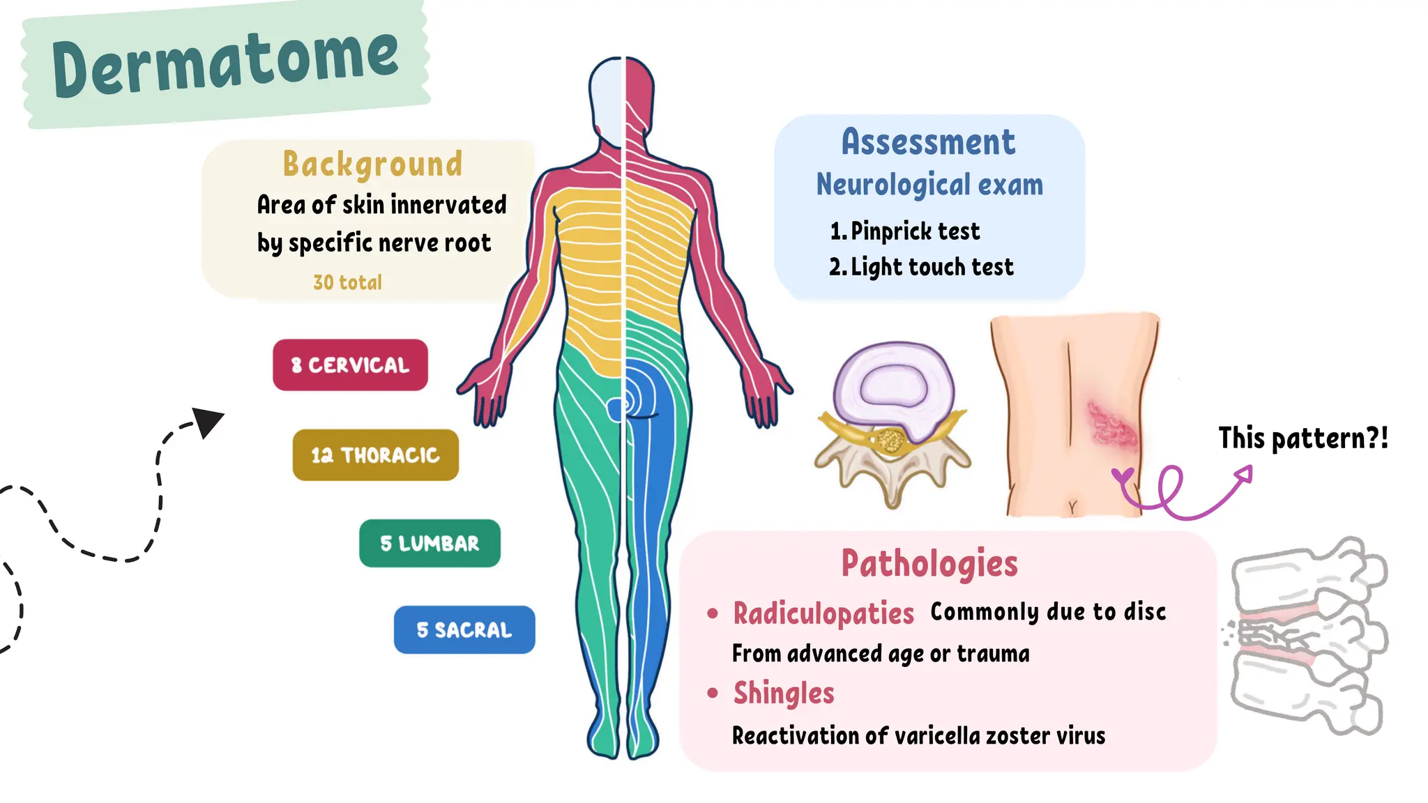
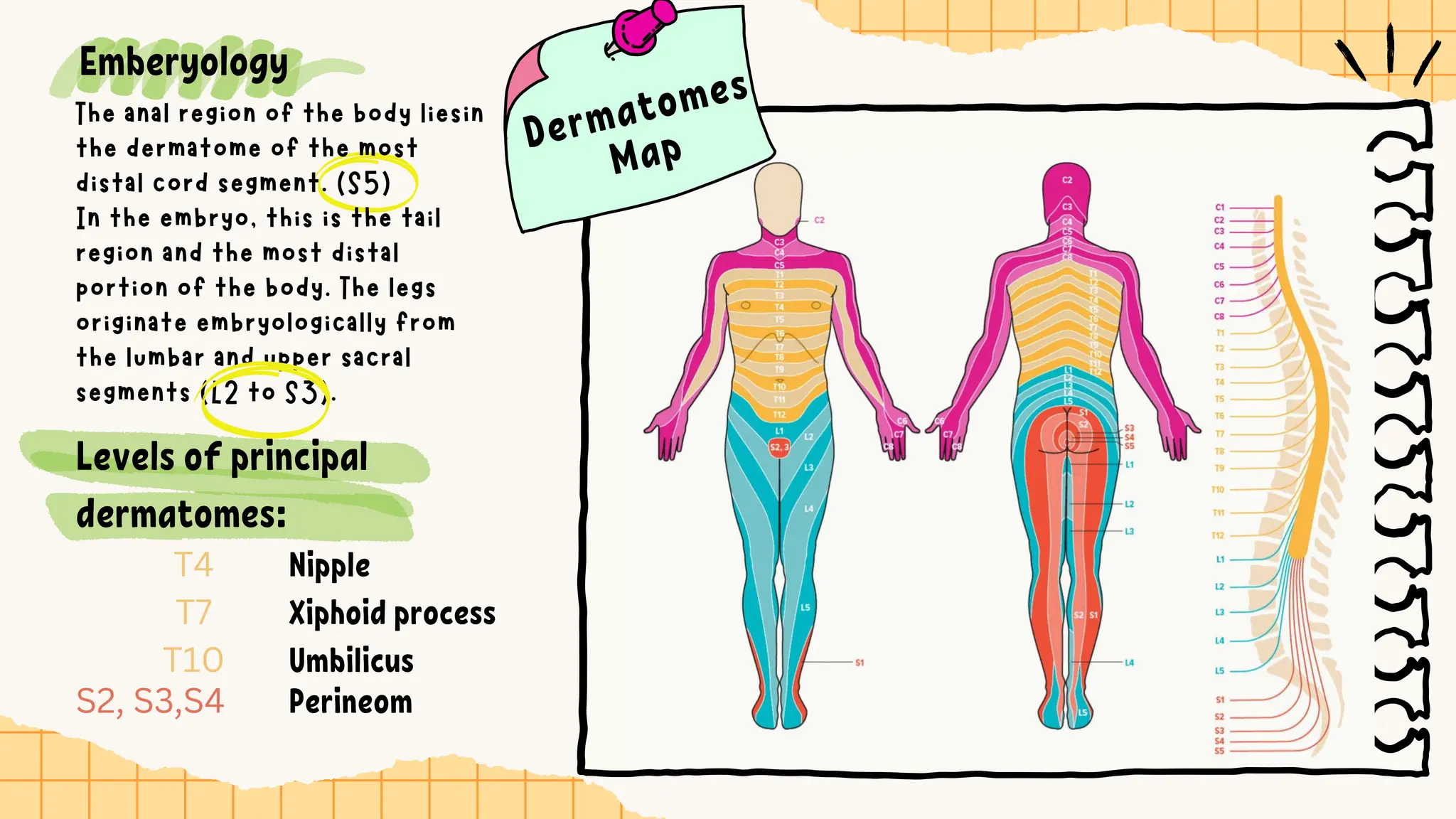
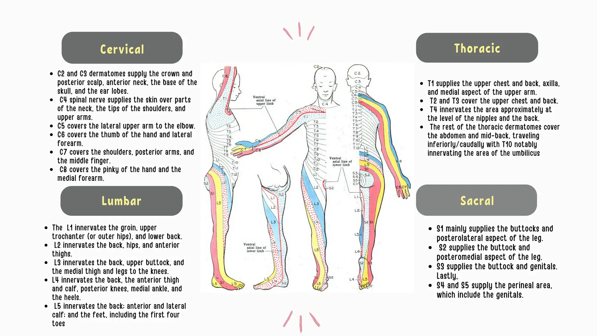
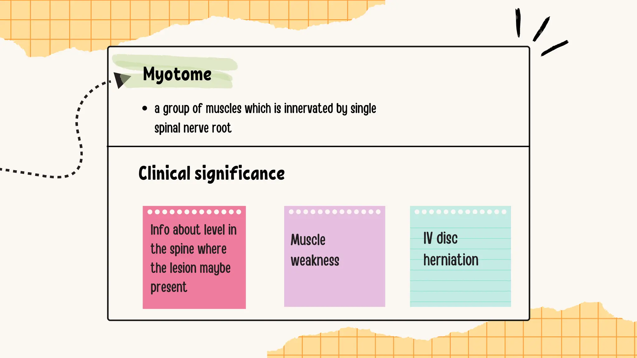
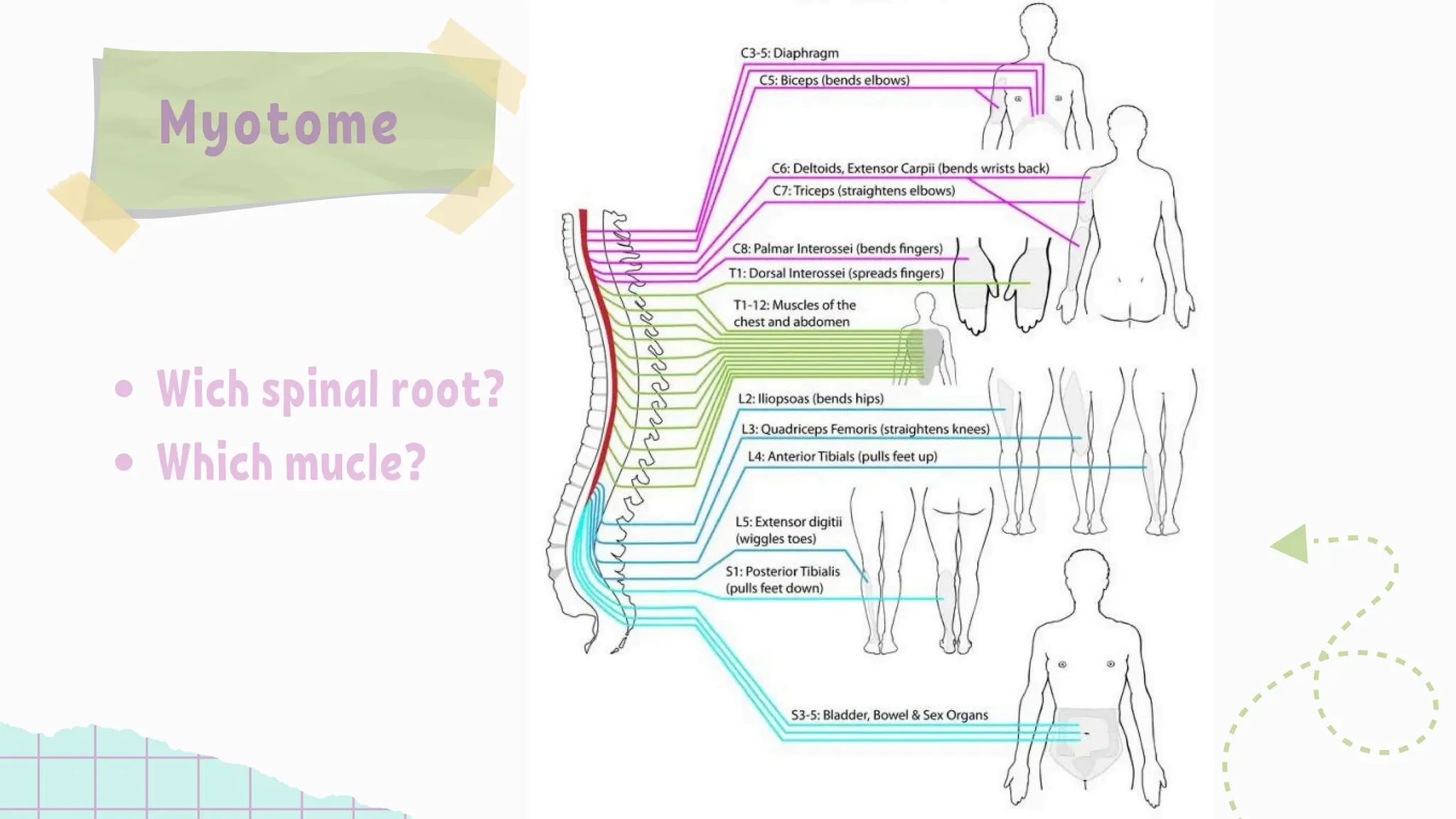
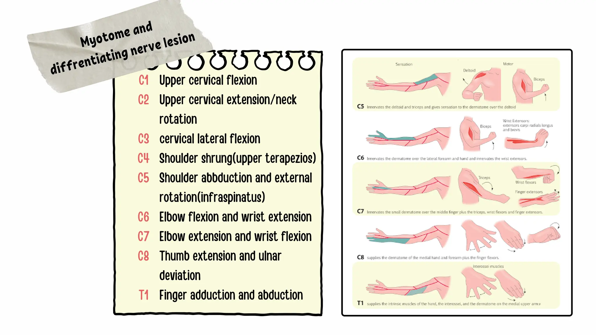
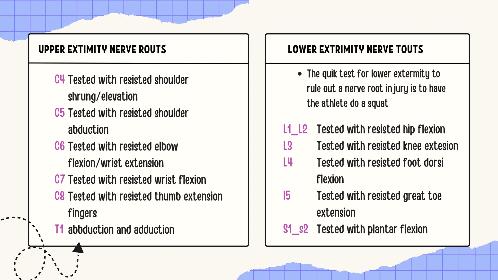
1. brachial plexus & its applied anatomy[1]MBBS IMS MSU
The brachial plexus is formed by the anterior rami of C5-C8 and T1 nerves. It originates in the neck, passes laterally over the first rib and enters the axilla. It has roots, trunks, divisions and cords that provide motor and sensory innervation to the upper limb. Injuries can occur from trauma, tumors or idiopathic causes. Presentations depend on the site of injury but can include weakness, sensory loss and pain. Diagnosis involves clinical exam, electrodiagnostics and imaging. Treatment focuses on pain management and rehabilitation to prevent complications. Prognosis is generally good with many patients recovering over 1-3 years.
Brachial Plexus Injury - An Introduction to the PhysiotherapistsJebarajFletcher
The document provides details about the brachial plexus, which is formed by the ventral rami of spinal nerves C5-T1. It innervates the upper limb and pectoral girdle. The brachial plexus consists of roots, trunks, divisions, cords, and branches. The trunks are the upper, middle, and lower trunks formed from C5/C6, C7, and C8/T1 respectively. The cords are the lateral, medial, and posterior cords. The major nerves include the axillary, radial, musculocutaneous, ulnar, and median nerves. Injuries to different parts of the brachial plexus can result
The document provides detailed information about the anatomy and function of the spinal cord. It can be summarized as follows:
The spinal cord is a cylindrical column of nervous tissue that extends from the brainstem and provides motor and sensory innervation to the body below the head. It is surrounded by protective meninges and terminates around the L1 vertebra in adults. The spinal cord is divided into regions that each give rise to pairs of spinal nerves which innervate different parts of the body. Injuries to the spinal cord can cause paralysis or other functional impairments depending on the level and severity of the injury.
thoracic outlet syndrome; one of the disorder affecting shoulder joint and neck movements due to limitation and pain. this slideshow describes about; the definition, types, causes and physiotherapy management for the same.
The document describes the anatomy and clinical presentations of spinal nerve and root lesions. It discusses:
- The anatomy of spinal nerves, with dorsal roots carrying sensory fibers and ventral roots carrying motor fibers.
- Common causes of spinal nerve and root lesions include disc herniations and diseases like Guillain-Barré syndrome.
- Signs and symptoms of lesions vary by level, and may include pain, weakness, or sensory changes in myotomal or dermatomal distributions, as well as depressed reflexes.
- Cervical lesions from C1-C7 are described, noting their sensory and motor involvement. Lumbar lesions from L1-L5 are also outlined.
The document discusses the brachial plexus, which is a network of nerves that supplies sensation and motor function to the upper extremity. It is formed from the lower cervical and upper thoracic spinal nerves. The document details the anatomy of the brachial plexus including its roots, trunks, divisions, cords and branches. It also discusses clinical conditions involving brachial plexus injury and techniques for brachial plexus nerve blocks such as interscalene and supraclavicular blocks.
Thoracic outlet syndrome is a condition that involves compression of the nerves or blood vessels that pass through the base of the neck. This can lead to disabling pain in the neck and shoulder, as well as pain, numbness, tingling and weakness in the hands and fingers.Thoracic outlet syndrome (TOS) is a term used to describe a group of disorders that occur when there is compression, injury, or irritation of the nerves and/or blood vessels (arteries and veins) in the lower neck and upper chest area. Thoracic outlet syndrome is named for the space (the thoracic outlet) between your lower neck and upper chest where this grouping of nerves and blood vessels is found.
Who is affected by thoracic outlet syndrome?
Thoracic outlet syndrome affects people of all ages and gender. The condition is common among athletes who participate in sports that require repetitive motions of the arm and shoulder, such as baseball, swimming, volleyball, and other sports.
Neurogenic TOS is the most common form of the disorder (95 percent of people with TOS have this form of the disorder) and generally affects middle-aged women.
Recent studies have shown that, in general, TOS is more common in women than men, particularly among those with poor muscular development, poor posture or both.
What are the symptoms?
Download a Free Guide on Thoracic Outlet Syndrome
The signs and symptoms of TOS include neck, shoulder, and arm pain, numbness or impaired circulation to the affected areas.
The pain of TOS is sometimes confused with the pain of angina (chest pain due to an inadequate supply of oxygen to the heart muscle), but the two conditions can be distinguished because the pain of thoracic outlet syndrome does not occur or increase when walking, while the pain of angina usually does. Additionally, the pain of TOS typically increases when raising the affected arm, which does not occur with angina.
Signs and symptoms of TOS help determine the type of disorder a patient has. Thoracic outlet syndrome disorders differ, depending on the part(s) of the body they affect. Thoracic outlet syndrome most commonly affects the nerves, but the condition can also affect the veins and arteries (least common type). In all types of TOS, the thoracic outlet space is narrowed, and there is scar formation around the structures.
Types of thoracic outlet syndrome disorders and related symptoms
Neurogenic thoracic outlet syndrome: This condition is related to abnormalities of bony and soft tissue in the lower neck region (which may include the cervical rib area) that compress and irritate the nerves of the brachial plexus, the complex of nerves that supply motor (movement) and sensory (feeling) function to the arm and hand. Symptoms include weakness or numbness of the hand; decreased size of hand muscles, which usually occurs on one side of the body; and/or pain, tingling, prickling, numbness and weakness of the neck, chest, and arms.
Venous thoracic outlet syndrome
Somites are embryonic structures that form segments along the neural tube and give rise to important tissues. They segment the embryo and are responsible for metamerism. Somites differentiate into sclerotome (axial skeleton), myotome (skeletal muscles), dermatome (skin and muscles), and syndotome (tendons). Dermatomes are dorsal portions of somites that form skin dermis and remain segmentally supplied by spinal nerves. Myotomes are ventral somite portions that form specific muscle groups. Dermatomes and myotomes have clinical significance as their patterns help locate spinal nerve involvement in conditions like shingles, sciatica, cervical radiculopathy, and cauda equina syndrome
This document provides an overview of the surgical anatomy of the neck, including:
1. The developmental anatomy of the neck structures derived from the pharyngeal arches.
2. The surface anatomy landmarks of the neck that can be palpated, including bones, muscles, vessels and glands.
3. The fascial layers of the neck, including the superficial cervical fascia, and three layers of the deep cervical fascia - the superficial investing layer, carotid sheath, and deep prevertebral layer.
4. The triangles used to compartmentalize the neck structures, including the anterior, posterior, and other subdivisions.
retract the muscles laterally
Thyroidectomy steps 4 – Delineating the gland
- After splitting the strap muscles, the thyroid gland comes into view.
- The gland is delineated from surrounding tissues by blunt dissection.
- The isthmus is identified and separated from trachea.
- The lateral lobes are separated from carotid sheath medially and strap muscles laterally.
- The upper pole is separated from cricothyroid membrane and lower pole from trachea.
Technique:
- Blunt dissection with artery forceps or fingers
- Always dissect in avascular plane between thyroid and surrounding structures
- Identify and preserve the external branch of SLN running over upper pole
The neck is divided into anterior and posterior triangles by the sternocleidomastoid muscle.
The anterior triangle contains the sternocleidomastoid muscle, which attaches to the sternum, clavicle, and mastoid process. Contracting one side tilts the head to that side and rotates it opposite, while contracting both flexes the neck and extends the head.
The posterior triangle is bounded anteriorly by the sternocleidomastoid, posteriorly by the trapezius muscle, and contains nerves like the brachial plexus and arteries like the subclavian. It is an important area for identifying structures during procedures like carotid endarterectomy.
This lecture give us an understanding about the pathway of the peripheral nerves that emerges from the brachial and cervical plexus. I also discuss about the motor and cutaneous innervation from these nerves and also some condition relate to peripheral nerve injury.
Localizaiton of level of lesion in paraplegiaAbino David
This document discusses spinal cord lesions and their effects. It describes how lesions can cause different types of paralysis or loss of function depending on the level and completeness of the lesion. It also discusses how lesions at different spinal levels correspond to specific vertebral levels and dermatomes. The document examines various ways to localize the level of a spinal cord lesion, including sensory loss, reflex changes, muscle weakness, and bladder/bowel dysfunction.
Surgical anatomy of upper limb nerves and plexusPirah Azadi
The document discusses the surgical anatomy of the upper limb nerves and brachial plexus. It begins by outlining the brachial plexus and its main branches, including the axillary, musculocutaneous, radial, median, and ulnar nerves. It then provides details on specific nerves, describing their motor and sensory innervation, clinical presentations of injuries, and dermatome patterns. Common nerve entrapment syndromes like carpal tunnel syndrome are also summarized.
This document discusses dermatomes and myotomes, which relate to the sensory and motor innervation of the body by spinal nerve roots. It provides detailed information on:
- The anatomy and distribution of dermatomes for each spinal nerve from C1 to S5.
- Clinical tests for dermatomes using pinprick and light touch at key points on the body.
- The muscles (myotomes) innervated by each spinal nerve root from C1 to S1.
- Clinical tests of myotomes through resisted movement exercises to evaluate motor function.
The vertebral column consists of 33 vertebrae that provide structural support, protect the spinal cord, and allow mobility. Each vertebra is composed of a vertebral body, processes, and joints that connect to adjacent vertebrae. The spinal cord runs through the vertebral column and is segmented into cervical, thoracic, lumbar, sacral and coccygeal regions. Surrounding structures include the ligaments, discs, dura mater, arachnoid mater, pia mater, epidural space, and subarachnoid space. Knowledge of the vertebral column's anatomy is essential for performing spinal and epidural procedures safely and effectively.
Presentation2, radiological imaging of brachial plexus pathology.Abdellah Nazeer
The document provides details on radiological imaging of brachial plexus pathology. It begins with an overview of brachial plexus anatomy including its origin, course, branches and functions. It then discusses various pathologies that can affect the brachial plexus including traumatic injuries, infections, inflammation, benign and malignant neoplasms, radiation plexopathy, compression and vascular abnormalities. For each pathology, it describes relevant imaging findings such as edema, enhancement patterns, masses, pseudomeningoceles and atrophy that help in diagnosis. Example images demonstrate various brachial plexus injuries and conditions.
This document discusses dermatomes and myotomes. It defines dermatomes as the sensory distribution of each nerve root, and myotomes as groups of muscles supplied by a single nerve root. It lists the dermatome levels from C1 to S4 and describes their sensory regions. The clinical significance of understanding dermatomes and myotomes is that they can help localize neurological levels in conditions like radiculopathy. Viral infections like herpes zoster also present in dermatomal patterns corresponding to the infected nerve root. Myotomes from C1 to S1 are listed along with the muscles each supplies and how weakness can indicate a spinal lesion level.
The median nerve innervates many of the flexor muscles of the forearm and hand. It passes through the cubital tunnel at the elbow and the carpal tunnel at the wrist. Carpal tunnel syndrome is a common entrapment of the median nerve in the wrist that causes pain, tingling, and numbness in the radial 3.5 fingers. The ulnar nerve supplies the small muscles of the hand and passes through the cubital tunnel at the elbow. It innervates muscles important for fine motor skills.
The parapharyngeal space is pyramidal in shape, located between the base of skull and hyoid bone. It has medial, posterior and lateral relations. Infection can occur from the pharynx, teeth, ear or other head and neck spaces. Clinical features depend on the involved compartment and include tonsillar prolapse, trismus, swelling and pain. Diagnosis is by CT or MRI. Abscesses are drained surgically under general anesthesia via an incision below the mandible. Complications include airway obstruction, thrombophlebitis, infection spread and carotid injury. Treatment involves intravenous antibiotics and surgical drainage.
The document discusses brachial plexus injuries, including anatomy, etiology, classification, clinical features, and investigations. The brachial plexus is formed by the anterior rami of C5-T1 nerves and provides sensation and motor function to the upper limb. Common causes of injury include motorcycle accidents, falls, and obstetric injuries. Injuries are classified based on the level and mechanism. Clinical features depend on the location and severity of injury. Investigations include imaging, EMG, and nerve conduction studies to evaluate the nature and extent of injury.
This document provides information about the muscles of the pectoral region, including the pectoralis major, pectoralis minor, serratus anterior, and subclavius. It describes the origin, insertion, innervation, and actions of each muscle. It also discusses the blood and nerve supply to the pectoral region, as well as the clinical relevance of structures like the clavipectoral fascia. Understanding the muscles of the pectoral region is important for identifying and describing them, and knowing their actions and clinical applications.
Osteoporosis - Definition , Evaluation and Management .pdfJim Jacob Roy
Osteoporosis is an increasing cause of morbidity among the elderly.
In this document , a brief outline of osteoporosis is given , including the risk factors of osteoporosis fractures , the indications for testing bone mineral density and the management of osteoporosis
Thoracic outlet syndrome is a condition that involves compression of the nerves or blood vessels that pass through the base of the neck. This can lead to disabling pain in the neck and shoulder, as well as pain, numbness, tingling and weakness in the hands and fingers.Thoracic outlet syndrome (TOS) is a term used to describe a group of disorders that occur when there is compression, injury, or irritation of the nerves and/or blood vessels (arteries and veins) in the lower neck and upper chest area. Thoracic outlet syndrome is named for the space (the thoracic outlet) between your lower neck and upper chest where this grouping of nerves and blood vessels is found.
Who is affected by thoracic outlet syndrome?
Thoracic outlet syndrome affects people of all ages and gender. The condition is common among athletes who participate in sports that require repetitive motions of the arm and shoulder, such as baseball, swimming, volleyball, and other sports.
Neurogenic TOS is the most common form of the disorder (95 percent of people with TOS have this form of the disorder) and generally affects middle-aged women.
Recent studies have shown that, in general, TOS is more common in women than men, particularly among those with poor muscular development, poor posture or both.
What are the symptoms?
Download a Free Guide on Thoracic Outlet Syndrome
The signs and symptoms of TOS include neck, shoulder, and arm pain, numbness or impaired circulation to the affected areas.
The pain of TOS is sometimes confused with the pain of angina (chest pain due to an inadequate supply of oxygen to the heart muscle), but the two conditions can be distinguished because the pain of thoracic outlet syndrome does not occur or increase when walking, while the pain of angina usually does. Additionally, the pain of TOS typically increases when raising the affected arm, which does not occur with angina.
Signs and symptoms of TOS help determine the type of disorder a patient has. Thoracic outlet syndrome disorders differ, depending on the part(s) of the body they affect. Thoracic outlet syndrome most commonly affects the nerves, but the condition can also affect the veins and arteries (least common type). In all types of TOS, the thoracic outlet space is narrowed, and there is scar formation around the structures.
Types of thoracic outlet syndrome disorders and related symptoms
Neurogenic thoracic outlet syndrome: This condition is related to abnormalities of bony and soft tissue in the lower neck region (which may include the cervical rib area) that compress and irritate the nerves of the brachial plexus, the complex of nerves that supply motor (movement) and sensory (feeling) function to the arm and hand. Symptoms include weakness or numbness of the hand; decreased size of hand muscles, which usually occurs on one side of the body; and/or pain, tingling, prickling, numbness and weakness of the neck, chest, and arms.
Venous thoracic outlet syndrome
Somites are embryonic structures that form segments along the neural tube and give rise to important tissues. They segment the embryo and are responsible for metamerism. Somites differentiate into sclerotome (axial skeleton), myotome (skeletal muscles), dermatome (skin and muscles), and syndotome (tendons). Dermatomes are dorsal portions of somites that form skin dermis and remain segmentally supplied by spinal nerves. Myotomes are ventral somite portions that form specific muscle groups. Dermatomes and myotomes have clinical significance as their patterns help locate spinal nerve involvement in conditions like shingles, sciatica, cervical radiculopathy, and cauda equina syndrome
This document provides an overview of the surgical anatomy of the neck, including:
1. The developmental anatomy of the neck structures derived from the pharyngeal arches.
2. The surface anatomy landmarks of the neck that can be palpated, including bones, muscles, vessels and glands.
3. The fascial layers of the neck, including the superficial cervical fascia, and three layers of the deep cervical fascia - the superficial investing layer, carotid sheath, and deep prevertebral layer.
4. The triangles used to compartmentalize the neck structures, including the anterior, posterior, and other subdivisions.
retract the muscles laterally
Thyroidectomy steps 4 – Delineating the gland
- After splitting the strap muscles, the thyroid gland comes into view.
- The gland is delineated from surrounding tissues by blunt dissection.
- The isthmus is identified and separated from trachea.
- The lateral lobes are separated from carotid sheath medially and strap muscles laterally.
- The upper pole is separated from cricothyroid membrane and lower pole from trachea.
Technique:
- Blunt dissection with artery forceps or fingers
- Always dissect in avascular plane between thyroid and surrounding structures
- Identify and preserve the external branch of SLN running over upper pole
The neck is divided into anterior and posterior triangles by the sternocleidomastoid muscle.
The anterior triangle contains the sternocleidomastoid muscle, which attaches to the sternum, clavicle, and mastoid process. Contracting one side tilts the head to that side and rotates it opposite, while contracting both flexes the neck and extends the head.
The posterior triangle is bounded anteriorly by the sternocleidomastoid, posteriorly by the trapezius muscle, and contains nerves like the brachial plexus and arteries like the subclavian. It is an important area for identifying structures during procedures like carotid endarterectomy.
This lecture give us an understanding about the pathway of the peripheral nerves that emerges from the brachial and cervical plexus. I also discuss about the motor and cutaneous innervation from these nerves and also some condition relate to peripheral nerve injury.
Localizaiton of level of lesion in paraplegiaAbino David
This document discusses spinal cord lesions and their effects. It describes how lesions can cause different types of paralysis or loss of function depending on the level and completeness of the lesion. It also discusses how lesions at different spinal levels correspond to specific vertebral levels and dermatomes. The document examines various ways to localize the level of a spinal cord lesion, including sensory loss, reflex changes, muscle weakness, and bladder/bowel dysfunction.
Surgical anatomy of upper limb nerves and plexusPirah Azadi
The document discusses the surgical anatomy of the upper limb nerves and brachial plexus. It begins by outlining the brachial plexus and its main branches, including the axillary, musculocutaneous, radial, median, and ulnar nerves. It then provides details on specific nerves, describing their motor and sensory innervation, clinical presentations of injuries, and dermatome patterns. Common nerve entrapment syndromes like carpal tunnel syndrome are also summarized.
This document discusses dermatomes and myotomes, which relate to the sensory and motor innervation of the body by spinal nerve roots. It provides detailed information on:
- The anatomy and distribution of dermatomes for each spinal nerve from C1 to S5.
- Clinical tests for dermatomes using pinprick and light touch at key points on the body.
- The muscles (myotomes) innervated by each spinal nerve root from C1 to S1.
- Clinical tests of myotomes through resisted movement exercises to evaluate motor function.
The vertebral column consists of 33 vertebrae that provide structural support, protect the spinal cord, and allow mobility. Each vertebra is composed of a vertebral body, processes, and joints that connect to adjacent vertebrae. The spinal cord runs through the vertebral column and is segmented into cervical, thoracic, lumbar, sacral and coccygeal regions. Surrounding structures include the ligaments, discs, dura mater, arachnoid mater, pia mater, epidural space, and subarachnoid space. Knowledge of the vertebral column's anatomy is essential for performing spinal and epidural procedures safely and effectively.
Presentation2, radiological imaging of brachial plexus pathology.Abdellah Nazeer
The document provides details on radiological imaging of brachial plexus pathology. It begins with an overview of brachial plexus anatomy including its origin, course, branches and functions. It then discusses various pathologies that can affect the brachial plexus including traumatic injuries, infections, inflammation, benign and malignant neoplasms, radiation plexopathy, compression and vascular abnormalities. For each pathology, it describes relevant imaging findings such as edema, enhancement patterns, masses, pseudomeningoceles and atrophy that help in diagnosis. Example images demonstrate various brachial plexus injuries and conditions.
This document discusses dermatomes and myotomes. It defines dermatomes as the sensory distribution of each nerve root, and myotomes as groups of muscles supplied by a single nerve root. It lists the dermatome levels from C1 to S4 and describes their sensory regions. The clinical significance of understanding dermatomes and myotomes is that they can help localize neurological levels in conditions like radiculopathy. Viral infections like herpes zoster also present in dermatomal patterns corresponding to the infected nerve root. Myotomes from C1 to S1 are listed along with the muscles each supplies and how weakness can indicate a spinal lesion level.
The median nerve innervates many of the flexor muscles of the forearm and hand. It passes through the cubital tunnel at the elbow and the carpal tunnel at the wrist. Carpal tunnel syndrome is a common entrapment of the median nerve in the wrist that causes pain, tingling, and numbness in the radial 3.5 fingers. The ulnar nerve supplies the small muscles of the hand and passes through the cubital tunnel at the elbow. It innervates muscles important for fine motor skills.
The parapharyngeal space is pyramidal in shape, located between the base of skull and hyoid bone. It has medial, posterior and lateral relations. Infection can occur from the pharynx, teeth, ear or other head and neck spaces. Clinical features depend on the involved compartment and include tonsillar prolapse, trismus, swelling and pain. Diagnosis is by CT or MRI. Abscesses are drained surgically under general anesthesia via an incision below the mandible. Complications include airway obstruction, thrombophlebitis, infection spread and carotid injury. Treatment involves intravenous antibiotics and surgical drainage.
The document discusses brachial plexus injuries, including anatomy, etiology, classification, clinical features, and investigations. The brachial plexus is formed by the anterior rami of C5-T1 nerves and provides sensation and motor function to the upper limb. Common causes of injury include motorcycle accidents, falls, and obstetric injuries. Injuries are classified based on the level and mechanism. Clinical features depend on the location and severity of injury. Investigations include imaging, EMG, and nerve conduction studies to evaluate the nature and extent of injury.
This document provides information about the muscles of the pectoral region, including the pectoralis major, pectoralis minor, serratus anterior, and subclavius. It describes the origin, insertion, innervation, and actions of each muscle. It also discusses the blood and nerve supply to the pectoral region, as well as the clinical relevance of structures like the clavipectoral fascia. Understanding the muscles of the pectoral region is important for identifying and describing them, and knowing their actions and clinical applications.
Similar to Dermatome and myotome test & pathology.pdf (20)
Osteoporosis - Definition , Evaluation and Management .pdfJim Jacob Roy
Osteoporosis is an increasing cause of morbidity among the elderly.
In this document , a brief outline of osteoporosis is given , including the risk factors of osteoporosis fractures , the indications for testing bone mineral density and the management of osteoporosis
Cell Therapy Expansion and Challenges in Autoimmune DiseaseHealth Advances
There is increasing confidence that cell therapies will soon play a role in the treatment of autoimmune disorders, but the extent of this impact remains to be seen. Early readouts on autologous CAR-Ts in lupus are encouraging, but manufacturing and cost limitations are likely to restrict access to highly refractory patients. Allogeneic CAR-Ts have the potential to broaden access to earlier lines of treatment due to their inherent cost benefits, however they will need to demonstrate comparable or improved efficacy to established modalities.
In addition to infrastructure and capacity constraints, CAR-Ts face a very different risk-benefit dynamic in autoimmune compared to oncology, highlighting the need for tolerable therapies with low adverse event risk. CAR-NK and Treg-based therapies are also being developed in certain autoimmune disorders and may demonstrate favorable safety profiles. Several novel non-cell therapies such as bispecific antibodies, nanobodies, and RNAi drugs, may also offer future alternative competitive solutions with variable value propositions.
Widespread adoption of cell therapies will not only require strong efficacy and safety data, but also adapted pricing and access strategies. At oncology-based price points, CAR-Ts are unlikely to achieve broad market access in autoimmune disorders, with eligible patient populations that are potentially orders of magnitude greater than the number of currently addressable cancer patients. Developers have made strides towards reducing cell therapy COGS while improving manufacturing efficiency, but payors will inevitably restrict access until more sustainable pricing is achieved.
Despite these headwinds, industry leaders and investors remain confident that cell therapies are poised to address significant unmet need in patients suffering from autoimmune disorders. However, the extent of this impact on the treatment landscape remains to be seen, as the industry rapidly approaches an inflection point.
- Video recording of this lecture in English language: https://youtu.be/Pt1nA32sdHQ
- Video recording of this lecture in Arabic language: https://youtu.be/uFdc9F0rlP0
- Link to download the book free: https://nephrotube.blogspot.com/p/nephrotube-nephrology-books.html
- Link to NephroTube website: www.NephroTube.com
- Link to NephroTube social media accounts: https://nephrotube.blogspot.com/p/join-nephrotube-on-social-media.html
DECLARATION OF HELSINKI - History and principlesanaghabharat01
This SlideShare presentation provides a comprehensive overview of the Declaration of Helsinki, a foundational document outlining ethical guidelines for conducting medical research involving human subjects.
The skin is the largest organ and its health plays a vital role among the other sense organs. The skin concerns like acne breakout, psoriasis, or anything similar along the lines, finding a qualified and experienced dermatologist becomes paramount.
Nano-gold for Cancer Therapy chemistry investigatory projectSIVAVINAYAKPK
chemistry investigatory project
The development of nanogold-based cancer therapy could revolutionize oncology by providing a more targeted, less invasive treatment option. This project contributes to the growing body of research aimed at harnessing nanotechnology for medical applications, paving the way for future clinical trials and potential commercial applications.
Cancer remains one of the leading causes of death worldwide, prompting the need for innovative treatment methods. Nanotechnology offers promising new approaches, including the use of gold nanoparticles (nanogold) for targeted cancer therapy. Nanogold particles possess unique physical and chemical properties that make them suitable for drug delivery, imaging, and photothermal therapy.
5-hydroxytryptamine or 5-HT or Serotonin is a neurotransmitter that serves a range of roles in the human body. It is sometimes referred to as the happy chemical since it promotes overall well-being and happiness.
It is mostly found in the brain, intestines, and blood platelets.
5-HT is utilised to transport messages between nerve cells, is known to be involved in smooth muscle contraction, and adds to overall well-being and pleasure, among other benefits. 5-HT regulates the body's sleep-wake cycles and internal clock by acting as a precursor to melatonin.
It is hypothesised to regulate hunger, emotions, motor, cognitive, and autonomic processes.
Co-Chairs, Val J. Lowe, MD, and Cyrus A. Raji, MD, PhD, prepared useful Practice Aids pertaining to Alzheimer’s disease for this CME/AAPA activity titled “Alzheimer’s Disease Case Conference: Gearing Up for the Expanding Role of Neuroradiology in Diagnosis and Treatment.” For the full presentation, downloadable Practice Aids, and complete CME/AAPA information, and to apply for credit, please visit us at https://bit.ly/3PvVY25. CME/AAPA credit will be available until June 28, 2025.
Test bank for karp s cell and molecular biology 9th edition by gerald karp.pdfrightmanforbloodline
Test bank for karp s cell and molecular biology 9th edition by gerald karp.pdf
NAVIGATING THE HORIZONS OF TIME LAPSE EMBRYO MONITORING.pdfRahul Sen
Time-lapse embryo monitoring is an advanced imaging technique used in IVF to continuously observe embryo development. It captures high-resolution images at regular intervals, allowing embryologists to select the most viable embryos for transfer based on detailed growth patterns. This technology enhances embryo selection, potentially increasing pregnancy success rates.
Breast cancer: Post menopausal endocrine therapyDr. Sumit KUMAR
Breast cancer in postmenopausal women with hormone receptor-positive (HR+) status is a common and complex condition that necessitates a multifaceted approach to management. HR+ breast cancer means that the cancer cells grow in response to hormones such as estrogen and progesterone. This subtype is prevalent among postmenopausal women and typically exhibits a more indolent course compared to other forms of breast cancer, which allows for a variety of treatment options.
Diagnosis and Staging
The diagnosis of HR+ breast cancer begins with clinical evaluation, imaging, and biopsy. Imaging modalities such as mammography, ultrasound, and MRI help in assessing the extent of the disease. Histopathological examination and immunohistochemical staining of the biopsy sample confirm the diagnosis and hormone receptor status by identifying the presence of estrogen receptors (ER) and progesterone receptors (PR) on the tumor cells.
Staging involves determining the size of the tumor (T), the involvement of regional lymph nodes (N), and the presence of distant metastasis (M). The American Joint Committee on Cancer (AJCC) staging system is commonly used. Accurate staging is critical as it guides treatment decisions.
Treatment Options
Endocrine Therapy
Endocrine therapy is the cornerstone of treatment for HR+ breast cancer in postmenopausal women. The primary goal is to reduce the levels of estrogen or block its effects on cancer cells. Commonly used agents include:
Selective Estrogen Receptor Modulators (SERMs): Tamoxifen is a SERM that binds to estrogen receptors, blocking estrogen from stimulating breast cancer cells. It is effective but may have side effects such as increased risk of endometrial cancer and thromboembolic events.
Aromatase Inhibitors (AIs): These drugs, including anastrozole, letrozole, and exemestane, lower estrogen levels by inhibiting the aromatase enzyme, which converts androgens to estrogen in peripheral tissues. AIs are generally preferred in postmenopausal women due to their efficacy and safety profile compared to tamoxifen.
Selective Estrogen Receptor Downregulators (SERDs): Fulvestrant is a SERD that degrades estrogen receptors and is used in cases where resistance to other endocrine therapies develops.
Combination Therapies
Combining endocrine therapy with other treatments enhances efficacy. Examples include:
Endocrine Therapy with CDK4/6 Inhibitors: Palbociclib, ribociclib, and abemaciclib are CDK4/6 inhibitors that, when combined with endocrine therapy, significantly improve progression-free survival in advanced HR+ breast cancer.
Endocrine Therapy with mTOR Inhibitors: Everolimus, an mTOR inhibitor, can be added to endocrine therapy for patients who have developed resistance to aromatase inhibitors.
Chemotherapy
Chemotherapy is generally reserved for patients with high-risk features, such as large tumor size, high-grade histology, or extensive lymph node involvement. Regimens often include anthracyclines and taxanes.
Travel vaccination in Manchester offers comprehensive immunization services for individuals planning international trips. Expert healthcare providers administer vaccines tailored to your destination, ensuring you stay protected against various diseases. Conveniently located clinics and flexible appointment options make it easy to get the necessary shots before your journey. Stay healthy and travel with confidence by getting vaccinated in Manchester. Visit us: www.nxhealthcare.co.uk
2. Background
Area of skin innervated
by specific nerve root
30 total
Pathologies
Radiculopaties
Shingles
Commonly due to disc
From advanced age or trauma
Reactivation of varicella zoster virus
Assessment
Neurological exam
Pinprick test
1.
Light touch test
2.
Dermatome
This pattern?!
3. Levels of principal
dermatomes:
Dermatomes
Map
T4
T7
T10
S2, S3,S4
Nipple
Xiphoid process
Umbilicus
Perineom
The anal region of the body liesin
the dermatome of the most
distal cord segment. (S5)
In the embryo, this is the tail
region and the most distal
portion of the body. The legs
originate embryologically from
the lumbar and upper sacral
segments (L2 to S3).
Emberyology
4. C2 and C3 dermatomes supply the crown and
posterior scalp, anterior neck, the base of the
skull, and the ear lobes.
C4 spinal nerve supplies the skin over parts
of the neck, the tips of the shoulders, and
upper arms.
C5 covers the lateral upper arm to the elbow.
C6 covers the thumb of the hand and lateral
forearm.
C7 covers the shoulders, posterior arms, and
the middle finger.
C8 covers the pinky of the hand and the
medial forearm.
The L1 innervates the groin, upper
trochanter (or outer hips), and lower back.
L2 innervates the back, hips, and anterior
thighs.
L3 innervates the back, upper buttock, and
the medial thigh and legs to the knees.
L4 innervates the back, the anterior thigh
and calf, posterior knees, medial ankle, and
the heels.
L5 innervates the back; anterior and lateral
calf; and the feet, including the first four
toes
Thoracic
Cervical
Sacral
Lumbar
T1 supplies the upper chest and back, axilla,
and medial aspect of the upper arm.
T2 and T3 cover the upper chest and back.
T4 innervates the area approximately at
the level of the nipples and the back.
The rest of the thoracic dermatomes cover
the abdomen and mid-back, traveling
inferiorly/caudally with T10 notably
innervating the area of the umbilicus
S1 mainly supplies the buttocks and
posterolateral aspect of the leg.
S2 supplies the buttock and
posteromedial aspect of the leg.
S3 supplies the buttock and genitals.
Lastly,
S4 and S5 supply the perineal area,
which include the genitals.
5. Myotome
Info about level in
the spine where
the lesion maybe
present
a group of muscles which is innervated by single
spinal nerve root
Muscle
weakness
IV disc
herniation
Clinical significance
7. Upper cervical flexion
Upper cervical extension/neck
rotation
cervical lateral flexion
Shoulder shrung(upper terapezios)
Shoulder abbduction and external
rotation(infraspinatus)
Elbow flexion and wrist extension
Elbow extension and wrist flexion
Thumb extension and ulnar
deviation
Finger adduction and abduction
Myotome and
diffrentiating nerve lesion
C1
C2
C3
C4
C5
C6
C7
C8
T1
8. Myotome and
diffrentiating nerve lesion
Hip flexion
Hip flexion(and also adduction and
medial rotasion)
Leg and knee extension
Dorsiflexion
Great/big toe extension
Ankle plantar flexion and
eversion/knee flexion
Ankle plantar flexion and knee
flexion
None
Bladder and rectome
L1
L2
L3
L4
L5
S1
S2
S3
S4
9. UPPER EXTIMITY NERVE ROUTS LOWER EXTRIMITY NERVE TOUTS
C4
C5
C6
C7
C8
T1
L1_L2
L3
L4
l5
S1_s2
Tested with resisted shoulder
shrung/elevation
Tested with resisted shoulder
abduction
Tested with resisted elbow
flexion/wrist extension
Tested with resisted wrist flexion
Tested with resisted thumb extension
fingers
abbduction and adduction
Tested with resisted hip flexion
Tested with resisted knee extesion
Tested with resisted foot dorsi
flexion
Tested with resisted great toe
extension
Tested with plantar flexion
The quik test for lower extermity to
rule out a nerve root injury is to have
the athlete do a squat