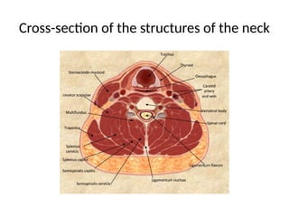
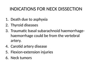
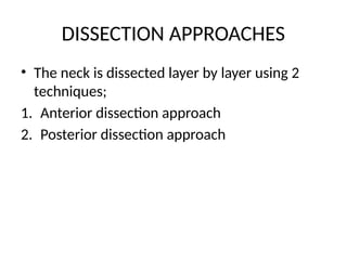
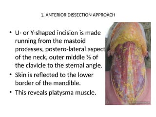
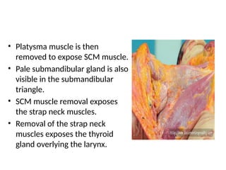
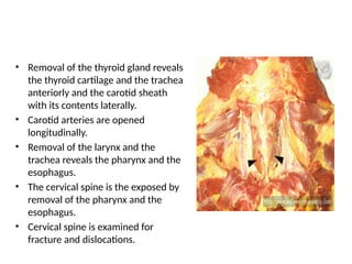
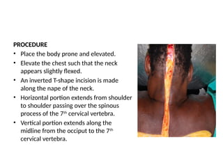
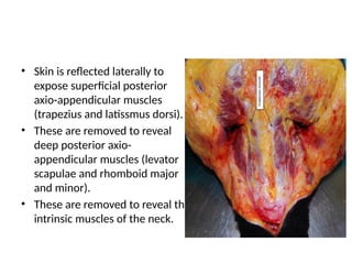
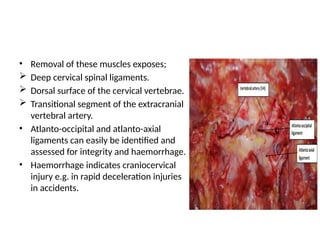
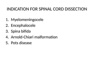
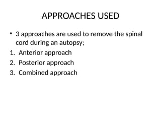
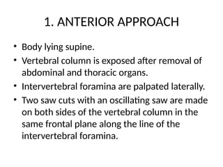
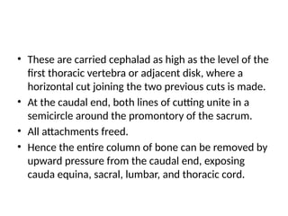
SPECIAL DISSECTION
TECHNIQUES
CHERUIYOT TOO
TM226-1800/2013
TYPES OF SPECIAL DISSECTION TECHNIQUES
AND THEIR INDICATIONS
1. Neck dissection
2. Pelvic dissection
3. Spinal cord dissection
4. Paediatric skull dissection
NECK DISSECTIONS
PRINCIPLES OF NECK DISSECTION
Drainage of vasculature should be done prior to
dissection.
• Aim- to avoid artefactual haemorrhage at the
beck of the larynx.
• How to achieve drainage- carrying out neck
dissection after removal of the brain and the
heart.
Cross-section of the structures of the neck
INDICATIONS FOR NECK DISSECTION
1. Death due to asphyxia
2. Thyroid diseases
3. Traumatic basal subarachnoid haemorrhage-
haemorrhage could be from the vertebral
artery.
4. Carotid artery disease
5. Flexion-extension injuries
6. Neck tumors
DISSECTION APPROACHES
• The neck is dissected layer by layer using 2
techniques;
1. Anterior dissection approach
2. Posterior dissection approach
1. ANTERIOR DISSECTION APPROACH
• U- or Y-shaped incision is made
running from the mastoid
processes, postero-lateral aspect
of the neck, outer middle ⅓ of
the clavicle to the sternal angle.
• Skin is reflected to the lower
border of the mandible.
• This reveals platysma muscle.
• Platysma muscle is then
removed to expose SCM muscle.
• Pale submandibular gland is also
visible in the submandibular
triangle.
• SCM muscle removal exposes
the strap neck muscles.
• Removal of the strap neck
muscles exposes the thyroid
gland overlying the larynx.
• Removal of the thyroid gland reveals
the thyroid cartilage and the trachea
anteriorly and the carotid sheath
with its contents laterally.
• Carotid arteries are opened
longitudinally.
• Removal of the larynx and the
trachea reveals the pharynx and the
esophagus.
• The cervical spine is the exposed by
removal of the pharynx and the
esophagus.
• Cervical spine is examined for
fracture and dislocations.
2. POSTERIOR DISSECTION APPROACH
• The goal is to;
Determine if any haemorrhage into the
subcutaneous tissue is present.
Examine the ligaments of the upper neck.
Visualize any ruptured vertebral arteries.
PROCEDURE
• Place the body prone and elevated.
• Elevate the chest such that the neck
appears slightly flexed.
• An inverted T-shape incision is made
along the nape of the neck.
• Horizontal portion extends from shoulder
to shoulder passing over the spinous
process of the 7th
cervical vertebra.
• Vertical portion extends along the
midline from the occiput to the 7th
cervical vertebra.
• Skin is reflected laterally to
expose superficial posterior
axio-appendicular muscles
(trapezius and latissmus dorsi).
• These are removed to reveal
deep posterior axio-
appendicular muscles (levator
scapulae and rhomboid major
and minor).
• These are removed to reveal the
intrinsic muscles of the neck.
• Removal of these muscles exposes;
 Deep cervical spinal ligaments.
 Dorsal surface of the cervical vertebrae.
 Transitional segment of the extracranial
vertebral artery.
• Atlanto-occipital and atlanto-axial
ligaments can easily be identified and
assessed for integrity and haemorrhage.
• Haemorrhage indicates craniocervical
injury e.g. in rapid deceleration injuries
in accidents.
PELVIC DISSECTION
INDICATIONS FOR PELVIC DISSECTION
1. Sexual assault
2. Pelvic malignancies
3. Pregnancy
4. Abortion
EXTERNAL AXAMINATION
• FEMALE- examine the inner aspect of the thighs,
labia majora and minora, clitoris, urethra,
periurethral tissues, hymen, hymenal opening,
the vestibule and posterior fourchette.
• MALE- examine the inner aspects of the thighs,
penile shaft, foreskin and glans and scrotum.
• BOTH- the perineum and anus should be
carefully examined under good light.
EXTERNAL GENITALIA
• Not usually removed as part of routine autopsy
but is important incase of urethral or penile
tumors, congenital posterior urethral valves
among others.
• The abdominal incision is extended to the base
of the penis in males and to the labia majora in
the females.
• Mid portion of the pubic arch is removed using
a saw.
• In males, the penis, scrotum and anus is
removed in continuity with the prostate,
bladder and rectum.
• In the females, after removal of the pubic
arch, the legs are separated to expose the
urogenital triangle, an elliptical incision is
made around the external genitalia and the
anus.
MALE LOWER GUT
• Open and inspect the rectum while still
attached to the genitourinary organ block.
• Prostate- measure size, make transverse
incisions and inspect for any suspicious nodules.
• Open proximal urethra and bladder, inspect the
urothelial mucosa.
• Probe the ureteral openings at the trigone and
make a longitudinal incision and inspect it.
• Make serial cuts into the seminal vesicle. Note
thickness of their walls and colour of the seminal fluid.
• Make several crosscuts along each vas deferens as well
and examine.
• Testes= differentiate left from right, cross cut any
attached spermatic cord, weigh the testes, cut in
sagittal plane and examine parenchyma.
• Use forceps, grasp a portion of the cut surface and
pull. Failure to string out easily is indicative of
testicular atrophy in postpubertal male.
FEMALE LOWER GUT
• Open and inspect the rectum, then separate it the
pelvic organs.
• Open bladder and examine, then separate it along
its attachments along the uterus.
• Inspect fallopian tubes and ovaries.
• Ovaries: measure in 3 dimensions, cut lengthwise
to expose parenchyma, examine it.
• Fallopian tubes: dissect longitudinally following
insertion of a probe or cut into serial cross sections.
• Proximal vagina: open along its lateral surface,
inspect the epithelium for any abnormality.
• Uterine cervix: inspect for any lesions,
erosions. Note the shape and greatest width
of the os.
• Uterus: open along its lateral aspect, measure
dimensions of the uterine cavity, note
thickness of the endometrium.
SPINAL CORD DISSECTION
INDICATION FOR SPINAL CORD DISSECTION
1. Myelomeningocele
2. Encephalocele
3. Spina bifida
4. Arnold-Chiari malformation
5. Pots disease
APPROACHES USED
• 3 approaches are used to remove the spinal
cord during an autopsy;
1. Anterior approach
2. Posterior approach
3. Combined approach
1. ANTERIOR APPROACH
• Body lying supine.
• Vertebral column is exposed after removal of
abdominal and thoracic organs.
• Intervertebral foramina are palpated laterally.
• Two saw cuts with an oscillating saw are made
on both sides of the vertebral column in the
same frontal plane along the line of the
intervertebral foramina.
• These are carried cephalad as high as the level of the
first thoracic vertebra or adjacent disk, where a
horizontal cut joining the two previous cuts is made.
• At the caudal end, both lines of cutting unite in a
semicircle around the promontory of the sacrum.
• All attachments freed.
• Hence the entire column of bone can be removed by
upward pressure from the caudal end, exposing
cauda equina, sacral, lumbar, and thoracic cord.
• Two parallel sagittal incisions are made with the saw
through the bodies of the lower cervical vertebra. (1-
1.5cm apart).
• A third cut is made at the most cephalad portion of
this block in order to remove it- exposes cervical
vertebra to the level of C3 or C4.
• The spinal cord and surrounding dura are now
removed or delivered in their entirety by cutting the
cauda equina.
• The cervical bone block may be replaced.
Advantages
Prevents leakage after embalming
Less mutilation visible
Course of peripheral nerves for any
length in contiguity from the spinal
cord can be accessed.
Disadvantages
Difficult to approach the proximal cervical
vertebrae
Conditions like myelomeningocele and occipital
encephalocele cannot be demonstrated.
Flexion-extension injuries to the back of the
neck or other injuries along the posterior
vertebral column cannot be demonstrated.
2. POSTERIOR APPROACH
• Body prone.
• Wooden blocks placed under both shoulders.
• Head is flexed.
• Midline incision is made over the spinous processes
and the back muscles are resected.
• Parallel saw-cuts are made through all the vertebral
laminae (laminectomy).
• Then the cauda equina is divided and lifted by Spencer
Wells forceps- not to twist or bend the spinal cord.
Advantages
Conditions like myelomeningocele and occipital
encephalocele can be easily demonstrated.
Dissection can be limited up to the desired level and
stopped.
Disadvantages
Course of peripheral nerves cannot be pursued along its
contiguity.
Embalming leakage.
More mutilation.
3. COMBINED APPROACH
• Body is turned back and incision is made
around the desired area then continued
anteriorly.
• Preferred approach for complete removal of
meningocele, myelomeningocele or other
midline fusion defect.
PAEDIATRIC HEAD DISSECTION
INDICATIONS FOR PAEDIATRIC HEAD
DISSECTION
• Hydrocephalus
• Stillbirths
INSPECTION
• The scalp is inspected externally, looking for edema,
caput, and bruising.
• The presence of localized swelling/mass may suggest
an underlying encephalocele.
• The head may be visibly enlarged in cases of severe
hydrocephalus.
• In intrapartum and neonatal deaths, any bruising,
hematomas, and forceps marks should be carefully
documented.
SCALP INCISION
• The scalp is reflected by making an incision
starting behind one ear and cutting posterior
to the crown of the head toward the other ear.
• The incision can then be extended toward the
neck and down the center of the back of the
neck to form the shape of a “question
mark”.
• This incision allows adequate access to the
brain and cervical spinal cord and satisfactory
reconstruction of the head.
• The scalp flaps are reflected forwards and
backwards and the inner aspect is examined
for the presence of edema and hemorrhage.
• The size of the fontanelles and width of the
sutures is then noted.
OPENING THE CRANIAL CAVITY
• Make parallel incisions in the anterior fontanelle away
from the midline. This is to ensure that the sagittal
sinus is not damaged.
• The incisions are then extended forwards and
backwards on either side of the midline.
• The fronto-parietal and parieto-occipital sutures are
then cut on each side.
• Care should be taken in all these steps that the
scissors point is kept up against the bone to
avoid damaging the brain.
• The parietal bones can then be opened in
order to expose the brain.
• The brain is then taken out underwater.
• This technique is useful in all perinatal
postmortems, especially small and macerated
fetuses, because the water will support the
brain and free the prosector’s hands to
separate the brain from the dura.

SPECIAL DISSECTION TECHNIQUES-d TOO.pptx

  • 1.
  • 2.
    TYPES OF SPECIALDISSECTION TECHNIQUES AND THEIR INDICATIONS 1. Neck dissection 2. Pelvic dissection 3. Spinal cord dissection 4. Paediatric skull dissection
  • 3.
  • 4.
    PRINCIPLES OF NECKDISSECTION Drainage of vasculature should be done prior to dissection. • Aim- to avoid artefactual haemorrhage at the beck of the larynx. • How to achieve drainage- carrying out neck dissection after removal of the brain and the heart.
  • 5.
    Cross-section of thestructures of the neck
  • 6.
    INDICATIONS FOR NECKDISSECTION 1. Death due to asphyxia 2. Thyroid diseases 3. Traumatic basal subarachnoid haemorrhage- haemorrhage could be from the vertebral artery. 4. Carotid artery disease 5. Flexion-extension injuries 6. Neck tumors
  • 7.
    DISSECTION APPROACHES • Theneck is dissected layer by layer using 2 techniques; 1. Anterior dissection approach 2. Posterior dissection approach
  • 8.
    1. ANTERIOR DISSECTIONAPPROACH • U- or Y-shaped incision is made running from the mastoid processes, postero-lateral aspect of the neck, outer middle ⅓ of the clavicle to the sternal angle. • Skin is reflected to the lower border of the mandible. • This reveals platysma muscle.
  • 9.
    • Platysma muscleis then removed to expose SCM muscle. • Pale submandibular gland is also visible in the submandibular triangle. • SCM muscle removal exposes the strap neck muscles. • Removal of the strap neck muscles exposes the thyroid gland overlying the larynx.
  • 10.
    • Removal ofthe thyroid gland reveals the thyroid cartilage and the trachea anteriorly and the carotid sheath with its contents laterally. • Carotid arteries are opened longitudinally. • Removal of the larynx and the trachea reveals the pharynx and the esophagus. • The cervical spine is the exposed by removal of the pharynx and the esophagus. • Cervical spine is examined for fracture and dislocations.
  • 11.
    2. POSTERIOR DISSECTIONAPPROACH • The goal is to; Determine if any haemorrhage into the subcutaneous tissue is present. Examine the ligaments of the upper neck. Visualize any ruptured vertebral arteries.
  • 12.
    PROCEDURE • Place thebody prone and elevated. • Elevate the chest such that the neck appears slightly flexed. • An inverted T-shape incision is made along the nape of the neck. • Horizontal portion extends from shoulder to shoulder passing over the spinous process of the 7th cervical vertebra. • Vertical portion extends along the midline from the occiput to the 7th cervical vertebra.
  • 13.
    • Skin isreflected laterally to expose superficial posterior axio-appendicular muscles (trapezius and latissmus dorsi). • These are removed to reveal deep posterior axio- appendicular muscles (levator scapulae and rhomboid major and minor). • These are removed to reveal the intrinsic muscles of the neck.
  • 14.
    • Removal ofthese muscles exposes;  Deep cervical spinal ligaments.  Dorsal surface of the cervical vertebrae.  Transitional segment of the extracranial vertebral artery. • Atlanto-occipital and atlanto-axial ligaments can easily be identified and assessed for integrity and haemorrhage. • Haemorrhage indicates craniocervical injury e.g. in rapid deceleration injuries in accidents.
  • 15.
  • 16.
    INDICATIONS FOR PELVICDISSECTION 1. Sexual assault 2. Pelvic malignancies 3. Pregnancy 4. Abortion
  • 17.
    EXTERNAL AXAMINATION • FEMALE-examine the inner aspect of the thighs, labia majora and minora, clitoris, urethra, periurethral tissues, hymen, hymenal opening, the vestibule and posterior fourchette. • MALE- examine the inner aspects of the thighs, penile shaft, foreskin and glans and scrotum. • BOTH- the perineum and anus should be carefully examined under good light.
  • 18.
    EXTERNAL GENITALIA • Notusually removed as part of routine autopsy but is important incase of urethral or penile tumors, congenital posterior urethral valves among others. • The abdominal incision is extended to the base of the penis in males and to the labia majora in the females. • Mid portion of the pubic arch is removed using a saw.
  • 19.
    • In males,the penis, scrotum and anus is removed in continuity with the prostate, bladder and rectum. • In the females, after removal of the pubic arch, the legs are separated to expose the urogenital triangle, an elliptical incision is made around the external genitalia and the anus.
  • 20.
    MALE LOWER GUT •Open and inspect the rectum while still attached to the genitourinary organ block. • Prostate- measure size, make transverse incisions and inspect for any suspicious nodules. • Open proximal urethra and bladder, inspect the urothelial mucosa. • Probe the ureteral openings at the trigone and make a longitudinal incision and inspect it.
  • 21.
    • Make serialcuts into the seminal vesicle. Note thickness of their walls and colour of the seminal fluid. • Make several crosscuts along each vas deferens as well and examine. • Testes= differentiate left from right, cross cut any attached spermatic cord, weigh the testes, cut in sagittal plane and examine parenchyma. • Use forceps, grasp a portion of the cut surface and pull. Failure to string out easily is indicative of testicular atrophy in postpubertal male.
  • 22.
    FEMALE LOWER GUT •Open and inspect the rectum, then separate it the pelvic organs. • Open bladder and examine, then separate it along its attachments along the uterus. • Inspect fallopian tubes and ovaries. • Ovaries: measure in 3 dimensions, cut lengthwise to expose parenchyma, examine it. • Fallopian tubes: dissect longitudinally following insertion of a probe or cut into serial cross sections.
  • 23.
    • Proximal vagina:open along its lateral surface, inspect the epithelium for any abnormality. • Uterine cervix: inspect for any lesions, erosions. Note the shape and greatest width of the os. • Uterus: open along its lateral aspect, measure dimensions of the uterine cavity, note thickness of the endometrium.
  • 24.
  • 25.
    INDICATION FOR SPINALCORD DISSECTION 1. Myelomeningocele 2. Encephalocele 3. Spina bifida 4. Arnold-Chiari malformation 5. Pots disease
  • 26.
    APPROACHES USED • 3approaches are used to remove the spinal cord during an autopsy; 1. Anterior approach 2. Posterior approach 3. Combined approach
  • 27.
    1. ANTERIOR APPROACH •Body lying supine. • Vertebral column is exposed after removal of abdominal and thoracic organs. • Intervertebral foramina are palpated laterally. • Two saw cuts with an oscillating saw are made on both sides of the vertebral column in the same frontal plane along the line of the intervertebral foramina.
  • 28.
    • These arecarried cephalad as high as the level of the first thoracic vertebra or adjacent disk, where a horizontal cut joining the two previous cuts is made. • At the caudal end, both lines of cutting unite in a semicircle around the promontory of the sacrum. • All attachments freed. • Hence the entire column of bone can be removed by upward pressure from the caudal end, exposing cauda equina, sacral, lumbar, and thoracic cord.
  • 29.
    • Two parallelsagittal incisions are made with the saw through the bodies of the lower cervical vertebra. (1- 1.5cm apart). • A third cut is made at the most cephalad portion of this block in order to remove it- exposes cervical vertebra to the level of C3 or C4. • The spinal cord and surrounding dura are now removed or delivered in their entirety by cutting the cauda equina. • The cervical bone block may be replaced.
  • 30.
    Advantages Prevents leakage afterembalming Less mutilation visible Course of peripheral nerves for any length in contiguity from the spinal cord can be accessed.
  • 31.
    Disadvantages Difficult to approachthe proximal cervical vertebrae Conditions like myelomeningocele and occipital encephalocele cannot be demonstrated. Flexion-extension injuries to the back of the neck or other injuries along the posterior vertebral column cannot be demonstrated.
  • 32.
    2. POSTERIOR APPROACH •Body prone. • Wooden blocks placed under both shoulders. • Head is flexed. • Midline incision is made over the spinous processes and the back muscles are resected. • Parallel saw-cuts are made through all the vertebral laminae (laminectomy). • Then the cauda equina is divided and lifted by Spencer Wells forceps- not to twist or bend the spinal cord.
  • 33.
    Advantages Conditions like myelomeningoceleand occipital encephalocele can be easily demonstrated. Dissection can be limited up to the desired level and stopped. Disadvantages Course of peripheral nerves cannot be pursued along its contiguity. Embalming leakage. More mutilation.
  • 34.
    3. COMBINED APPROACH •Body is turned back and incision is made around the desired area then continued anteriorly. • Preferred approach for complete removal of meningocele, myelomeningocele or other midline fusion defect.
  • 35.
  • 36.
    INDICATIONS FOR PAEDIATRICHEAD DISSECTION • Hydrocephalus • Stillbirths
  • 37.
    INSPECTION • The scalpis inspected externally, looking for edema, caput, and bruising. • The presence of localized swelling/mass may suggest an underlying encephalocele. • The head may be visibly enlarged in cases of severe hydrocephalus. • In intrapartum and neonatal deaths, any bruising, hematomas, and forceps marks should be carefully documented.
  • 38.
    SCALP INCISION • Thescalp is reflected by making an incision starting behind one ear and cutting posterior to the crown of the head toward the other ear. • The incision can then be extended toward the neck and down the center of the back of the neck to form the shape of a “question mark”.
  • 39.
    • This incisionallows adequate access to the brain and cervical spinal cord and satisfactory reconstruction of the head. • The scalp flaps are reflected forwards and backwards and the inner aspect is examined for the presence of edema and hemorrhage. • The size of the fontanelles and width of the sutures is then noted.
  • 40.
    OPENING THE CRANIALCAVITY • Make parallel incisions in the anterior fontanelle away from the midline. This is to ensure that the sagittal sinus is not damaged. • The incisions are then extended forwards and backwards on either side of the midline. • The fronto-parietal and parieto-occipital sutures are then cut on each side. • Care should be taken in all these steps that the scissors point is kept up against the bone to avoid damaging the brain.
  • 41.
    • The parietalbones can then be opened in order to expose the brain. • The brain is then taken out underwater. • This technique is useful in all perinatal postmortems, especially small and macerated fetuses, because the water will support the brain and free the prosector’s hands to separate the brain from the dura.