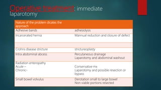
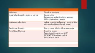
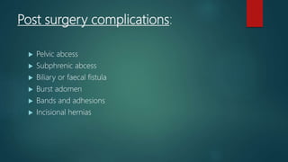
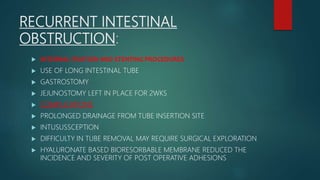
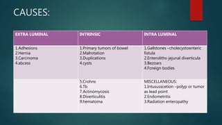
CAUSES:
EXTRA LUMINAL INTRINSIC INTRA LUMINAL
1.Adhesions
2.Hernia
3.Carcinoma
4.abcess
1.Primary tumors of bowel
2.Malrotation
3.Duplications
4.cysts
1.Gallstones –cholecystoenteric
fistula
2.Enteroliths-jejunal diverticula
3.Bezoars
4.Foreign bodies
5.Crohns
6.Tb
7.Actinomycosis
8.Diverticulitis
9.hematoma
MISCELLANEOUS:
1.Intususscetion –polyp or tumor
as lead point
2.Endometritis
3.Radiation enteropathy
 ADHESIONS:
 60% ;preponderance to lower abdominal procedures because the bowel
more mobile in pelvis.
 MALIGNANT TUMORS :
1. 20%;Most are metastatic secondary to peritoneal implants.
2. large intra abdominal tumors –extrinsic compression
3. primary smallbowel tumors can cause obstruction but are
exceedingly RARE.
 Hernias
1.10%; typically ventral or inguinal.
2. internal hernias related to prior abdominal surgery.
 CROHN’S DISEASE:
1. 5%; acute inflammation and edema – resolve by conservative mx.
2. chronic cases strictures develop – resection and reanastomosis
or sticturoplasty.
 ABCESS:
1.ruptured appendix or diverticulum
2.dehiscence of intestinal anastomosis
3.small bowel is a wall of abcess cavity - kinking
PATHOPHYSIOLOGY:
Obstruction
Increase in peristalsis proximal
and distal to obstruction
diarrhea Early in the course
Bowel gets fatigued and dilates Water and electrolytes accumulate
Third space fluid loss
DEHYDRATION AND HYPOVOLEMIA
Later in the course
PATHOPHYSIOLOGY :
PROXIMAL
• DEHYDRATION
HYPOCHLOREMI
A
HYPOKALEMIA
METABOLIC
ALKALOSIS
• ASSOCIATED
DISTAL
• DEHYDRATIO
N
OLIGURIA
AZOTEMIA
HEMOCONCE
NTRATION
HYPOTENSIO
N AND
SHOCK
CLOSED LOOP
• INCREASE
INTRALUMINA
L PRESSURE
ARTERIAL
OCCLUSION
AND
ISCHEMIA
• PERFORATION
OTHER FEATURES
• INCREASED IAP
DECREASED VR
ELEVATION
DIAPHRAGHM
COMPROMISING
VENTILATION
BACTERIAL
TRANSLOCATION
• E.COLI,
STREPTOCOCCUS
FAECALIS,KLEBSIEL
LA
• 10’ 9 TO 10 ’10/ML
• AMPLIFIES LOCAL
RESPONS
CARDINAL SYMPTOMS:
• PAROXYSMS 4 TO 5 MIN INTERVALS
• OCCUR LESS FREQUENT IN DISTAL OBSTRUCTION
• INITIAL AND MOST PROMINENT
COLICKY ABDOMINAL PAIN
• MORE COMMON WITH HIGHER OBSTRUCTION
• FECULENT IN CHARACTER INDICATES AN LATE AND ESTABLISHED
INTESTINAL OBSTRUCTION
NAUSEA AND VOMITING
• LATE SYMPTOM AS DISEASE PROGRESS
• OBSTIPATION IS DIFFICULTY TO PASS BOTH FLATUS AND STOOL.
• SEVERE FORM OF CONSTIPATION
DISTENSION AND
OBSTIPATION
• MALIGNANT DISEASE
• INTUSUSSCEPTION
• INFARCTION
• HYPERACTIVE BOWEL SOUNDS
• VIGOROUS PERISTALSIS
• MINIMALOR NO BOWEL SOUNDS LATELY
• DISTENDED ABDOMEN
• PREVIOUS SCARS
• RULE OUT INCARCERATED HERNIAS
GROIN,FEMORAL TRIANGLE & OBTURATOR
• FEVER
• LOCALIZED
TENDERNESS
• REBOUND
• GUARDING
• TACHYCARDIA
• HYPOTENSION
SEVERE
DEHYDRATION
STRANGULATION
AND PERITONITIS
EXAMINE STOOL
FOR OCCULT
BLOOD
ON ABDOMINAL
EXAMINATION
COMPLICATIONS
 PERITONITIS
 HYPOVOLEMIC SEPTIC SHOCK
 RENALFAILURE
 ARDS
 INTRA ABDOMINAL ABCESS
FORMATION
 MORIBUND STATUS
DIFFERENTIAL DIAGNOSIS
 PARALYTIC OBSTRUCTION
 PSEUDO OBSTRUCTION
 ASCITES
Plain radiographs
• Accuracy 60%
• Confirm suspiscion and
site of obstruction &
Cause like FB ,gall stones.
• SUPINE :
• DILATED LOOPS >3CM
WITHOUT COLONIC
DISTENSION
• UPRIGHT :
• MULTIPLE AIR FLUID
LEVELS IN STEP WISE
PATTERN
Ct scan
• Particularly sensitive –high
grade obstruction
• Determine location and cause
• h/o abdominal
malignant disease,post
surgical pt,no h/o sx
symptom +
• Typical ct findings :
• Dilated small bowel loops
filled with fluid and
decompressed ascending and
descending colon
• In case of strangulation
irreversible ischemia and
enteroclysis
• Baso4 and
methylcellulose
• Double contrast
procedure
• Degree and defines
obstructed segment
• Uncertain low grade
intermittent obstruction
• Recurring obstruction
• Disadvantages :
• Nasoenteric
intubation
Serum sodium
Chloride
Potssium & creatinine levels checked for
ADEQUACY OF RESUSCITATION
HEMOCRIT VALUE FOR HEMOCONCENTRATION
SUPERCONDUCTING QUANTUM INTER FERENCE
DEVICE [SQUID ] MAGNETOMETER –MESENTERIC
ISCHEMIA
BER CHANGES – INTESTINA ISCHEMIA
ELEVATION OR ABSENCE OF LEUCOCYTOSIS
DOES NOT R/O STRANGULATION
LABORATORY TESTS
TREATMENT:
PLAN 0F RX TO BE MONITORED
Fluid resuscitation with ringer lactate
aggressively
Sodium, chloride and potassium
After adequate urine formed add
potassium chloride to the infusion
Urine output
Prophylactic antibiotics no beneficial
evidence in nontoxic pt.
but Administered preoperatively.
For older patients central venous
assesment
Nasogastric suction- reduces
aspiration risk
FOR Select cases placement of swan
ganz catheter
Partial obstruction can be treated
conservatively alone
SURGERY FOR SMALL INTESTINAL
OBSTRUCTION:
 General anaesthesia + airway protection
 Significant distension makes laparoscopy difficult
 Distended Loops of small bowel displaced to left and caecum is inspected.
 If caecum collapsed – small bowel obstruction
 If caecum distened –large bowel obstruction
 Searched in right illiac fossa and pelvis for loops of collapsed bowel
 if Collapsed caecum but not distal ileum –obstruction is closed to ileocaecalvalve
mostly carcinoma caecum in older pt.
 When collapsed iileum is identified it is traced proximally until obstruction reached
 Distended loops retained within abdomen
 If not possible cover with a warm moist pack and supported to prevent
undue traction on the mesentry.
 In loop of strangulated bowl Mesentric vessels to the loop clamped before
release of constriction or untwisting volvulus.
 Clamping prevents restoration of circulation and toxic metabolites release.
 Milking back small bowel contents into stomach and aspirated.
 Needle aspiration is another alternative
Viability of bowel:
1.Bowel completely released and placed in a warm ,saline moistened sponge for 15 to 20 mins
WITH 100 % OXYGEN.
2. normal colour returned and peristalsis is evident safe to return
bowel.
3.Doppler or administration of fluorescein –intraoperative discrimination of viability.
4.intraoperative near infra red angiography.
5.second look laparotomy 18 to24 hrs after initial procedure if condition is deteriorating
6.if not viable resection and re anastamosis
 Abdomen is closed using non absorbable sutures.often tension sutures
required.
 Smallbowel decompressed using savagesdecompressor within a purse string
suture.
Laparoscopic management
Indications:
 Mild abdominal distension allowing adequate visualization.
 Proximal obstruction
 Partial obstruction
 Single band obstruction
SURGICAL TECHNIQUE LIMITING ADHESIONS:
 GENTLE HANDLING OF BOWEL-REDUCE SEROSAL TRAUMA.
 AVOID UNNECESSARY DISSECTION.
 EXCLUSION OF FB MATERIAL FROM PERITONEAL CAVITY.
 ADEQUATE IRRIGATION AND DRAINAGE
 REMOVAL OF INFECTIOUS AND DEBRIS
 PRESERVATION AND USE OF OMENTUM AROUND SITE OFSURGERY
Operative treatment: immediate
laparotomy
Nature of the problem dicates the
approach
Adhesive bands adhesiolysis
Incarcerated hernia Mannual reduction and closure of defect
Crohns disease stricture strictureoplasty
Intra abdominal abcess Percutaneous drainage
Laparotomy and abdominal washout
Radiation enteropathy
Acute –
Chronic-
Conservative mx
Laparotomy and possible resection or
bypass
Small bowel volvulus Derotation small to large bowel
Non viable portions resected
Gallstone Simple enterotomy
Ascaris lumbricoides bolus of worms Conservative
Deworming and enterotomy avoided
Milking bolus into caecum
malignant adhesions radical resections of primary tumor enbloc
with involved loop of small bowel
Peritoneal deposits One or more side to side anastamosis
Small bowel tumors Intestinal bypass
Resection with generous V OF
MESENTERY to obtain radical
lymphadenectomy
Post surgery complications:
 Pelvic abcess
 Subphrenic abcess
 Biliary or faecal fistula
 Burst adomen
 Bands and adhesions
 Incisional hernias
RECURRENT INTESTINAL
OBSTRUCTION:
 INTERNAL FIXATION AND STENTING PROCEDURES
 USE OF LONG INTESTINAL TUBE
 GASTROSTOMY
 JEJUNOSTOMY LEFT IN PLACE FOR 2WKS
 COMPLICATIONS
 PROLONGED DRAINAGE FROM TUBE INSERTION SITE
 INTUSUSSCEPTION
 DIFFICULTY IN TUBE REMOVAL MAY REQUIRE SURGICAL EXPLORATION
 HYALURONATE BASED BIORESORBABLE MEMBRANE REDUCED THE
INCIDENCE AND SEVERITY OF POST OPERATIVE ADHESIONS
ACUTE POST OPERATIVE
OBSTRUCTION:
 90 % EARLY POST OPERATIVE OBSTRUCTIONS ARE PARTIAL AND
RESOLVE SPONTANEOUSLY.
 BOWEL REST,FLUID AND ELECTROLYTE RESUSCITATION, AND
PARENTERAL NUTRITION.
 SIGNS OF STRANGULATION –REOPERATE
 POSTLAPAROSCOPY – DEFINITIVE OBSTRUCTION POINT – DUE TO
PORT SITE HERNIA OR INTERNAL HERNIA-OPERATIVE INTERVENTION

Small bowel obstruction obstruction

  • 1.
    CAUSES: EXTRA LUMINAL INTRINSICINTRA LUMINAL 1.Adhesions 2.Hernia 3.Carcinoma 4.abcess 1.Primary tumors of bowel 2.Malrotation 3.Duplications 4.cysts 1.Gallstones –cholecystoenteric fistula 2.Enteroliths-jejunal diverticula 3.Bezoars 4.Foreign bodies 5.Crohns 6.Tb 7.Actinomycosis 8.Diverticulitis 9.hematoma MISCELLANEOUS: 1.Intususscetion –polyp or tumor as lead point 2.Endometritis 3.Radiation enteropathy
  • 2.
     ADHESIONS:  60%;preponderance to lower abdominal procedures because the bowel more mobile in pelvis.  MALIGNANT TUMORS : 1. 20%;Most are metastatic secondary to peritoneal implants. 2. large intra abdominal tumors –extrinsic compression 3. primary smallbowel tumors can cause obstruction but are exceedingly RARE.
  • 3.
     Hernias 1.10%; typicallyventral or inguinal. 2. internal hernias related to prior abdominal surgery.  CROHN’S DISEASE: 1. 5%; acute inflammation and edema – resolve by conservative mx. 2. chronic cases strictures develop – resection and reanastomosis or sticturoplasty.  ABCESS: 1.ruptured appendix or diverticulum 2.dehiscence of intestinal anastomosis 3.small bowel is a wall of abcess cavity - kinking
  • 4.
    PATHOPHYSIOLOGY: Obstruction Increase in peristalsisproximal and distal to obstruction diarrhea Early in the course Bowel gets fatigued and dilates Water and electrolytes accumulate Third space fluid loss DEHYDRATION AND HYPOVOLEMIA Later in the course
  • 5.
    PATHOPHYSIOLOGY : PROXIMAL • DEHYDRATION HYPOCHLOREMI A HYPOKALEMIA METABOLIC ALKALOSIS •ASSOCIATED DISTAL • DEHYDRATIO N OLIGURIA AZOTEMIA HEMOCONCE NTRATION HYPOTENSIO N AND SHOCK CLOSED LOOP • INCREASE INTRALUMINA L PRESSURE ARTERIAL OCCLUSION AND ISCHEMIA • PERFORATION OTHER FEATURES • INCREASED IAP DECREASED VR ELEVATION DIAPHRAGHM COMPROMISING VENTILATION BACTERIAL TRANSLOCATION • E.COLI, STREPTOCOCCUS FAECALIS,KLEBSIEL LA • 10’ 9 TO 10 ’10/ML • AMPLIFIES LOCAL RESPONS
  • 6.
    CARDINAL SYMPTOMS: • PAROXYSMS4 TO 5 MIN INTERVALS • OCCUR LESS FREQUENT IN DISTAL OBSTRUCTION • INITIAL AND MOST PROMINENT COLICKY ABDOMINAL PAIN • MORE COMMON WITH HIGHER OBSTRUCTION • FECULENT IN CHARACTER INDICATES AN LATE AND ESTABLISHED INTESTINAL OBSTRUCTION NAUSEA AND VOMITING • LATE SYMPTOM AS DISEASE PROGRESS • OBSTIPATION IS DIFFICULTY TO PASS BOTH FLATUS AND STOOL. • SEVERE FORM OF CONSTIPATION DISTENSION AND OBSTIPATION
  • 7.
    • MALIGNANT DISEASE •INTUSUSSCEPTION • INFARCTION • HYPERACTIVE BOWEL SOUNDS • VIGOROUS PERISTALSIS • MINIMALOR NO BOWEL SOUNDS LATELY • DISTENDED ABDOMEN • PREVIOUS SCARS • RULE OUT INCARCERATED HERNIAS GROIN,FEMORAL TRIANGLE & OBTURATOR • FEVER • LOCALIZED TENDERNESS • REBOUND • GUARDING • TACHYCARDIA • HYPOTENSION SEVERE DEHYDRATION STRANGULATION AND PERITONITIS EXAMINE STOOL FOR OCCULT BLOOD ON ABDOMINAL EXAMINATION
  • 8.
    COMPLICATIONS  PERITONITIS  HYPOVOLEMICSEPTIC SHOCK  RENALFAILURE  ARDS  INTRA ABDOMINAL ABCESS FORMATION  MORIBUND STATUS DIFFERENTIAL DIAGNOSIS  PARALYTIC OBSTRUCTION  PSEUDO OBSTRUCTION  ASCITES
  • 9.
    Plain radiographs • Accuracy60% • Confirm suspiscion and site of obstruction & Cause like FB ,gall stones. • SUPINE : • DILATED LOOPS >3CM WITHOUT COLONIC DISTENSION • UPRIGHT : • MULTIPLE AIR FLUID LEVELS IN STEP WISE PATTERN Ct scan • Particularly sensitive –high grade obstruction • Determine location and cause • h/o abdominal malignant disease,post surgical pt,no h/o sx symptom + • Typical ct findings : • Dilated small bowel loops filled with fluid and decompressed ascending and descending colon • In case of strangulation irreversible ischemia and enteroclysis • Baso4 and methylcellulose • Double contrast procedure • Degree and defines obstructed segment • Uncertain low grade intermittent obstruction • Recurring obstruction • Disadvantages : • Nasoenteric intubation
  • 10.
    Serum sodium Chloride Potssium &creatinine levels checked for ADEQUACY OF RESUSCITATION HEMOCRIT VALUE FOR HEMOCONCENTRATION SUPERCONDUCTING QUANTUM INTER FERENCE DEVICE [SQUID ] MAGNETOMETER –MESENTERIC ISCHEMIA BER CHANGES – INTESTINA ISCHEMIA ELEVATION OR ABSENCE OF LEUCOCYTOSIS DOES NOT R/O STRANGULATION LABORATORY TESTS
  • 11.
    TREATMENT: PLAN 0F RXTO BE MONITORED Fluid resuscitation with ringer lactate aggressively Sodium, chloride and potassium After adequate urine formed add potassium chloride to the infusion Urine output Prophylactic antibiotics no beneficial evidence in nontoxic pt. but Administered preoperatively. For older patients central venous assesment Nasogastric suction- reduces aspiration risk FOR Select cases placement of swan ganz catheter Partial obstruction can be treated conservatively alone
  • 12.
    SURGERY FOR SMALLINTESTINAL OBSTRUCTION:  General anaesthesia + airway protection  Significant distension makes laparoscopy difficult  Distended Loops of small bowel displaced to left and caecum is inspected.  If caecum collapsed – small bowel obstruction  If caecum distened –large bowel obstruction  Searched in right illiac fossa and pelvis for loops of collapsed bowel  if Collapsed caecum but not distal ileum –obstruction is closed to ileocaecalvalve mostly carcinoma caecum in older pt.  When collapsed iileum is identified it is traced proximally until obstruction reached
  • 13.
     Distended loopsretained within abdomen  If not possible cover with a warm moist pack and supported to prevent undue traction on the mesentry.  In loop of strangulated bowl Mesentric vessels to the loop clamped before release of constriction or untwisting volvulus.  Clamping prevents restoration of circulation and toxic metabolites release.  Milking back small bowel contents into stomach and aspirated.  Needle aspiration is another alternative
  • 15.
    Viability of bowel: 1.Bowelcompletely released and placed in a warm ,saline moistened sponge for 15 to 20 mins WITH 100 % OXYGEN. 2. normal colour returned and peristalsis is evident safe to return bowel. 3.Doppler or administration of fluorescein –intraoperative discrimination of viability. 4.intraoperative near infra red angiography. 5.second look laparotomy 18 to24 hrs after initial procedure if condition is deteriorating 6.if not viable resection and re anastamosis
  • 17.
     Abdomen isclosed using non absorbable sutures.often tension sutures required.  Smallbowel decompressed using savagesdecompressor within a purse string suture.
  • 18.
    Laparoscopic management Indications:  Mildabdominal distension allowing adequate visualization.  Proximal obstruction  Partial obstruction  Single band obstruction
  • 19.
    SURGICAL TECHNIQUE LIMITINGADHESIONS:  GENTLE HANDLING OF BOWEL-REDUCE SEROSAL TRAUMA.  AVOID UNNECESSARY DISSECTION.  EXCLUSION OF FB MATERIAL FROM PERITONEAL CAVITY.  ADEQUATE IRRIGATION AND DRAINAGE  REMOVAL OF INFECTIOUS AND DEBRIS  PRESERVATION AND USE OF OMENTUM AROUND SITE OFSURGERY
  • 20.
    Operative treatment: immediate laparotomy Natureof the problem dicates the approach Adhesive bands adhesiolysis Incarcerated hernia Mannual reduction and closure of defect Crohns disease stricture strictureoplasty Intra abdominal abcess Percutaneous drainage Laparotomy and abdominal washout Radiation enteropathy Acute – Chronic- Conservative mx Laparotomy and possible resection or bypass Small bowel volvulus Derotation small to large bowel Non viable portions resected
  • 21.
    Gallstone Simple enterotomy Ascarislumbricoides bolus of worms Conservative Deworming and enterotomy avoided Milking bolus into caecum malignant adhesions radical resections of primary tumor enbloc with involved loop of small bowel Peritoneal deposits One or more side to side anastamosis Small bowel tumors Intestinal bypass Resection with generous V OF MESENTERY to obtain radical lymphadenectomy
  • 22.
    Post surgery complications: Pelvic abcess  Subphrenic abcess  Biliary or faecal fistula  Burst adomen  Bands and adhesions  Incisional hernias
  • 23.
    RECURRENT INTESTINAL OBSTRUCTION:  INTERNALFIXATION AND STENTING PROCEDURES  USE OF LONG INTESTINAL TUBE  GASTROSTOMY  JEJUNOSTOMY LEFT IN PLACE FOR 2WKS  COMPLICATIONS  PROLONGED DRAINAGE FROM TUBE INSERTION SITE  INTUSUSSCEPTION  DIFFICULTY IN TUBE REMOVAL MAY REQUIRE SURGICAL EXPLORATION  HYALURONATE BASED BIORESORBABLE MEMBRANE REDUCED THE INCIDENCE AND SEVERITY OF POST OPERATIVE ADHESIONS
  • 24.
    ACUTE POST OPERATIVE OBSTRUCTION: 90 % EARLY POST OPERATIVE OBSTRUCTIONS ARE PARTIAL AND RESOLVE SPONTANEOUSLY.  BOWEL REST,FLUID AND ELECTROLYTE RESUSCITATION, AND PARENTERAL NUTRITION.  SIGNS OF STRANGULATION –REOPERATE  POSTLAPAROSCOPY – DEFINITIVE OBSTRUCTION POINT – DUE TO PORT SITE HERNIA OR INTERNAL HERNIA-OPERATIVE INTERVENTION