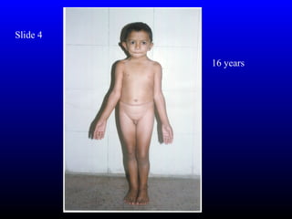
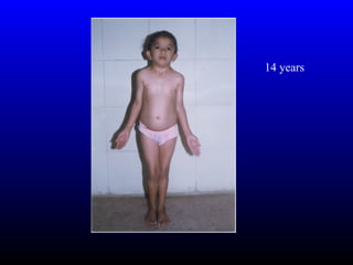
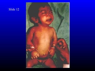
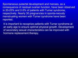
Picture Quiz For Medical
  Officers and trainees

By Dr. Varsha Atul Shah
Slide 1
A 10-year-old boy presented with a gradually progressive
swelling over his right arm for last 7 years. Examination
revealed a soft, diffuse swelling measuring 10 × 12 cm
over posterior aspect of right arm. The lesion was non-
tender and freely mobile over the underlying tissues. In
addition, he had multiple hyperpigmented macules with
serrated margins over the trunk (cafe au lait macules) and
multiple freckle like macules (axillary freckles) in both
axillae. He also had multiple soft nodules in the skin
(mollusca fibrosa) which were widely dispersed over trunk
and limbs.
Q.A What is the most likely diagnosis?
Q.B What other systemic examination is essential?
Slide 2
Slide 2
A male infant aged 3 months was brought
with a maculopapular, erythematous rash
with scaling at the edges distributed
symmetrically over the malar region,
forehead, chin and forearms. Heart rate was
60/min. Rest of the clinical examination was
normal.
Q. 1 What is the probable diagnosis?
Slide 3


      Before   After
Slide 3
A two-month-old male infant presented with progressive swelling
over both sides of scrotum, moderate to high fever and listlessness
since five days. The scrotal skin was reddened, edematous, tense,
and shiny. There was an ulcerated lesion of size 5 × 5 centimeters
present over both sides of the scrotum. The edges of the ulcer were
edematous, reddened, and irregular. The floor had slough, with a
purulent, fowl smelling discharge. Both the testes were exposed.
Perineal and perianal area was otherwise normal.
QA What is the most likely diagnosis?
Q B.What is the treatment of choice?
Slide 4

          16 years
14 years
Slide 4

Q 1. Describe the 5 gross features.

Q 2 What is likely diagnosis?
Two siblings, a girl aged 16 years and a boy aged 14
years presented with short stature. On examination the
height of the female child was 105 cms (<5th centile) and
that of male child was 104 cms (<5th centile). Both
children had typical leonin facies (prominent eyes,
anteverted nares, antimongoloid slant, depressed nasal
bridge and posteriorly placed prominent ears), low hair
line, short and webbed neck, shield chest, small widely
placed nipples, cubitus valgus with limited extension of
the elbows, mild scoliosis, knock knee, flat feet. Boy had
features of hypogonadism. Their mental development
was normal and had satisfactory scholastic performance.
Other systems were normal. These features are typical of
those seen in Noonan syndrome.
Slide 5
Slide 5
An 8-year-old girl presented with complaints of inability to stand from
sitting position for 3 months and inability to lift her head for one month.
Her past, history was insignificant. Examination revealed periorbital
puffiness, hypertrichosis, papular lesions over skin of hands, wrist and
elbow bilaterally. There was genralized hypotonia with a positive
Gower’s sign. She had burning sensation and redness on tips of fingers.
The dorsum of hands had Gottron’s papules and scaling eczematoid
dermatosis. Other systems were normal. Her CPK levels were 953IU,
LDH was 4496 IU, SGOT was 450 IU and ESR was 75 mm/hr. ANA was
negative. The muscle biopsy showed inflammatory changes with
perivascular infiltration and occlusion of microvasculture with
perifascicular atrophy.
Q 1What is the diagnosis?
Q 2 What is the treatment ?
Slide 5
Answer 1
A diagnosis of Juvenile Dermatomyositis was made.
Answer 2
The patient was treated with prednsiolone and physiotherapy.
Slide 6
Slide 6
Q.A six-year-old female child born out of
nonconsanguineous parents with no adverse perinatal
events was noticed to be having abnormal facies with
microcephaly (head circumference 43 cms; < -3SD),
bushy eyebrows meeting in the midline (synophrys)
(Fig.1), down-turned upperlips and hypertrichosis and
marked short stature (87.0 cm; < 80% of expected). Her
upper segment to lower segment ratio was 1.5 and bone
age was also retarded (3 years). There was no limb
shortening. Child also had global retardation with recurrent
seizures. Examination also revealed hirsuitism and
contractures in all the four limbs. X-ray skull excluded
craniosynostosis. Growth hormone assay was not done.
Slide 6

Q 1 Likely diagnosis?
Slide 6 Answers
Ans 1:The condition is characterized by microcephaly,
bushy eyebrows meeting in the midline (synophrys),
hypertrichosis, marked short stature and variable
mental retardation and limb abnormality. Hirsuitism is
usually seen in most of the patients. There is no fixed
pattern of inheritance described. However, in some of
these patients duplication of long arm of chromosome 3
has been described.
Ans 2: Clinical diagnosis of Cornelia De Lange
Syndrome was entertained.
Slide 7
Slide 7 Q
A 7 days old male neonate from an orphanage, presented
with loose stools. On examination, he had flattened
occiput, prominent coronal suture, hypertelorism,
depressed nasal bridge, antimongoloid slant, maxillary
hypoplasia and small nose (Fig.1). Examination of oral
cavity revealed cleft of soft palate (Fig. 2). Syndactyly of
both hands with complete fusion of 2nd, 3rd, 4th and 5th
fingers with broad distal phalanx of thumb and syndactyly
of all toes were other prominent features. The fused
fingers and toes had separate nails. There were no other
skeletal deformities and his fundus examination was
normal. Cardiovascular system examination revealed a
systolic murmur over the pulmonary area. His
neurosonogram and ultrasonogram abdomen were normal
but echo cardiogram showed an atrial septal defect.
Slide 7

Q 1 Describe 5 features

Q 2 Diagnosis
Apert syndrome was first described by Eugene Apert in 1906
as a triad of craniosynostosis, syndactyly and maxillary
hypoplasia. It is an autosomal dominant disorder but majority
of cases are sporadic and associated with older paternal age.
Apert Syndrome is characterized by irregular
craniosynostosis, mid facial hypoplasia, syndactyly, broad
distal phalanx of thumb and big toe. Mutations in the fibroblast
growth factor receptor 2 gene which maps to chromosome
10q 25 – 10q 26 cause Apert Syndrome. Majority of patients
have mental retardation. Agenesis of corpus callosum,
progressive hydrocephalus and hippocampal abnormalities
can occur in children with Apert Syndrome. Early surgery for
craniosynostosis is indicated when there is evidence of
increased intracranial pressure. When the thumb is
immobilized, early surgery should be done to facilitate pincer
grasp.
Slide 8
A 7 year old male child was brought with a large
cephalhematoma following a trivial injury. It occupied right
occipito-parietal region of the scalp. He had hyperextensible
skin and hypermobile joints.The thin elastic skin had cigarette
paper scars evident over the knee joint and shin of the tibia
(Fig.1). He had marfanoid features, arachnodactyly, widely
spaced eyes, a prominant nasal bridge, epicanthal folds,
frontal bossing, but no cataract or lens dislocation. There was
no gastrointestinal bleeding and the cardiovascular system
was normal. There was no similar family history or history of
consanguinity.
Slide 8
Q 1. What is the diagnosis
Ehler-Danlos syndrome (EDS) is a group of generalized
disorder of connective tissue characterized by fragility of the
skin and blood vessels, hyperextensibility of the skin and
joint hypermobility and poor wound healing with atrophic
scarring. At least 10 types have been defined. The
syndrome may be inherited as an autosomal dominant,
recessive or X linked recessive trait. This child had the
characteristics of Type VI which presents with marfanoid
features, bleeding from wounds, scoliosis and delayed
motor development.
Genetic counseling is important for this inherited disorder.
Ascorbic acid diminishes bruising, improves wound healing
and muscular strength in some types such as types II and
IV. Bleeding is managed conservatively. Cosmetic surgery
may help for ugly scars. The patient should be protected
from sun exposure and trauma.
Slide 9
A 4-year-old boy born to non-consanguineous parents
was diagnosed to have achondroplasia by antenatal
ultrasound. At birth, clinical examination revealed a term
baby with large head, prominent forehead, depressed
nasal bridge and short upper and lower limbs. His birth
weight was 3 kg, length 47 cm and head circumference 40
cm. He also had mild hypotonia and short thoracic cage at
birth. He had been followed up with serial head
circumference measurements during infancy (Fig.1). At
the age of 8 months he had rapidly increasing head
circumference with wide and bulging anterior fontanel. A
cranial ultrasound performed at that time revealed dilated
lateral and third ventricles. A ventriculo peritoneal shunt
was done at 8-months of age. Now, at the age of 4 years,
his height is 75 cm (height age 1 year) and head
circumference is 52 cm. His mental development and
intelligence is normal for his age.
Slide 9


Q 1 Describe 3 features and complicatios.
Q 2 How it is inherited?
Achondroplasia is the most common chondrodysplasia occurring with the
frequency of 1 in 15,000. It is an autosomal dominant disorder, but
majority of the cases represent a fresh gene mutation. Older paternal age
is a contributory factor. Children with achondroplasia have short stature,
megalencephaly, small foramen magnum, depressed nasal bridge,
prominent forehead and short limbs. The limbs shortening is greatest in
the proximal segments and the fingers display a trident configuration.
They have mild hypotonia and slow motor development but intelligence is
usually normal. Prominent skeletal features are small cuboid shaped
vertebral bodies with progressive narrowing of lumbar interpedicular
distance and short tubular bones especially humerii. Hydrocephalus is
one of the neurological complications that can occur in these children.
Hence it is recommended that head circumference should be measured
throughout infancy and plotted against norms published for
achondroplastic individuals. Ultrasound studies of the brain should be
considered if the fontanel size is large and head circumference increases
rapidly. Mutation in the gene encoding fibroblast growth factor receptor 3
located at 4p16.3 has been shown to cause achondroplasia.
Slide 10
           A 4-month-old product of non-
           consanguineous normal parents
           presented with three days history of
           recurrent seizures. Convulsions
           were of mixed type consisting of
           three to four episodes of
           generalized seizures interspersed
           with fifteen to twenty episodes of
           infantile spasms in a day.
           Examination revealed a normal
           weight baby with normal head
           circumference. Skin examination
           revealed three hypopigmented
           macules resembling ash leaves.
           Two were on low back and one
           was on anterior aspect of right
           shoulder.
Slide 10
Q 1 What are the lesions called?


Q 2 What is the most likely diagnosis?


Q 3 How it is inherited?
CT scan head revealed multiple subependymal tubers
involving wall of frontal horn and body of lateral ventricle.
There was evidence of calcification in the wall of lateral
ventricle. EEG was suggestive of mixed seizure disorder. USG
abdomen and Echocardiography were normal. Child was
started on Sodium Valproate with significant control in
seizures.
Tuberous sclerosis is an autosomal dominant neurocutaneous
syndrome with an incidence of 1:30,000. Bourneville and
Brissaud first described it in 1880. Hypopigmented macules
are seen in 50% of <2 year old. Rest of the clinical features like
adenoma sebaceum, shagreen patches and sub ungual
fibromas make their appearance latter in childhood. Prognosis
for seizure control and mental retardation is guarded. More the
number of tubers as was in this child, worse is the intellectual
development.
Slide 11
Slide 11

Q.1 What is the diagnosis?
Hyperpigmented lesions like giant con-genital nevus (GCN) (the
largest diameter greater than 20 cm) are very rare. This baby
had many nevi on various parts of the skin covering about 30%
of surface area. The largest nevus covered large part of left
cheek, left ear pinna, left temporal area of scalp, neck, anterior
part of chest, and a large portion of back and measured 32 cm in
circumference and 20 cm in highest width. There were many
satellite lesions scattered over scalp, trunk, and limbs (Fig. 1).
All lesions were black colored and had well defined, and raised
edges. The surface of the largest lesion was rough and nodular
at places. Some leisons had smooth surfaces also. Hair were
present at surface of some lesions at the back. Other physical
examination was normal except a lacerated wound in front of left
ear that resulted from application of forceps. There was no
evidence of clubfoot, meningomyelocele, or any other congenital
anomaly. Histopathological find-ings were consistent with giant
congenital melanocytic nevus (compound type).
The management of this condition is controversial. The best is
to have complete excision but when such a large part of the
skin is involved, partial removal of superficial nevus cells by
dermabrasion, laser therapy, curettage, or shave excision is
less traumatic than excision surgery and produces acceptable
cosmetic result. However, presence of neural melanosis
makes these efforts futile. Further, none of these techniques
or excision of GCN to superficial fat completely removes the
risk of malignant melanoma transformation. The overall
incidence of malignant melanoma arising out of GCN is
estimated to be approximately 5-10% and 50% of these
transformations occur by the age of 5 years.
Slide 12
Slide 12

Q1.What is seen in picture?

Q2. Likely diagnosis.
A 2-month-old girl was brought with swelling on the
dorsum of both hands and feet. She was born to
non-consanguineous parents, by normal vaginal
delivery. There were no adverse antenatal or
perinatal events. The salient features on
examination were non-pitting edema of distal parts
of the extremities, webbing of the neck, a low hair
line and widely spaced nipples. These classical
manifestations prompted a clinical diagnosis of
Turner syndrome. Chromosomal studies confirmed
that the propositus had a 45, X0 karyotype.
Investigations excluded coexistent cardiac or renal
anomalies.
The incidence of Turner syndrome is estimated
to be 1 in 8000 live births. Poor development of
lymphatic channels in fetal life is implicated in
the causation of abnormalities of the ears, shield
chest, lymphedema and coarctation of arota.
Bicuspid aortic value (50%) and coarctation of
aorta (20.6%) are the common cardiac
anomalies associated with Turner syndrome.
Other abnormalities that have been described
include ectopic kidney, abnormal renal
vasculature, double collecting system, primary
hypothyroidism (10-30%) and scoliosis.
Spontaneous pubertal development and menses, as a
consequence of residual ovarian function, have been observed
in 20-25% and 2-5% of patients with Turner syndrome,
respectively. Nearly 30 pregnancies in sponta-neously
menstruating women with Turner syndrome have been
reported.
It is important to recognize patients with Turner syndrome at
an early age to ensure optimal physical growth. Development
of secondary sexual characteristics can be improved with
hormone replacement therapy.
Slide 13
Slide 13

Q.1 What is the diagnosis?

Q.2 What is high risk factors ?
A full term male newborn was delivered vaginally with lower
limb deformities. The mother was a non-diabetic primigravida.
The deformities were in the form of hyperextension of the
lower limbs, club foot, hypoplastic femur and tibia . The baby
had motor and sensory neurological deficit in the form of lower
motor neuron paraplegia and absent anal reflex. There was no
imperforate anus but it was malposed. There were two sinuses
on either sides of the buttocks. There was no other neural tube
defect or renal abnormalities on sonography.
Caudal regression syndrome, also known as caudal
dysplasia or sacral agenesis syndrome is a congenital
malformation characterized by various degrees of
developmental failure involving legs, lumbar, sacral and
coccygeal vertebra, and corresponding segments of spinal
cord due to defect in neuralisation. This results in motor and
sensory deficits. Other congenital anomalies such as
gastrointestinal, genito-urinary or congenital heart disease
may be present. A history of maternal diabetes is obtained in
16% of such infants.

Slide show for paediatric trainees

  • 1.
    Picture Quiz ForMedical Officers and trainees By Dr. Varsha Atul Shah
  • 2.
  • 3.
    A 10-year-old boypresented with a gradually progressive swelling over his right arm for last 7 years. Examination revealed a soft, diffuse swelling measuring 10 × 12 cm over posterior aspect of right arm. The lesion was non- tender and freely mobile over the underlying tissues. In addition, he had multiple hyperpigmented macules with serrated margins over the trunk (cafe au lait macules) and multiple freckle like macules (axillary freckles) in both axillae. He also had multiple soft nodules in the skin (mollusca fibrosa) which were widely dispersed over trunk and limbs. Q.A What is the most likely diagnosis? Q.B What other systemic examination is essential?
  • 4.
  • 5.
    Slide 2 A maleinfant aged 3 months was brought with a maculopapular, erythematous rash with scaling at the edges distributed symmetrically over the malar region, forehead, chin and forearms. Heart rate was 60/min. Rest of the clinical examination was normal. Q. 1 What is the probable diagnosis?
  • 6.
    Slide 3 Before After
  • 7.
    Slide 3 A two-month-oldmale infant presented with progressive swelling over both sides of scrotum, moderate to high fever and listlessness since five days. The scrotal skin was reddened, edematous, tense, and shiny. There was an ulcerated lesion of size 5 × 5 centimeters present over both sides of the scrotum. The edges of the ulcer were edematous, reddened, and irregular. The floor had slough, with a purulent, fowl smelling discharge. Both the testes were exposed. Perineal and perianal area was otherwise normal. QA What is the most likely diagnosis? Q B.What is the treatment of choice?
  • 8.
    Slide 4 16 years
  • 9.
  • 10.
    Slide 4 Q 1.Describe the 5 gross features. Q 2 What is likely diagnosis?
  • 11.
    Two siblings, agirl aged 16 years and a boy aged 14 years presented with short stature. On examination the height of the female child was 105 cms (<5th centile) and that of male child was 104 cms (<5th centile). Both children had typical leonin facies (prominent eyes, anteverted nares, antimongoloid slant, depressed nasal bridge and posteriorly placed prominent ears), low hair line, short and webbed neck, shield chest, small widely placed nipples, cubitus valgus with limited extension of the elbows, mild scoliosis, knock knee, flat feet. Boy had features of hypogonadism. Their mental development was normal and had satisfactory scholastic performance. Other systems were normal. These features are typical of those seen in Noonan syndrome.
  • 12.
  • 13.
    Slide 5 An 8-year-oldgirl presented with complaints of inability to stand from sitting position for 3 months and inability to lift her head for one month. Her past, history was insignificant. Examination revealed periorbital puffiness, hypertrichosis, papular lesions over skin of hands, wrist and elbow bilaterally. There was genralized hypotonia with a positive Gower’s sign. She had burning sensation and redness on tips of fingers. The dorsum of hands had Gottron’s papules and scaling eczematoid dermatosis. Other systems were normal. Her CPK levels were 953IU, LDH was 4496 IU, SGOT was 450 IU and ESR was 75 mm/hr. ANA was negative. The muscle biopsy showed inflammatory changes with perivascular infiltration and occlusion of microvasculture with perifascicular atrophy. Q 1What is the diagnosis? Q 2 What is the treatment ?
  • 14.
    Slide 5 Answer 1 Adiagnosis of Juvenile Dermatomyositis was made. Answer 2 The patient was treated with prednsiolone and physiotherapy.
  • 15.
  • 16.
    Slide 6 Q.A six-year-oldfemale child born out of nonconsanguineous parents with no adverse perinatal events was noticed to be having abnormal facies with microcephaly (head circumference 43 cms; < -3SD), bushy eyebrows meeting in the midline (synophrys) (Fig.1), down-turned upperlips and hypertrichosis and marked short stature (87.0 cm; < 80% of expected). Her upper segment to lower segment ratio was 1.5 and bone age was also retarded (3 years). There was no limb shortening. Child also had global retardation with recurrent seizures. Examination also revealed hirsuitism and contractures in all the four limbs. X-ray skull excluded craniosynostosis. Growth hormone assay was not done.
  • 17.
    Slide 6 Q 1Likely diagnosis?
  • 18.
    Slide 6 Answers Ans1:The condition is characterized by microcephaly, bushy eyebrows meeting in the midline (synophrys), hypertrichosis, marked short stature and variable mental retardation and limb abnormality. Hirsuitism is usually seen in most of the patients. There is no fixed pattern of inheritance described. However, in some of these patients duplication of long arm of chromosome 3 has been described. Ans 2: Clinical diagnosis of Cornelia De Lange Syndrome was entertained.
  • 19.
  • 21.
    Slide 7 Q A7 days old male neonate from an orphanage, presented with loose stools. On examination, he had flattened occiput, prominent coronal suture, hypertelorism, depressed nasal bridge, antimongoloid slant, maxillary hypoplasia and small nose (Fig.1). Examination of oral cavity revealed cleft of soft palate (Fig. 2). Syndactyly of both hands with complete fusion of 2nd, 3rd, 4th and 5th fingers with broad distal phalanx of thumb and syndactyly of all toes were other prominent features. The fused fingers and toes had separate nails. There were no other skeletal deformities and his fundus examination was normal. Cardiovascular system examination revealed a systolic murmur over the pulmonary area. His neurosonogram and ultrasonogram abdomen were normal but echo cardiogram showed an atrial septal defect.
  • 22.
    Slide 7 Q 1Describe 5 features Q 2 Diagnosis
  • 23.
    Apert syndrome wasfirst described by Eugene Apert in 1906 as a triad of craniosynostosis, syndactyly and maxillary hypoplasia. It is an autosomal dominant disorder but majority of cases are sporadic and associated with older paternal age. Apert Syndrome is characterized by irregular craniosynostosis, mid facial hypoplasia, syndactyly, broad distal phalanx of thumb and big toe. Mutations in the fibroblast growth factor receptor 2 gene which maps to chromosome 10q 25 – 10q 26 cause Apert Syndrome. Majority of patients have mental retardation. Agenesis of corpus callosum, progressive hydrocephalus and hippocampal abnormalities can occur in children with Apert Syndrome. Early surgery for craniosynostosis is indicated when there is evidence of increased intracranial pressure. When the thumb is immobilized, early surgery should be done to facilitate pincer grasp.
  • 24.
  • 25.
    A 7 yearold male child was brought with a large cephalhematoma following a trivial injury. It occupied right occipito-parietal region of the scalp. He had hyperextensible skin and hypermobile joints.The thin elastic skin had cigarette paper scars evident over the knee joint and shin of the tibia (Fig.1). He had marfanoid features, arachnodactyly, widely spaced eyes, a prominant nasal bridge, epicanthal folds, frontal bossing, but no cataract or lens dislocation. There was no gastrointestinal bleeding and the cardiovascular system was normal. There was no similar family history or history of consanguinity.
  • 26.
    Slide 8 Q 1.What is the diagnosis
  • 27.
    Ehler-Danlos syndrome (EDS)is a group of generalized disorder of connective tissue characterized by fragility of the skin and blood vessels, hyperextensibility of the skin and joint hypermobility and poor wound healing with atrophic scarring. At least 10 types have been defined. The syndrome may be inherited as an autosomal dominant, recessive or X linked recessive trait. This child had the characteristics of Type VI which presents with marfanoid features, bleeding from wounds, scoliosis and delayed motor development. Genetic counseling is important for this inherited disorder. Ascorbic acid diminishes bruising, improves wound healing and muscular strength in some types such as types II and IV. Bleeding is managed conservatively. Cosmetic surgery may help for ugly scars. The patient should be protected from sun exposure and trauma.
  • 28.
  • 29.
    A 4-year-old boyborn to non-consanguineous parents was diagnosed to have achondroplasia by antenatal ultrasound. At birth, clinical examination revealed a term baby with large head, prominent forehead, depressed nasal bridge and short upper and lower limbs. His birth weight was 3 kg, length 47 cm and head circumference 40 cm. He also had mild hypotonia and short thoracic cage at birth. He had been followed up with serial head circumference measurements during infancy (Fig.1). At the age of 8 months he had rapidly increasing head circumference with wide and bulging anterior fontanel. A cranial ultrasound performed at that time revealed dilated lateral and third ventricles. A ventriculo peritoneal shunt was done at 8-months of age. Now, at the age of 4 years, his height is 75 cm (height age 1 year) and head circumference is 52 cm. His mental development and intelligence is normal for his age.
  • 30.
    Slide 9 Q 1Describe 3 features and complicatios. Q 2 How it is inherited?
  • 31.
    Achondroplasia is themost common chondrodysplasia occurring with the frequency of 1 in 15,000. It is an autosomal dominant disorder, but majority of the cases represent a fresh gene mutation. Older paternal age is a contributory factor. Children with achondroplasia have short stature, megalencephaly, small foramen magnum, depressed nasal bridge, prominent forehead and short limbs. The limbs shortening is greatest in the proximal segments and the fingers display a trident configuration. They have mild hypotonia and slow motor development but intelligence is usually normal. Prominent skeletal features are small cuboid shaped vertebral bodies with progressive narrowing of lumbar interpedicular distance and short tubular bones especially humerii. Hydrocephalus is one of the neurological complications that can occur in these children. Hence it is recommended that head circumference should be measured throughout infancy and plotted against norms published for achondroplastic individuals. Ultrasound studies of the brain should be considered if the fontanel size is large and head circumference increases rapidly. Mutation in the gene encoding fibroblast growth factor receptor 3 located at 4p16.3 has been shown to cause achondroplasia.
  • 32.
    Slide 10 A 4-month-old product of non- consanguineous normal parents presented with three days history of recurrent seizures. Convulsions were of mixed type consisting of three to four episodes of generalized seizures interspersed with fifteen to twenty episodes of infantile spasms in a day. Examination revealed a normal weight baby with normal head circumference. Skin examination revealed three hypopigmented macules resembling ash leaves. Two were on low back and one was on anterior aspect of right shoulder.
  • 33.
    Slide 10 Q 1What are the lesions called? Q 2 What is the most likely diagnosis? Q 3 How it is inherited?
  • 34.
    CT scan headrevealed multiple subependymal tubers involving wall of frontal horn and body of lateral ventricle. There was evidence of calcification in the wall of lateral ventricle. EEG was suggestive of mixed seizure disorder. USG abdomen and Echocardiography were normal. Child was started on Sodium Valproate with significant control in seizures. Tuberous sclerosis is an autosomal dominant neurocutaneous syndrome with an incidence of 1:30,000. Bourneville and Brissaud first described it in 1880. Hypopigmented macules are seen in 50% of <2 year old. Rest of the clinical features like adenoma sebaceum, shagreen patches and sub ungual fibromas make their appearance latter in childhood. Prognosis for seizure control and mental retardation is guarded. More the number of tubers as was in this child, worse is the intellectual development.
  • 35.
  • 36.
    Slide 11 Q.1 Whatis the diagnosis?
  • 37.
    Hyperpigmented lesions likegiant con-genital nevus (GCN) (the largest diameter greater than 20 cm) are very rare. This baby had many nevi on various parts of the skin covering about 30% of surface area. The largest nevus covered large part of left cheek, left ear pinna, left temporal area of scalp, neck, anterior part of chest, and a large portion of back and measured 32 cm in circumference and 20 cm in highest width. There were many satellite lesions scattered over scalp, trunk, and limbs (Fig. 1). All lesions were black colored and had well defined, and raised edges. The surface of the largest lesion was rough and nodular at places. Some leisons had smooth surfaces also. Hair were present at surface of some lesions at the back. Other physical examination was normal except a lacerated wound in front of left ear that resulted from application of forceps. There was no evidence of clubfoot, meningomyelocele, or any other congenital anomaly. Histopathological find-ings were consistent with giant congenital melanocytic nevus (compound type).
  • 38.
    The management ofthis condition is controversial. The best is to have complete excision but when such a large part of the skin is involved, partial removal of superficial nevus cells by dermabrasion, laser therapy, curettage, or shave excision is less traumatic than excision surgery and produces acceptable cosmetic result. However, presence of neural melanosis makes these efforts futile. Further, none of these techniques or excision of GCN to superficial fat completely removes the risk of malignant melanoma transformation. The overall incidence of malignant melanoma arising out of GCN is estimated to be approximately 5-10% and 50% of these transformations occur by the age of 5 years.
  • 39.
  • 40.
    Slide 12 Q1.What isseen in picture? Q2. Likely diagnosis.
  • 41.
    A 2-month-old girlwas brought with swelling on the dorsum of both hands and feet. She was born to non-consanguineous parents, by normal vaginal delivery. There were no adverse antenatal or perinatal events. The salient features on examination were non-pitting edema of distal parts of the extremities, webbing of the neck, a low hair line and widely spaced nipples. These classical manifestations prompted a clinical diagnosis of Turner syndrome. Chromosomal studies confirmed that the propositus had a 45, X0 karyotype. Investigations excluded coexistent cardiac or renal anomalies.
  • 42.
    The incidence ofTurner syndrome is estimated to be 1 in 8000 live births. Poor development of lymphatic channels in fetal life is implicated in the causation of abnormalities of the ears, shield chest, lymphedema and coarctation of arota. Bicuspid aortic value (50%) and coarctation of aorta (20.6%) are the common cardiac anomalies associated with Turner syndrome. Other abnormalities that have been described include ectopic kidney, abnormal renal vasculature, double collecting system, primary hypothyroidism (10-30%) and scoliosis.
  • 43.
    Spontaneous pubertal developmentand menses, as a consequence of residual ovarian function, have been observed in 20-25% and 2-5% of patients with Turner syndrome, respectively. Nearly 30 pregnancies in sponta-neously menstruating women with Turner syndrome have been reported. It is important to recognize patients with Turner syndrome at an early age to ensure optimal physical growth. Development of secondary sexual characteristics can be improved with hormone replacement therapy.
  • 44.
  • 45.
    Slide 13 Q.1 Whatis the diagnosis? Q.2 What is high risk factors ?
  • 46.
    A full termmale newborn was delivered vaginally with lower limb deformities. The mother was a non-diabetic primigravida. The deformities were in the form of hyperextension of the lower limbs, club foot, hypoplastic femur and tibia . The baby had motor and sensory neurological deficit in the form of lower motor neuron paraplegia and absent anal reflex. There was no imperforate anus but it was malposed. There were two sinuses on either sides of the buttocks. There was no other neural tube defect or renal abnormalities on sonography.
  • 47.
    Caudal regression syndrome,also known as caudal dysplasia or sacral agenesis syndrome is a congenital malformation characterized by various degrees of developmental failure involving legs, lumbar, sacral and coccygeal vertebra, and corresponding segments of spinal cord due to defect in neuralisation. This results in motor and sensory deficits. Other congenital anomalies such as gastrointestinal, genito-urinary or congenital heart disease may be present. A history of maternal diabetes is obtained in 16% of such infants.