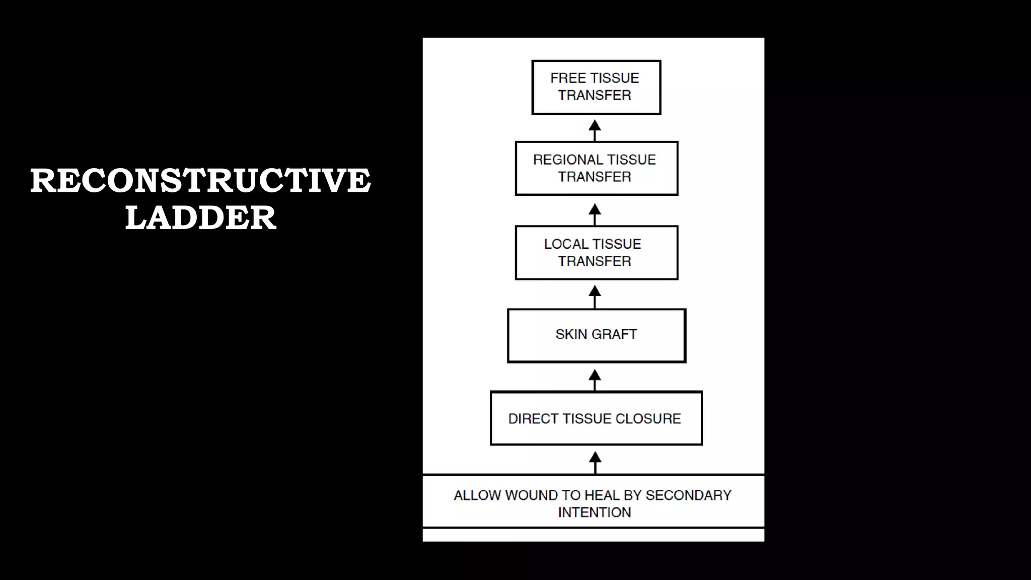
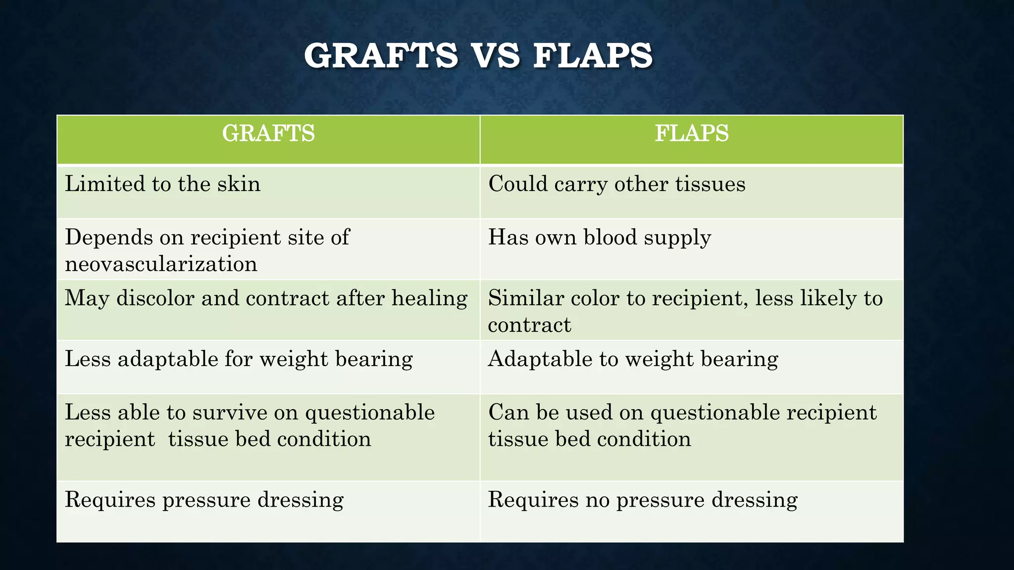
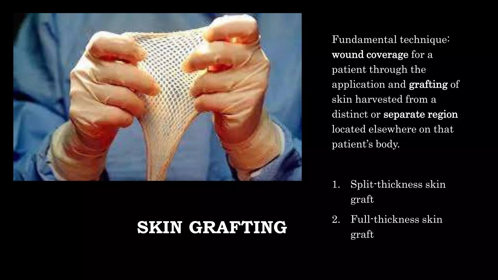
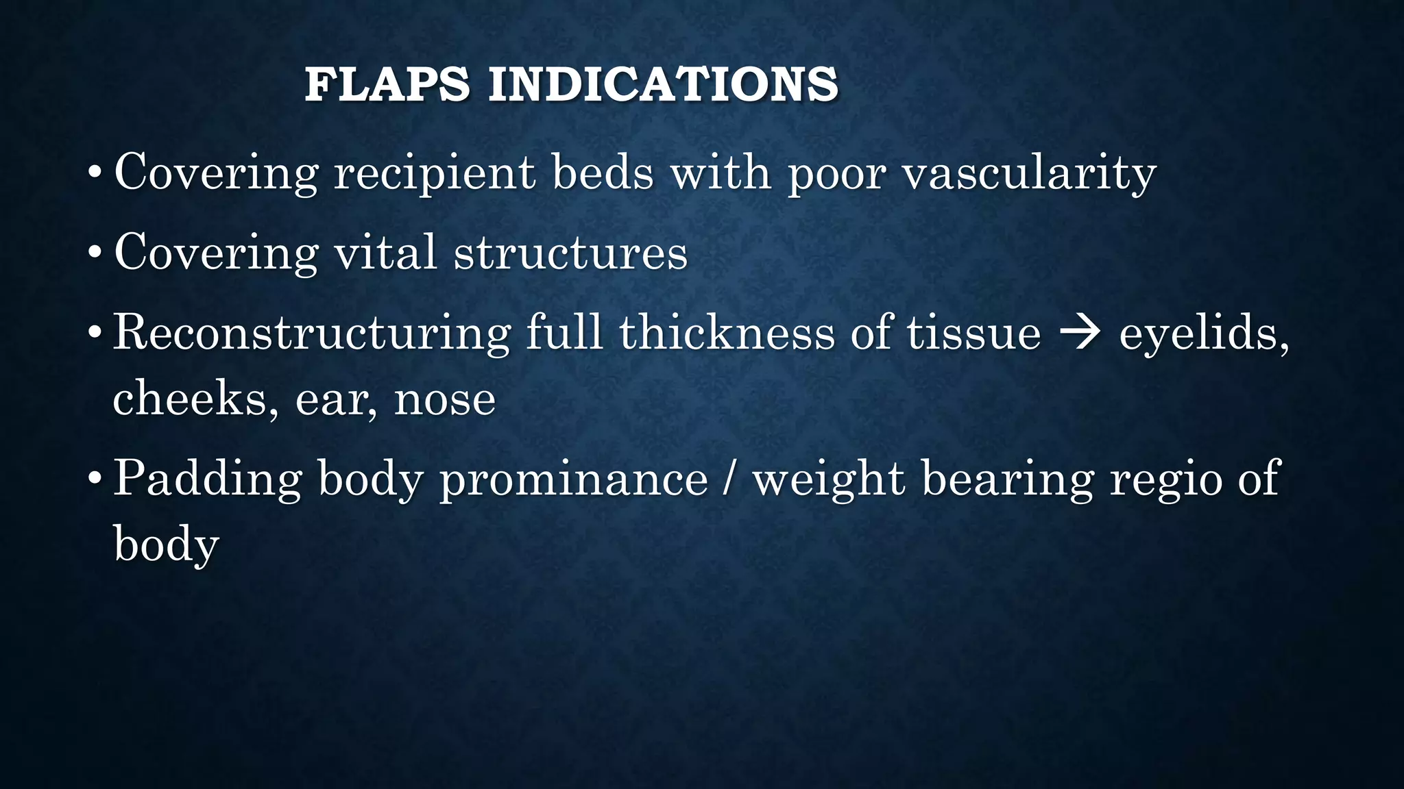
SKIN GRAFT & FLAP
dr. Shandy Vama Putra
Supervisor:
dr.Sylvia E Nuruth, SpBP-RE
dr. Muhammad Samiadji, SpBP-RE
THE SKIN
The skin is the largest organ in
the body, accounting for 15% of
total adult body weight.
Layered structures:
• Epidermis
Stratified squamous
epithelium, primarily
composed of keratinocytes,
no blood vessels
• Dermis
Collagen, capillaries,
elastic fibers, fibroblasts,
nerve endings, etc.
FLAP BLOOD SUPPLY
• The skin, because of its role in
thermoregulation to maintain
homeostasis and its immunological
function, has a rich blood supply, far
greater than what is needed for its
own inherent metabolic demand
• The blood reaches the skin from
deeper named vessels, via multiple
perforating arteries and their
accompanying veins that course
through overlying muscles, septa, and
fascia
RECONSTRUCTIVE
LADDER
GRAFTS VS FLAPS
GRAFTS FLAPS
Limited to the skin Could carry other tissues
Depends on recipient site of
neovascularization
Has own blood supply
May discolor and contract after healing Similar color to recipient, less likely to
contract
Less adaptable for weight bearing Adaptable to weight bearing
Less able to survive on questionable
recipient tissue bed condition
Can be used on questionable recipient
tissue bed condition
Requires pressure dressing Requires no pressure dressing
SKIN GRAFTING
Fundamental technique:
wound coverage for a
patient through the
application and grafting of
skin harvested from a
distinct or separate region
located elsewhere on that
patient’s body.
1. Split-thickness skin
graft
2. Full-thickness skin
graft
SKIN GRAFTS
Split Thickness Skin Graft (STSG),
yaitu skin graft yang terdiri atas
epidermis dan sebagian dermis,
dibagi lagi menjadi
 Thick : Epidermis + ¾ bagian
lapisan dermis
 Medium : Epidermis + ½ bagian
lapisan dermis
 Thin: Epidermis + ¼ bagian lapisan
dermis
Full Thickness Skin Graft (FTSG),
yaitu skin graft yang terdiri atas
epidermis dan seluruh bagian tebal
dermis
Asal Skin Graft
• 1. Autograft: Graft berasal dari individu yang sama
• 2. Homograft: berasal dari individu lain yang sama spesiesnya
• 3. Heterograft (xenograft): berasal dari mahluk lain yang berbeda spesies
SPLIT THICKNESS SKIN GRAFT
Keuntungan
1. Kemungkinan take lebih besar
2. Dapat dipakai untuk menutup defek yang luas
3. Donor dapat diambil dari daerah tubuh mana
saja
4. Daerah donor dapat sembuh sendiri/epitelisasi
Kerugian
a. Punya kecenderungan kontraksi lebih besar
b. Punya kecenderungan terjadi perubahan warna
c. Permukaan kulit mengkilat
d. Secara estetik kurang baik
DONOR SITES FOR SKIN
GRAFT
STSG
Donor site may be used
again for subsequent STSG
• Scalp
• Thigh
• Buttocks
• Abdominal wall
FTSG
No remaining epithelial
structures for re-
epithelization
• Postauricular area
• Upper eyelid
• Groin
• Supraclavicular
TEKNIK STSG
GRAFT HARVESTING
• Humby knife
• Padgette Drum-type
dermatome
• Brown-electrical
dermatome
TEKNIK STSG
MESHED AND SHEET GRAFTS
Meshed skin graft
• Multiple mechanical
incisions result 
immediate expansion of
the graft
• Covers a larger area per
and
• Allows drainage through
the numerous holes
Sheet skin graft
• Continuous interrupted
surface
TEKNIK STSG
Perawatan daerah donor
• dapat digunakan occlusive dressings,
semiocclusive dressings, semiopen dressings,
atau open dressings. Biasanya dibuka setelah 2-
3 minggu.
Perawatan daerah resipien
• penutup yang tidak menempel, cukup lembab,
dan memberikan tekanan yang merata. Penutup
dibiarkan selama 5 hari pertama, sedangkan
kasa lemak (tulle) atau penutup yang tidak
menempel bisa dipertahankan lebih lama agar
tidak menggeser graftnya.
SKIN GRAFT
HEALING
Imbibition (0 – 48 hrs)
• Grafts are initially nourished by fluid from the wound bed
prior to restoration of perfusion (time dependent to wound
bed perfusion)
• Plasma fibrinogen converted into fibrin  adherence to
wound bed
Inosculation (48 – 72 hrs)
• Connections between the wound bed vessels and graft occur
through the process of angiogenesis and new vessel
ingrowth in a unidirectional fashion from the wound bed
into the graft
Revascularization (>96 hrs)
• Reperfusion by angiogenesis from the wound bed – it is
hypothesized that recipient-derived endothelial cells
migrating into the graft use the skin graft’s intrinsic
preexisting vascular network as a conduit for ingrowth, with
eventual replacement of the graft’s endothelial structure.
SYARAT TAKE
Vaskularisasi resipien yang baik
Kontak yang akurat antara skin graft dengan resipien
Immobilisasi
Tidak infeksi
Tidak ada perdarahan atau hematom
FULL THICKNESS SKIN GRAFT
Keuntungan
o Kecenderungan untuk terjadi kontraksi lebih kecil
o Kecenderungan untuk berubah warna lebih kecil
o Kecenderungan permukaan kulit mengkilat lebih
kecil
o Secara estetik lebih baik dari split thickness skin
graft
Kerugian
o Kemungkinan take lebih kecil dibandingkan STSG
o Hanya dapat menutup defek yang tidak terlalu luas
o Donor harus dijahit atau sebagian ditutup oleh
STSG bila luka donor agak luas sehingga tidak
dapat ditutup primer
o Donor terbatas pada tempat-tempat tertentu seperti
inguinal, supraklavikular, retroaurikular.
TEKNIK FTSG
a. Persiapan luka: pembersihan, debridement, dan hemostasis
b. Pengambilan: jaringan lemak dipisahkan dari kulit agar jaringan dapat bertahan melalui imbibisi di
daerah resipien
c. Perawatan luka: di daerah donor ditutup secara primer, di daerah resipien diberikan penutup
dengan tekanan yang merata. Biasa dibantu dengan jahitan pada graft ke dasarnya atau memakai tie
over untuk memfiksasi.
d. Tissue Expansion di daerah donor yang dilakukan sebelum pengambilan dapat meningkatkan luas
daerah donor dan memungkinkan penutupan secara primer
SKIN
FLAP
• Flaps are usually required for covering
recipient beds that have poor vascularity;
covering vital structures; reconstructing
the full thickness of the eyelids, lips, ears,
nose, and cheeks; and padding body
prominences.
• A skin flap consists of skin and subcutaneous
tissue that are transferred from one part of the
body to another with a vascular pedicle or
attachment to the body being maintained for
nourishment.
• Proper planning of a flap is essential to the
success of the operation.
FLAPS INDICATIONS
• Covering recipient beds with poor vascularity
• Covering vital structures
• Reconstructuring full thickness of tissue  eyelids,
cheeks, ear, nose
• Padding body prominance / weight bearing regio of
body
PRINCIPLES OF
REPARATION
USING SKIN
FLAPS
1. Planning: type and method of transfer
2. Size of flap
3. Closure of donor area
4. Prevention of failure
• Tension
• Venous congestion
• Hematoma
JENIS FLAP
Vaskularisasi
JENIS FLAP
Cara Berpindah
 Rotasi dengan Pivot Point: Rotasi (flap berbentuk semisirkuler yang
dirotasikan terhadap suatu pivot point untuk menutup suatu defek),
Transposisi, Interpolasi
 Advancement Flap: Single pedicle, V-Y advancement, Y-V
advancement, Bipedicle advancement
 Flap langsung atau tubed melibatkan flap yang terhubung ke situs
donor dan penerima secara bersamaan.
JENIS FLAP
Jarak dari Defek
• Local flap  immediately adjacent to defect
• Regional flap  moved form adjacent
region
• Distant flap  move from remote anatomic
area
• Free flap
LOCAL FLAPS
Local Flaps
Pivot Point
Rotational
Transpositional
Z plasty
Rhomboid flap
Interposition
Bilobed
Advancement
Single Pedicle /
Simple
Advancement
Bipedicle
V-Y / Y-V Flap
BASED ON CONFIGURATION (DESIGN AND
METHOD OF TRANSFER) (2)
Advancement flap Rhamboid flap
Transpositional flap
Rotational flap
Z-PLASTY
• Z-plasties can be applied to revise and redirect
existing scars or to provide additional length in
the setting of scar contracture
• The principle involves the transposition of two
triangular flaps
• The classic Z-plasty has an angle of 60 degrees
and provides a 75% theoretical gain in length of
the central limb by recruiting lateral tissue.
• Gain in length is in the direction of the central
limb of the Z and depends on the angle used and
the length of the central limb.
RHOMBOID FLAP
• The Limberg flap is a type of
transposition flap. This flap
depends on the looseness of
adjacent skin
• A Limberg flap is designed for
rhomboid defects with angles of 60
and 120 degrees, but most wounds
can be made rhomboid, or imagined
as rhomboid, so the principle is
applicable to most facial wounds
• The flap is designed with sides that
are the same length as the short
axis of the rhomboid defect
BIPEDICLE
ADVANCEMENT
FLAPS
V-Y ADVANCEMENT FLAP
• Rather, a V-shaped incision is
made in the skin, after which the
skin on each side of the V is
advanced and the incision is closed
as a Y
• This V-Y technique can be used to
lengthen such structures as the
nasal columella, eliminate minor
notches of the lip, and, in certain
instances, close the donor site of a
skin flap
DIRECT FLAP
• Direct or tubed flaps involve having
the flap connected to both the donor
and recipient sites simultaneously,
forming a bridge.
• This allows blood to be supplied by
the donor site while a new blood
supply from the recipient site is
formed.
• Once this happens, the "bridge" can
be disconnected from the donor site
if necessary, completing the
transfer.
FREE FLAP
A free flap has the blood
supply cut and then
reattached micro
surgically to a new blood
supply at the recipient site.
FREE FLAPS
• Advantages:
• Being able to select exactly the best tissue to reconstruct defect
• Can be used in a questionable donor site bed tissue
• Disadvantages:
• Complex surgical technique
• Usually takes longer operative time
• Failure of donor mean a total loss of all transfer tissue (double defect
of donor and recipient site)
JENIS FLAP
Jaringan yang Dimiliki
• Cutaneous
• Fasciocutaneous
• Musculocutaneous
• osteomusculocutaneous
Flap musculocutaneous
Perdarahan Axial , interpolasi.
THANK YOU

Skin graft and Flap surgery

  • 1.
    SKIN GRAFT &FLAP dr. Shandy Vama Putra Supervisor: dr.Sylvia E Nuruth, SpBP-RE dr. Muhammad Samiadji, SpBP-RE
  • 2.
    THE SKIN The skinis the largest organ in the body, accounting for 15% of total adult body weight. Layered structures: • Epidermis Stratified squamous epithelium, primarily composed of keratinocytes, no blood vessels • Dermis Collagen, capillaries, elastic fibers, fibroblasts, nerve endings, etc.
  • 3.
    FLAP BLOOD SUPPLY •The skin, because of its role in thermoregulation to maintain homeostasis and its immunological function, has a rich blood supply, far greater than what is needed for its own inherent metabolic demand • The blood reaches the skin from deeper named vessels, via multiple perforating arteries and their accompanying veins that course through overlying muscles, septa, and fascia
  • 4.
  • 5.
    GRAFTS VS FLAPS GRAFTSFLAPS Limited to the skin Could carry other tissues Depends on recipient site of neovascularization Has own blood supply May discolor and contract after healing Similar color to recipient, less likely to contract Less adaptable for weight bearing Adaptable to weight bearing Less able to survive on questionable recipient tissue bed condition Can be used on questionable recipient tissue bed condition Requires pressure dressing Requires no pressure dressing
  • 6.
    SKIN GRAFTING Fundamental technique: woundcoverage for a patient through the application and grafting of skin harvested from a distinct or separate region located elsewhere on that patient’s body. 1. Split-thickness skin graft 2. Full-thickness skin graft
  • 7.
    SKIN GRAFTS Split ThicknessSkin Graft (STSG), yaitu skin graft yang terdiri atas epidermis dan sebagian dermis, dibagi lagi menjadi  Thick : Epidermis + ¾ bagian lapisan dermis  Medium : Epidermis + ½ bagian lapisan dermis  Thin: Epidermis + ¼ bagian lapisan dermis Full Thickness Skin Graft (FTSG), yaitu skin graft yang terdiri atas epidermis dan seluruh bagian tebal dermis
  • 8.
    Asal Skin Graft •1. Autograft: Graft berasal dari individu yang sama • 2. Homograft: berasal dari individu lain yang sama spesiesnya • 3. Heterograft (xenograft): berasal dari mahluk lain yang berbeda spesies
  • 9.
    SPLIT THICKNESS SKINGRAFT Keuntungan 1. Kemungkinan take lebih besar 2. Dapat dipakai untuk menutup defek yang luas 3. Donor dapat diambil dari daerah tubuh mana saja 4. Daerah donor dapat sembuh sendiri/epitelisasi Kerugian a. Punya kecenderungan kontraksi lebih besar b. Punya kecenderungan terjadi perubahan warna c. Permukaan kulit mengkilat d. Secara estetik kurang baik
  • 10.
    DONOR SITES FORSKIN GRAFT STSG Donor site may be used again for subsequent STSG • Scalp • Thigh • Buttocks • Abdominal wall FTSG No remaining epithelial structures for re- epithelization • Postauricular area • Upper eyelid • Groin • Supraclavicular
  • 11.
    TEKNIK STSG GRAFT HARVESTING •Humby knife • Padgette Drum-type dermatome • Brown-electrical dermatome
  • 12.
    TEKNIK STSG MESHED ANDSHEET GRAFTS Meshed skin graft • Multiple mechanical incisions result  immediate expansion of the graft • Covers a larger area per and • Allows drainage through the numerous holes Sheet skin graft • Continuous interrupted surface
  • 13.
    TEKNIK STSG Perawatan daerahdonor • dapat digunakan occlusive dressings, semiocclusive dressings, semiopen dressings, atau open dressings. Biasanya dibuka setelah 2- 3 minggu. Perawatan daerah resipien • penutup yang tidak menempel, cukup lembab, dan memberikan tekanan yang merata. Penutup dibiarkan selama 5 hari pertama, sedangkan kasa lemak (tulle) atau penutup yang tidak menempel bisa dipertahankan lebih lama agar tidak menggeser graftnya.
  • 14.
    SKIN GRAFT HEALING Imbibition (0– 48 hrs) • Grafts are initially nourished by fluid from the wound bed prior to restoration of perfusion (time dependent to wound bed perfusion) • Plasma fibrinogen converted into fibrin  adherence to wound bed Inosculation (48 – 72 hrs) • Connections between the wound bed vessels and graft occur through the process of angiogenesis and new vessel ingrowth in a unidirectional fashion from the wound bed into the graft Revascularization (>96 hrs) • Reperfusion by angiogenesis from the wound bed – it is hypothesized that recipient-derived endothelial cells migrating into the graft use the skin graft’s intrinsic preexisting vascular network as a conduit for ingrowth, with eventual replacement of the graft’s endothelial structure.
  • 15.
    SYARAT TAKE Vaskularisasi resipienyang baik Kontak yang akurat antara skin graft dengan resipien Immobilisasi Tidak infeksi Tidak ada perdarahan atau hematom
  • 16.
    FULL THICKNESS SKINGRAFT Keuntungan o Kecenderungan untuk terjadi kontraksi lebih kecil o Kecenderungan untuk berubah warna lebih kecil o Kecenderungan permukaan kulit mengkilat lebih kecil o Secara estetik lebih baik dari split thickness skin graft Kerugian o Kemungkinan take lebih kecil dibandingkan STSG o Hanya dapat menutup defek yang tidak terlalu luas o Donor harus dijahit atau sebagian ditutup oleh STSG bila luka donor agak luas sehingga tidak dapat ditutup primer o Donor terbatas pada tempat-tempat tertentu seperti inguinal, supraklavikular, retroaurikular.
  • 17.
    TEKNIK FTSG a. Persiapanluka: pembersihan, debridement, dan hemostasis b. Pengambilan: jaringan lemak dipisahkan dari kulit agar jaringan dapat bertahan melalui imbibisi di daerah resipien c. Perawatan luka: di daerah donor ditutup secara primer, di daerah resipien diberikan penutup dengan tekanan yang merata. Biasa dibantu dengan jahitan pada graft ke dasarnya atau memakai tie over untuk memfiksasi. d. Tissue Expansion di daerah donor yang dilakukan sebelum pengambilan dapat meningkatkan luas daerah donor dan memungkinkan penutupan secara primer
  • 18.
    SKIN FLAP • Flaps areusually required for covering recipient beds that have poor vascularity; covering vital structures; reconstructing the full thickness of the eyelids, lips, ears, nose, and cheeks; and padding body prominences. • A skin flap consists of skin and subcutaneous tissue that are transferred from one part of the body to another with a vascular pedicle or attachment to the body being maintained for nourishment. • Proper planning of a flap is essential to the success of the operation.
  • 19.
    FLAPS INDICATIONS • Coveringrecipient beds with poor vascularity • Covering vital structures • Reconstructuring full thickness of tissue  eyelids, cheeks, ear, nose • Padding body prominance / weight bearing regio of body
  • 20.
    PRINCIPLES OF REPARATION USING SKIN FLAPS 1.Planning: type and method of transfer 2. Size of flap 3. Closure of donor area 4. Prevention of failure • Tension • Venous congestion • Hematoma
  • 21.
  • 22.
    JENIS FLAP Cara Berpindah Rotasi dengan Pivot Point: Rotasi (flap berbentuk semisirkuler yang dirotasikan terhadap suatu pivot point untuk menutup suatu defek), Transposisi, Interpolasi  Advancement Flap: Single pedicle, V-Y advancement, Y-V advancement, Bipedicle advancement  Flap langsung atau tubed melibatkan flap yang terhubung ke situs donor dan penerima secara bersamaan.
  • 23.
    JENIS FLAP Jarak dariDefek • Local flap  immediately adjacent to defect • Regional flap  moved form adjacent region • Distant flap  move from remote anatomic area • Free flap
  • 24.
    LOCAL FLAPS Local Flaps PivotPoint Rotational Transpositional Z plasty Rhomboid flap Interposition Bilobed Advancement Single Pedicle / Simple Advancement Bipedicle V-Y / Y-V Flap
  • 25.
    BASED ON CONFIGURATION(DESIGN AND METHOD OF TRANSFER) (2) Advancement flap Rhamboid flap Transpositional flap Rotational flap
  • 26.
    Z-PLASTY • Z-plasties canbe applied to revise and redirect existing scars or to provide additional length in the setting of scar contracture • The principle involves the transposition of two triangular flaps • The classic Z-plasty has an angle of 60 degrees and provides a 75% theoretical gain in length of the central limb by recruiting lateral tissue. • Gain in length is in the direction of the central limb of the Z and depends on the angle used and the length of the central limb.
  • 27.
    RHOMBOID FLAP • TheLimberg flap is a type of transposition flap. This flap depends on the looseness of adjacent skin • A Limberg flap is designed for rhomboid defects with angles of 60 and 120 degrees, but most wounds can be made rhomboid, or imagined as rhomboid, so the principle is applicable to most facial wounds • The flap is designed with sides that are the same length as the short axis of the rhomboid defect
  • 28.
  • 29.
    V-Y ADVANCEMENT FLAP •Rather, a V-shaped incision is made in the skin, after which the skin on each side of the V is advanced and the incision is closed as a Y • This V-Y technique can be used to lengthen such structures as the nasal columella, eliminate minor notches of the lip, and, in certain instances, close the donor site of a skin flap
  • 30.
    DIRECT FLAP • Director tubed flaps involve having the flap connected to both the donor and recipient sites simultaneously, forming a bridge. • This allows blood to be supplied by the donor site while a new blood supply from the recipient site is formed. • Once this happens, the "bridge" can be disconnected from the donor site if necessary, completing the transfer.
  • 31.
    FREE FLAP A freeflap has the blood supply cut and then reattached micro surgically to a new blood supply at the recipient site.
  • 32.
    FREE FLAPS • Advantages: •Being able to select exactly the best tissue to reconstruct defect • Can be used in a questionable donor site bed tissue • Disadvantages: • Complex surgical technique • Usually takes longer operative time • Failure of donor mean a total loss of all transfer tissue (double defect of donor and recipient site)
  • 33.
    JENIS FLAP Jaringan yangDimiliki • Cutaneous • Fasciocutaneous • Musculocutaneous • osteomusculocutaneous
  • 34.
  • 35.