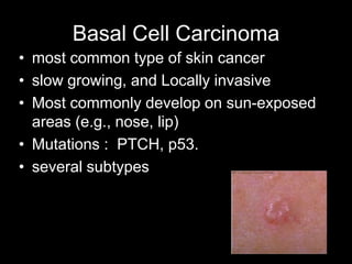
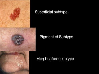
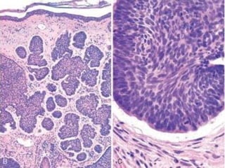
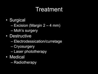
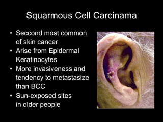
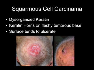
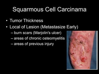
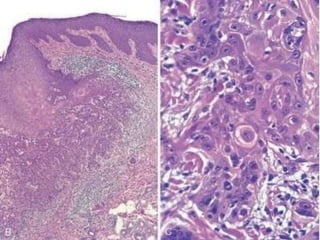
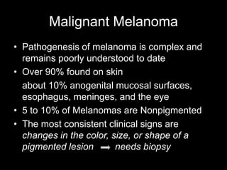
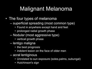
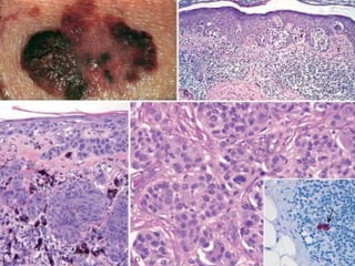
Skin Cancer
Group A 4th year
Surgery Department
Chiangrai Prachanukroh Hospital
Physiologic Function
• Squamous epithelial cells
(keratinocytes)
• Melanocytes
• Dendritic cells & Langerhans cells
• Neural end organs
• Adnexal components
Malignant Tumors of the Skin
• frequently subject to epidermal tumors
• arising from cells of the dermis or adnexal
structures are relatively uncommon
• UV radiation, albino individuals of dark
skinned races is associated with an
increased development of skin cancer
Type of Skin Cancer
• Non – Melanomas
– Basal Cell Carcinoma
– Squarmous cell Carcinoma
• Melanomas
• Others
– Merkel Cell Carcinoma
– Kaposi's Sarcoma
– Extramammary Paget's Disease
– Angiosarcoma
– Dermatofibrosarcoma Protuberans
– Fibrosarcoma
– Liposarcoma
Basal Cell Carcinoma
• most common type of skin cancer
• slow growing, and Locally invasive
• Most commonly develop on sun-exposed
areas (e.g., nose, lip)
• Mutations : PTCH, p53.
• several subtypes
Basal Cell Carcinoma
• Nodulocystic Subtype
– Found on 70% of BCC
– pearly papules often containing prominent,
dilated subepidermal blood vessels
– Rodent Ulcer
(Rat Tooth)
Basal Cell Carcinoma
• Superficial subtype
– commonly occur on the trunk
– Flat red, scaling lesion (Bowenoid Lesion)
• Pigmented subtype
– tan to black in color must DDx with early
Malignant Melanomas
• Others : morpheaform, micronodular,
infiltrative
Superficial subtype
Pigmented Subtype
Morpheaform subtype
Treatment
• Surgical
– Excision (Margin 2 – 4 mm)
– Moh’s surgery
• Destructive
– Electrodessication/curretage
– Cryosurgery
– Laser phototherapy
• Medical
– Radiotherapy
Squarmous Cell Carcinama
• Seccond most common
of skin cancer
• Arise from Epidermal
Keratinocytes
• More invasiveness and
tendency to metastasize
than BCC
• Sun-exposed sites
in older people
Squarmous Cell Carcinama
• Dysorganized Keratin
• Keratin Horns on fleshy tumorous base
• Surface tends to ulcerate
Squarmous Cell Carcinama
• Tumor Thickness
• Local of Lesion (Metastasize Early)
– burn scars (Marjolin's ulcer)
– areas of chronic osteomyelitis
– areas of previous injury
Treatment
• Surgical
– Excision (Margin 1 mm)
• lymph node (LN) dissection remains topic of debate
– Moh’s surgery
• Destructive
– Electrodessication/curretage
– Cryosurgery
– Laser phototherapy
• Medical
– Radiotherapy
Malignant Melanoma
• Pathogenesis of melanoma is complex and
remains poorly understood to date
• Over 90% found on skin
about 10% anogenital mucosal surfaces,
esophagus, meninges, and the eye
• 5 to 10% of Melanomas are Nonpigmented
• The most consistent clinical signs are
changes in the color, size, or shape of a
pigmented lesion needs biopsy
Malignant Melanoma
– A (Asymmetry) one portion of the mole does
not match the other
• Radial : Grows horizontally; not associated with
metastases.
• Vertical : Grows downward into the dermis; is
associated with metastases.
– B (Border & Bleeding) edges are irregular,
notched, or blurred
– C (Color) different shades of black or brown,
patchy colors
– D (Diameter) spot is 6 millimeters across, or
growing larger
Malignant Melanoma
• The four types of melanoma
– superficial spreading (most common type)
• Found in anywhere except hand and feet
• prolonged radial growth phase
– Nodular (most aggressive type)
• vertical growth phase
– lentigo maligna
• the best prognosis
• indolent lesion on the face of older men
– acral lentiginous
• Unrelated to sun exposure (soles,palms, subungal)
• Hutchinson's sign
Additional Skin Malignancies
• Merkel Cell Carcinoma
• Kaposi's Sarcoma
• Extramammary Paget's Disease
• Angiosarcoma
• Dermatofibrosarcoma Protuberans
• Fibrosarcoma
• Liposarcoma
Tumors of Skin Migrants
• Mycosis Fungoides (Cutaneous T-Cell
Lymphomas)
• Mastocytosis
Syndromic Skin Malignancies
• Basal Cell Nevus (Gorlin's syndrome )
• Nevus Sebaceus of Jadassohn
• Epidermodysplasia verruciformis
• Epidermolysis Bullosus and Lupus
Erythematosus
• Xeroderma pigmentosum
• Familial dysplastic nevus syndrome
Preventions
• “Slip! Slop! Slap! Wrap!”
– Slip on a Shirt
– Slop on SPF 15+ Sunscreen
– Slap on Hat
– Wrap on Sunglasses
Reference

Skin cancer

  • 1.
    Skin Cancer Group A4th year Surgery Department Chiangrai Prachanukroh Hospital
  • 3.
    Physiologic Function • Squamousepithelial cells (keratinocytes) • Melanocytes • Dendritic cells & Langerhans cells • Neural end organs • Adnexal components
  • 4.
    Malignant Tumors ofthe Skin • frequently subject to epidermal tumors • arising from cells of the dermis or adnexal structures are relatively uncommon • UV radiation, albino individuals of dark skinned races is associated with an increased development of skin cancer
  • 5.
    Type of SkinCancer • Non – Melanomas – Basal Cell Carcinoma – Squarmous cell Carcinoma • Melanomas • Others – Merkel Cell Carcinoma – Kaposi's Sarcoma – Extramammary Paget's Disease – Angiosarcoma – Dermatofibrosarcoma Protuberans – Fibrosarcoma – Liposarcoma
  • 6.
    Basal Cell Carcinoma •most common type of skin cancer • slow growing, and Locally invasive • Most commonly develop on sun-exposed areas (e.g., nose, lip) • Mutations : PTCH, p53. • several subtypes
  • 7.
    Basal Cell Carcinoma •Nodulocystic Subtype – Found on 70% of BCC – pearly papules often containing prominent, dilated subepidermal blood vessels – Rodent Ulcer (Rat Tooth)
  • 8.
    Basal Cell Carcinoma •Superficial subtype – commonly occur on the trunk – Flat red, scaling lesion (Bowenoid Lesion) • Pigmented subtype – tan to black in color must DDx with early Malignant Melanomas • Others : morpheaform, micronodular, infiltrative
  • 9.
  • 11.
    Treatment • Surgical – Excision(Margin 2 – 4 mm) – Moh’s surgery • Destructive – Electrodessication/curretage – Cryosurgery – Laser phototherapy • Medical – Radiotherapy
  • 12.
    Squarmous Cell Carcinama •Seccond most common of skin cancer • Arise from Epidermal Keratinocytes • More invasiveness and tendency to metastasize than BCC • Sun-exposed sites in older people
  • 13.
    Squarmous Cell Carcinama •Dysorganized Keratin • Keratin Horns on fleshy tumorous base • Surface tends to ulcerate
  • 14.
    Squarmous Cell Carcinama •Tumor Thickness • Local of Lesion (Metastasize Early) – burn scars (Marjolin's ulcer) – areas of chronic osteomyelitis – areas of previous injury
  • 16.
    Treatment • Surgical – Excision(Margin 1 mm) • lymph node (LN) dissection remains topic of debate – Moh’s surgery • Destructive – Electrodessication/curretage – Cryosurgery – Laser phototherapy • Medical – Radiotherapy
  • 17.
    Malignant Melanoma • Pathogenesisof melanoma is complex and remains poorly understood to date • Over 90% found on skin about 10% anogenital mucosal surfaces, esophagus, meninges, and the eye • 5 to 10% of Melanomas are Nonpigmented • The most consistent clinical signs are changes in the color, size, or shape of a pigmented lesion needs biopsy
  • 18.
    Malignant Melanoma – A(Asymmetry) one portion of the mole does not match the other • Radial : Grows horizontally; not associated with metastases. • Vertical : Grows downward into the dermis; is associated with metastases. – B (Border & Bleeding) edges are irregular, notched, or blurred – C (Color) different shades of black or brown, patchy colors – D (Diameter) spot is 6 millimeters across, or growing larger
  • 19.
    Malignant Melanoma • Thefour types of melanoma – superficial spreading (most common type) • Found in anywhere except hand and feet • prolonged radial growth phase – Nodular (most aggressive type) • vertical growth phase – lentigo maligna • the best prognosis • indolent lesion on the face of older men – acral lentiginous • Unrelated to sun exposure (soles,palms, subungal) • Hutchinson's sign
  • 24.
    Additional Skin Malignancies •Merkel Cell Carcinoma • Kaposi's Sarcoma • Extramammary Paget's Disease • Angiosarcoma • Dermatofibrosarcoma Protuberans • Fibrosarcoma • Liposarcoma
  • 25.
    Tumors of SkinMigrants • Mycosis Fungoides (Cutaneous T-Cell Lymphomas) • Mastocytosis
  • 26.
    Syndromic Skin Malignancies •Basal Cell Nevus (Gorlin's syndrome ) • Nevus Sebaceus of Jadassohn • Epidermodysplasia verruciformis • Epidermolysis Bullosus and Lupus Erythematosus • Xeroderma pigmentosum • Familial dysplastic nevus syndrome
  • 27.
    Preventions • “Slip! Slop!Slap! Wrap!” – Slip on a Shirt – Slop on SPF 15+ Sunscreen – Slap on Hat – Wrap on Sunglasses
  • 28.