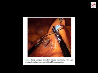
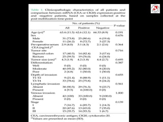
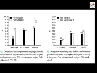
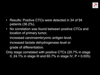
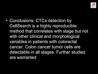
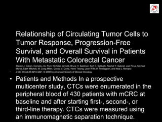
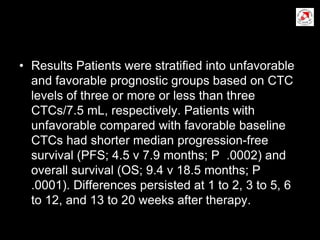
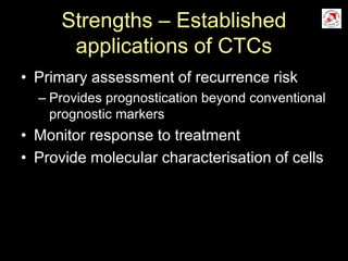
Circulating Tumor Cells in
colorectal cancer
Discussion for today
• What are CTC
• Biology of cancer metastasis
• Facts and hypothesis
• How do we measure CTCs?
• Clinical applications and studies
• Summary of the World CTC Summit
• A SWOT analysis
Glossary
• CTC Circulating tumor cells
• DTC Disseminated tumor cells
• EpCAM Epithelial Cell Adhesion Molecule
• CK Cytokeratin
• EMT Epithelial Mesenchymal Transition
• MET Mesenchymal Epithelial Transition
What are CTCs?
• Tumor cells in circulation!
• CTCs are biomarkers
(substance/s used as an indicator of a biological
state which can be measured objectively)
• They can be used as a Indicator of
1. Normal or pathogenic biological processes
2. Responses to a therapeutic intervention
3. Prognostic vs predictive biomarkers
The biology of cancer metastasis
Some facts…
• Disseminated tumor cells – MC seen in
Bone Marrow of patients with varied ca.
• Seen in 20-40% pts who are clinical N0 M0
• Seen even in cancers which conventionally
have low rate of bone metastases
• Significant correlation between detection of
DTCs and metastatic relapse
• Not all patients with DTCs relapse (50%)
Some hypotheses
• A fraction of DTCs in early stage cancers
represent putative metastatic stem cells
• DTCs can be used to monitor response to
systemic (anti-metastatic) therapy
• The ‘lead interval’ between biomarker
detection and overt metastases provides
an intermediate window between early and
metastatic disease, enabling treatment
How do we measure CTCs?
Enrichment
Identification
Methods for CTC detection
• Physical and biological properties
• Capture and enrichment – morphological
and phenotypic characteristics of tumor
cells
– Size, Density and protein expression
• CTC identification
– Immunocytochemistry (protein expression)
– Nucleic acid characteristics (RT-PCR)
Enrichment methods
• Isolation by Size of Epithelial Tumor
(ISET) Cells
– Cells > 8 micron
• Density gradient (Tumor cells have lower
density than other cells)
– Ficoll-Hypaque
• Based on immunomagnetic techniques
– Separation based on specific surface markers
Immunomagnetic separation
• Most commonly used
• MACS, RARE, AdnaGen, CellSearch
• Differentiate tumor cells from other cells by
specific surface markers
• Positive selection – cytokeratin
• Negative selection – CD-45
• More sensitive than size and density
based methods
CTC Identification
• Immunocytochemistry
– Uses monoclonal antibodies against specific
tumor types (CK, Mg, CEA)
– Identifies intact cells (preserves the cell)
• RT-PCR
– Assess DNA/RNA changes specific to tumor
cells
– Amplifies minute quantities of tumor RNA
– More sensitive (false +ve: non-CTC mRNA)
The CellSearch
• Only FDA approved method
• 7.5 ml blood centrifuged
• Plasma and buffer coat removed
• CTCs captured using EpCAM labelled
ferrofluid
• Differentiated from WBCs by panel of
monoclonal antibodies
– Pan-CK, anti CD-45, nucleic acid dye
The CellSearch
• Labelled sample loaded into a cassette
and analysed (CellTracks) – an automated
fluorescence detection system
• CTCs detected as
– Nucleated cells
– CK +ve
– CD-45 -ve
• Highly reproducible
Different markers
• K- ras and p53
• Expression of CEA mRNA.
• Expression of CK20 mRNA.
• Expression of CK19 mRNA.
Clinical evaluation of CTCs in
CRC
• Circulating Tumor Cell Clusters in the
Peripheral Blood of Colorectal Cancer
Patients
Bela Molnar,1 Andras Ladanyi, Lenke Tanko, Lydia Sre´ter, and Zsolt Tulassay II.
Department of Medicine, Semmelweis University, Budapest 1088, Hungary
• Results: Of 20 healthy samples, 2 contained one cytok- eratin-
positive cell. Of 32 single samples from malignant cases, 24 showed
cytokeratin-positive cells. Tumor cell clus- ters, mixed-cell doublets
(one cytokeratin-positive and -neg- ative cell), and mixed-cell
clusters were detected in 22 of 24 patients. In six cases, cytokeratin-
positive dendritic-like cells were detected. Follow-up data indicate
that chemotherapy cannot destroy all of the circulating tumor cell
clusters.
• Conclusions: Using the methods
presented, we could detect circulating
colon cancer cells and cell clusters in
colon carcinoma patients. Similar cellular
structures were de- scribed previously only
in rats. Present data prove that such
structures are present in human colorectal
cancer, too.
Circulating tumor cells in colorectal cancer:
correlation with clinical and pathological
variables
J. Sastre1*, M. L. Maestro2, J. Puente1, S. Veganzones2, R. Alfonso1, S. Rafael2, J. A. Garcı ´a-Saenz1, M. Vidaurreta 2,
M. Martı ´n1, M. Arroyo2, M. T. Sanz-Casla 2 & E. Dı ´az-Rubio1 1Department of Medical Oncology, 2Department of Central
Laboratory, Hospital Clı´nico San Carlos de Madrid, Madrid, Spain
Received 24 September 2007; revised 26 November 2007; accepted 30 November 2007
• Results: Positive CTCs were detected in 34 of 94
patients (36.2%).
• No correlation was found between positive CTCs and
location of primary tumor,
increased carcinoembryonic antigen level,
increased lactate dehydrogenase level or
grade of differentiation.
Only stage correlated with positive CTCs (20.7% in stage
II, 24.1% in stage III and 60.7% in stage IV, P = 0.005).
• Conclusions: CTCs detection by
CellSearch is a highly reproducible
method that correlates with stage but not
with other clinical and morphological
variables in patients with colorectal
cancer. Colon cancer tumor cells are
detectable in all stages. Further studies
are warranted
Relationship of Circulating Tumor Cells to
Tumor Response, Progression-Free
Survival, and Overall Survival in Patients
With Metastatic Colorectal Cancer
Steven J. Cohen, Cornelis J.A. Punt, Nicholas Iannotti, Bruce H. Saidman, Kert D. Sabbath, Nashat Y. Gabrail, Joel Picus, Michael
Morse, Edith Mitchell, M. Craig Miller, Gerald V. Doyle, Henk Tissing, Leon W.M.M. Terstappen, and Neal J. Meropol
• J Clin Oncol 26:3213-3221. © 2008 by American Society of Clinical Oncology
• Patients and Methods In a prospective
multicenter study, CTCs were enumerated in the
peripheral blood of 430 patients with mCRC at
baseline and after starting first-, second-, or
third-line therapy. CTCs were measured using
an immunomagnetic separation technique.
• Results Patients were stratified into unfavorable
and favorable prognostic groups based on CTC
levels of three or more or less than three
CTCs/7.5 mL, respectively. Patients with
unfavorable compared with favorable baseline
CTCs had shorter median progression-free
survival (PFS; 4.5 v 7.9 months; P .0002) and
overall survival (OS; 9.4 v 18.5 months; P
.0001). Differences persisted at 1 to 2, 3 to 5, 6
to 12, and 13 to 20 weeks after therapy.
• Conversion of baseline unfavorable CTCs to
favorable at 3 to 5 weeks was associated with
significantly longer PFS and OS compared with
patients with unfavorable CTCs at both time
points (PFS, 6.2 v 1.6 months; P .02; OS, 11.0 v
3.7 months; P .0002). Among nonprogressing
patients, favorable compared with unfavorable
CTCs within 1 month of imaging was associated
with longer survival (18.8 v 7.1 months; P
.0001). Baseline and follow-up CTC levels
remained strong predictors of PFS and OS after
adjustment for clinically significant factors.
• Conclusion The number of CTCs before
and during treatment is an independent
predictor of PFS and OS in patients with
metastatic colorectal cancer. CTCs
provide prognostic information in addition
to that of imaging studies.
advantages
• Can test both the hypotheses in single pt
• Each person acts as his own control – excludes
bias
• No of trial pts will increase
• Easy to perform
• If IMV is clamped at the beginning if surgery the
tumor cells released in portal circulation can be
trapped and if systemic tumor cells are less at
the end of surgery support our hypothesis of
tumor cell release into systemic circulation
Strengths – Established
applications of CTCs
• Primary assessment of recurrence risk
– Provides prognostication beyond conventional
prognostic markers
• Monitor response to treatment
• Provide molecular characterisation of cells
Weaknesses - Challenges in
CTC detection
• Isolating, quantifying and molecularly
characterising CTCs is extremely
challenging
• Very little standardisation and automation
of processes
– Laborious sample preparation procedures
– High intra and inter lab differences
– Reproducibility
Opportunities - the promise of
CTCs
• Prospective validation of CTCs against
conventional biomarkers
• Markers assessment to guide choice of
personalized therapy
• Identify new therapeutic targets
• Mutation analysis of CTCs can enable
targeted therapy
• Alternative to invasive biopsies
Threats
• Has to compete with other biomarkers for
reliability, feasibility of performance
• Expensive technology
• Labour intensive
Summary
• CTCs are promising biomarkers with
independent prognostic ability
• Clearly prognostic in varied advanced
cancers
• Methodology is challenging and expensive
• Has promise in varied advanced
applications
• Not everything that can be counted
counts, and not everything that counts can
be counted

Circulating tumor cells in crc

  • 1.
    Circulating Tumor Cellsin colorectal cancer
  • 2.
    Discussion for today •What are CTC • Biology of cancer metastasis • Facts and hypothesis • How do we measure CTCs? • Clinical applications and studies • Summary of the World CTC Summit • A SWOT analysis
  • 3.
    Glossary • CTC Circulatingtumor cells • DTC Disseminated tumor cells • EpCAM Epithelial Cell Adhesion Molecule • CK Cytokeratin • EMT Epithelial Mesenchymal Transition • MET Mesenchymal Epithelial Transition
  • 4.
    What are CTCs? •Tumor cells in circulation! • CTCs are biomarkers (substance/s used as an indicator of a biological state which can be measured objectively) • They can be used as a Indicator of 1. Normal or pathogenic biological processes 2. Responses to a therapeutic intervention 3. Prognostic vs predictive biomarkers
  • 5.
    The biology ofcancer metastasis
  • 6.
    The biology ofcancer metastasis
  • 7.
    Some facts… • Disseminatedtumor cells – MC seen in Bone Marrow of patients with varied ca. • Seen in 20-40% pts who are clinical N0 M0 • Seen even in cancers which conventionally have low rate of bone metastases • Significant correlation between detection of DTCs and metastatic relapse • Not all patients with DTCs relapse (50%)
  • 8.
    Some hypotheses • Afraction of DTCs in early stage cancers represent putative metastatic stem cells • DTCs can be used to monitor response to systemic (anti-metastatic) therapy • The ‘lead interval’ between biomarker detection and overt metastases provides an intermediate window between early and metastatic disease, enabling treatment
  • 9.
    How do wemeasure CTCs?
  • 10.
    How do wemeasure CTCs? Enrichment Identification
  • 11.
    Methods for CTCdetection • Physical and biological properties • Capture and enrichment – morphological and phenotypic characteristics of tumor cells – Size, Density and protein expression • CTC identification – Immunocytochemistry (protein expression) – Nucleic acid characteristics (RT-PCR)
  • 12.
    Enrichment methods • Isolationby Size of Epithelial Tumor (ISET) Cells – Cells > 8 micron • Density gradient (Tumor cells have lower density than other cells) – Ficoll-Hypaque • Based on immunomagnetic techniques – Separation based on specific surface markers
  • 13.
    Immunomagnetic separation • Mostcommonly used • MACS, RARE, AdnaGen, CellSearch • Differentiate tumor cells from other cells by specific surface markers • Positive selection – cytokeratin • Negative selection – CD-45 • More sensitive than size and density based methods
  • 14.
    CTC Identification • Immunocytochemistry –Uses monoclonal antibodies against specific tumor types (CK, Mg, CEA) – Identifies intact cells (preserves the cell) • RT-PCR – Assess DNA/RNA changes specific to tumor cells – Amplifies minute quantities of tumor RNA – More sensitive (false +ve: non-CTC mRNA)
  • 15.
    The CellSearch • OnlyFDA approved method • 7.5 ml blood centrifuged • Plasma and buffer coat removed • CTCs captured using EpCAM labelled ferrofluid • Differentiated from WBCs by panel of monoclonal antibodies – Pan-CK, anti CD-45, nucleic acid dye
  • 16.
    The CellSearch • Labelledsample loaded into a cassette and analysed (CellTracks) – an automated fluorescence detection system • CTCs detected as – Nucleated cells – CK +ve – CD-45 -ve • Highly reproducible
  • 19.
    Different markers • K-ras and p53 • Expression of CEA mRNA. • Expression of CK20 mRNA. • Expression of CK19 mRNA.
  • 20.
  • 21.
    • Circulating TumorCell Clusters in the Peripheral Blood of Colorectal Cancer Patients Bela Molnar,1 Andras Ladanyi, Lenke Tanko, Lydia Sre´ter, and Zsolt Tulassay II. Department of Medicine, Semmelweis University, Budapest 1088, Hungary • Results: Of 20 healthy samples, 2 contained one cytok- eratin- positive cell. Of 32 single samples from malignant cases, 24 showed cytokeratin-positive cells. Tumor cell clus- ters, mixed-cell doublets (one cytokeratin-positive and -neg- ative cell), and mixed-cell clusters were detected in 22 of 24 patients. In six cases, cytokeratin- positive dendritic-like cells were detected. Follow-up data indicate that chemotherapy cannot destroy all of the circulating tumor cell clusters.
  • 22.
    • Conclusions: Usingthe methods presented, we could detect circulating colon cancer cells and cell clusters in colon carcinoma patients. Similar cellular structures were de- scribed previously only in rats. Present data prove that such structures are present in human colorectal cancer, too.
  • 23.
    Circulating tumor cellsin colorectal cancer: correlation with clinical and pathological variables J. Sastre1*, M. L. Maestro2, J. Puente1, S. Veganzones2, R. Alfonso1, S. Rafael2, J. A. Garcı ´a-Saenz1, M. Vidaurreta 2, M. Martı ´n1, M. Arroyo2, M. T. Sanz-Casla 2 & E. Dı ´az-Rubio1 1Department of Medical Oncology, 2Department of Central Laboratory, Hospital Clı´nico San Carlos de Madrid, Madrid, Spain Received 24 September 2007; revised 26 November 2007; accepted 30 November 2007
  • 24.
    • Results: PositiveCTCs were detected in 34 of 94 patients (36.2%). • No correlation was found between positive CTCs and location of primary tumor, increased carcinoembryonic antigen level, increased lactate dehydrogenase level or grade of differentiation. Only stage correlated with positive CTCs (20.7% in stage II, 24.1% in stage III and 60.7% in stage IV, P = 0.005).
  • 25.
    • Conclusions: CTCsdetection by CellSearch is a highly reproducible method that correlates with stage but not with other clinical and morphological variables in patients with colorectal cancer. Colon cancer tumor cells are detectable in all stages. Further studies are warranted
  • 26.
    Relationship of CirculatingTumor Cells to Tumor Response, Progression-Free Survival, and Overall Survival in Patients With Metastatic Colorectal Cancer Steven J. Cohen, Cornelis J.A. Punt, Nicholas Iannotti, Bruce H. Saidman, Kert D. Sabbath, Nashat Y. Gabrail, Joel Picus, Michael Morse, Edith Mitchell, M. Craig Miller, Gerald V. Doyle, Henk Tissing, Leon W.M.M. Terstappen, and Neal J. Meropol • J Clin Oncol 26:3213-3221. © 2008 by American Society of Clinical Oncology • Patients and Methods In a prospective multicenter study, CTCs were enumerated in the peripheral blood of 430 patients with mCRC at baseline and after starting first-, second-, or third-line therapy. CTCs were measured using an immunomagnetic separation technique.
  • 27.
    • Results Patientswere stratified into unfavorable and favorable prognostic groups based on CTC levels of three or more or less than three CTCs/7.5 mL, respectively. Patients with unfavorable compared with favorable baseline CTCs had shorter median progression-free survival (PFS; 4.5 v 7.9 months; P .0002) and overall survival (OS; 9.4 v 18.5 months; P .0001). Differences persisted at 1 to 2, 3 to 5, 6 to 12, and 13 to 20 weeks after therapy.
  • 28.
    • Conversion ofbaseline unfavorable CTCs to favorable at 3 to 5 weeks was associated with significantly longer PFS and OS compared with patients with unfavorable CTCs at both time points (PFS, 6.2 v 1.6 months; P .02; OS, 11.0 v 3.7 months; P .0002). Among nonprogressing patients, favorable compared with unfavorable CTCs within 1 month of imaging was associated with longer survival (18.8 v 7.1 months; P .0001). Baseline and follow-up CTC levels remained strong predictors of PFS and OS after adjustment for clinically significant factors.
  • 29.
    • Conclusion Thenumber of CTCs before and during treatment is an independent predictor of PFS and OS in patients with metastatic colorectal cancer. CTCs provide prognostic information in addition to that of imaging studies.
  • 43.
    advantages • Can testboth the hypotheses in single pt • Each person acts as his own control – excludes bias • No of trial pts will increase • Easy to perform • If IMV is clamped at the beginning if surgery the tumor cells released in portal circulation can be trapped and if systemic tumor cells are less at the end of surgery support our hypothesis of tumor cell release into systemic circulation
  • 44.
    Strengths – Established applicationsof CTCs • Primary assessment of recurrence risk – Provides prognostication beyond conventional prognostic markers • Monitor response to treatment • Provide molecular characterisation of cells
  • 45.
    Weaknesses - Challengesin CTC detection • Isolating, quantifying and molecularly characterising CTCs is extremely challenging • Very little standardisation and automation of processes – Laborious sample preparation procedures – High intra and inter lab differences – Reproducibility
  • 46.
    Opportunities - thepromise of CTCs • Prospective validation of CTCs against conventional biomarkers • Markers assessment to guide choice of personalized therapy • Identify new therapeutic targets • Mutation analysis of CTCs can enable targeted therapy • Alternative to invasive biopsies
  • 47.
    Threats • Has tocompete with other biomarkers for reliability, feasibility of performance • Expensive technology • Labour intensive
  • 48.
    Summary • CTCs arepromising biomarkers with independent prognostic ability • Clearly prognostic in varied advanced cancers • Methodology is challenging and expensive • Has promise in varied advanced applications
  • 49.
    • Not everythingthat can be counted counts, and not everything that counts can be counted