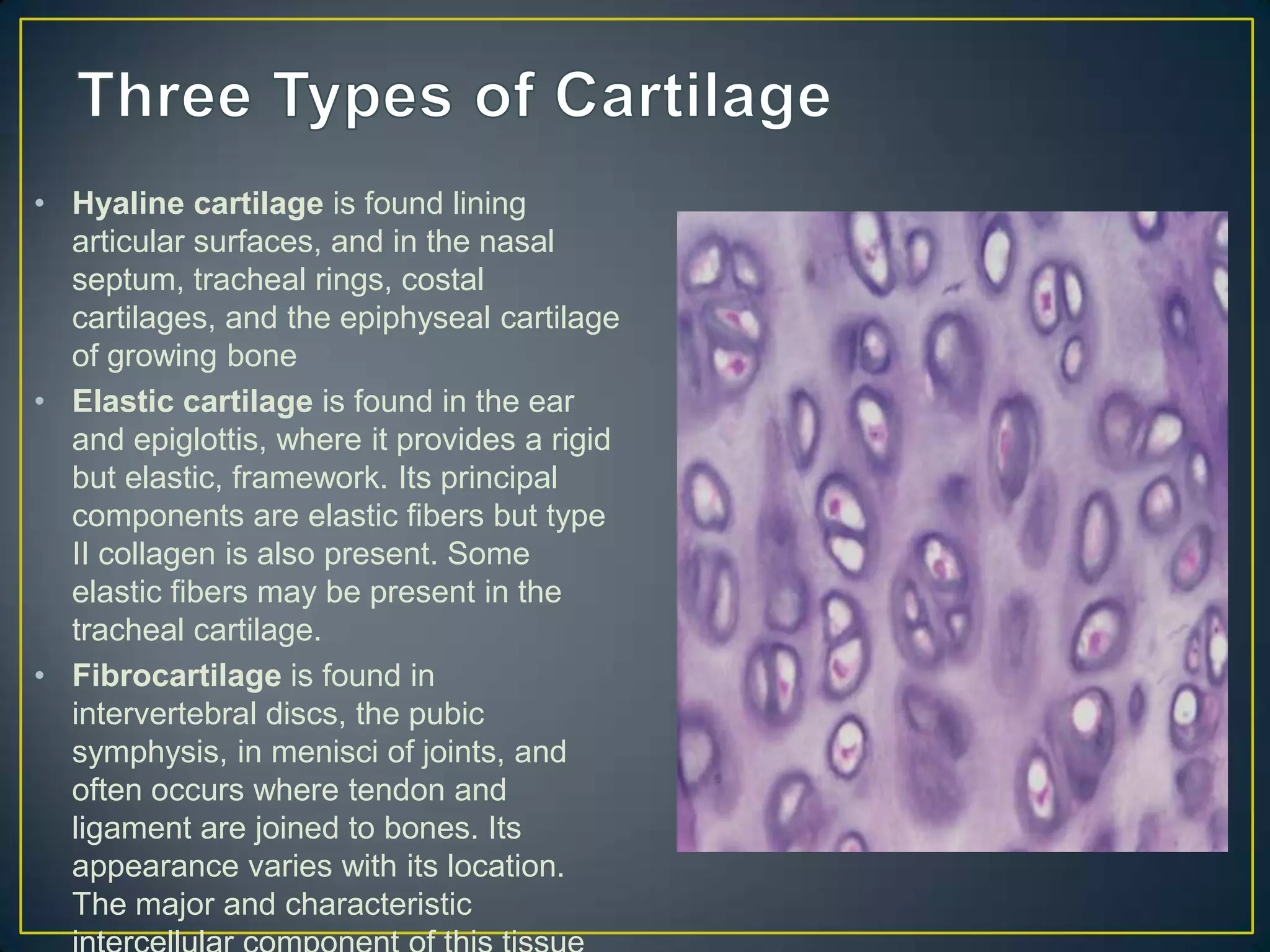
• Cartilage is a specialized form of connective tissue
  containing chondrocytes which secrete, and are
  surrounded by, an extensive intercellular matrix.
  Chondrocytes occur singly or in isogenous
  groups, composed of 2-8 cells derived by mitosis from a
  single chondrocyte. The cells are in the lacunae (cavities)
  within the matrix. Matrix stains more intensely
  immediately adjacent to the lacunae and the dark
  staining zone is called the capsule. The strength and
  durability of cartilage are properties of the matrix, which
  is an interlaced network of collagenous and/or elastic
  fibers in a ground substance, a gel of complex
  proteoglycans.
• Hyaline cartilage is found lining
  articular surfaces, and in the nasal
  septum, tracheal rings, costal
  cartilages, and the epiphyseal cartilage
  of growing bone
• Elastic cartilage is found in the ear
  and epiglottis, where it provides a rigid
  but elastic, framework. Its principal
  components are elastic fibers but type
  II collagen is also present. Some
  elastic fibers may be present in the
  tracheal cartilage.
• Fibrocartilage is found in
  intervertebral discs, the pubic
  symphysis, in menisci of joints, and
  often occurs where tendon and
  ligament are joined to bones. Its
  appearance varies with its location.
  The major and characteristic
Bone                                Cartilage
• Bones grow longer over time        • Cartilage forms from initial
  but they also get thicker.           condensation directly from
  There are mechanism that             mesenchyme and then grows by a
  allow for bones to grow in           combination of apositional growth
  both length and width.             • Interstitial growth - Chondroblasts
• To make a bone longer just           within the existing cartilage divide
  add bone tissue to the ends.         and form small groups of cells,
  If tissue is added at the end of     isogenous groups, which produce
  the bone the skeleton could          matrix to become separated from
  not move properly. It's like         each other by a thin partition of
  closing both lanes on an             matrix. Interstitial growth occurs
  interstate until the bridge is       mainly in immature cartilage.
  repaired.                          • Appositional growth - Mesenchymal
• Bone tissue must be added            cells surrounding the cartilage in
  below the joint somewhere            the deep part of the perichondrium
  along the length of the bone.        (or the chondrogenic layer)
  This occurs at the epiphyseal        differentiate into chondroblasts.
  plate, or growth plate. Here         Appositional growth occurs also in
  chondrocytes first produce           mature cartilage.
• Flexion/ extension.
• Elbow and knee.
• Rotation of one bone around another.
• Neck.
• Flexion, extension, abduction.
• Shoulder and hip.
• Flexion, extension, adduction, abduction, circumduction.
• Thumb.
• Flexion/Extension/Adduction/ Abduction/Circumduction.
• Wrist joint.
• Gliding movement.
• Intercarpal joints.
• Osteoporosis is when bone density is decreased,
  normally the process of matrix remodeling keeps bones
  at the correct density but Osteoporosis slows this down.
• Cancer treatment can destroy bone marrow which is
  where red blood cells are naturally replaced. Without the
  bone marrow patients are more susceptible to sickness.
  Bone marrow transplants replace the bone marrow with
  healthy normal marrow.
Intramembranous ossification: when cells of an embryo
transforms into bone. In early development, the embryo has
three cell layers: the ectoderm which is on the outside,
mesoderm in the middle, and endoderm on the inside of the
embryo. Bones of the skull come directly from the mesenchyme
cells by intramembranous ossification.
Endochondral ossification: this is the gradual replacement of
cartilage by the bone. This process is responsible for forming
most of the skeleton of vertebrates. Osteoblasts arise in
regions of cartilage called ossification centers. (9)
1. Inflammation: in a bone fracture, white blood cells move in to the area to
clean up debris created by the break. Inflammation triggers the growth new
blood cells.
2. Soft callus: as the blood cells divide and multiply near the break, the new
blood vessels develop to fuel the repair process; the body also creates
simply fibrous tissue cartilage around the bone fracture to bridge the gap.
3. Hard callus: later on, the body replaces the soft callus with a hard callus,
connecting the fragments of the bone more solidly. It creates a bulge at the
site of the fracture.
4. Remodeling: the body replaces the old bone with a new bone in a
continual process called remodeling; it makes the bone stronger and
compact and blood circulation in the bone improves.(8)
8       8


    8       8
Bone: the types of bones are divided into long, short, irregular, flat,
sesamoid and sutural bones. Their function is to protect the body from
mechanical damage, to provide support, assist in movement, to store
minerals, produce red blood and white blood cells, and to be the
framework and shape for the body. The bones are made up of
osteoblasts, osteocytes, osteoclasts, and bone lining cells.
Cartilage: the types of cartilage are hyaline cartilage, fibrocartilage and
elastic cartilage. The main functions is to reduce the friction at the
joints, support tracheal and bronchial tubes, they act as shock
absorbers between the vertebrae, also maintaining the shape and
flexibility of ear, nose, along with others. Cartilage is made up of
chondroblasts, chondrocytes, and dense matrix that is composed of
collagen and elastic fibres. (10)
Main Parts
•Haversian Canal
•Lucanae
•Canaliculi
•Osteon
•Periosteum
•Lamellae
•Trabeculae
• Osteoblasts- These are bone forming cells within the bone.

• Osteocytes- These are involved in the formation of the bone, maintenance of
  the matrix, and homeostasis of the calcium.

• Osteoclasts- The cells are responsible for bone resorption and remodeling.
•   Maintain shape and structure of the bone.
•   Strengthening the bone and adding tissue.
•   Remodeling broken bones.
•   Calcium from bones being used in other parts of body.
•   Mineral reservoir, contains many minerals that the body
    can use.
• Femur,
• Carpals
• vertebrae
• Bones of the skull
• Spongy
• Compact
• Is made up of concentric rings of matrix that surround
  central canals which contain blood vessels.
• Embedded in this bone tissue are small cave-like spaces
  called lacunae, which are connected to each other
  through small tunnels called canalicula.
• The lacunae contain osteocytes cells. As just discussed,
  osteocytes help maintain healthy bone tissue and are
  involved in the bone remodeling process that will be
  outlined later in this lesson.
• Looks like an irregular latticework (or sponge) with lots of
  spaces throughout.
• These spaces are filled with red bone marrow which is
  the site of hemopoesis or formation of blood cells
• During the aging process, adults face the issue of a
  changing skeletal framework.
• As humans age, the force-generating capacity (strength)
  of their skeletal muscles is reduced. As a result, many
  older people experience difficulty in performing their
  activities of daily living. The loss of force production in
  older people is primarily to the result of muscle atrophy
  and alterations in the percentage of contractile tissue
  within muscle rather than deficits in muscle.
Growth plate                            In growing children,
                                          sprains and strains often
  fractures                               result in potentially
                                          serious growth plate
                                          fractures and physeal
                                          fractures. These same
                                          sprains and strains in
                                          active adults are
                                          relatively benign injuries.
                                          This article discusses
                                          some of the important
                                          orthopedic history
                                          relative to the physes,
                                          relevant anatomy,
                                          classification systems,
                                          and some details of
                                          physeal fractures in
                                          specific areas of the
An image depicting growth plate fractures can be seen below.
                                          body.
• Growth plate (physeal)
  fractures. Clinical appearance
  of the knee of a patient with a
  minimally displaced Salter-
  Harris I fracture of the distal
  femur. Impressive swelling
  was noted adjacent to the
  joint, but no evidence of intra-
  articular swelling was
  present. The patient was
  markedly tender to palpation
  about the distal femoral
  physis.

Skelphysll

  • 3.
    • Cartilage isa specialized form of connective tissue containing chondrocytes which secrete, and are surrounded by, an extensive intercellular matrix. Chondrocytes occur singly or in isogenous groups, composed of 2-8 cells derived by mitosis from a single chondrocyte. The cells are in the lacunae (cavities) within the matrix. Matrix stains more intensely immediately adjacent to the lacunae and the dark staining zone is called the capsule. The strength and durability of cartilage are properties of the matrix, which is an interlaced network of collagenous and/or elastic fibers in a ground substance, a gel of complex proteoglycans.
  • 4.
    • Hyaline cartilageis found lining articular surfaces, and in the nasal septum, tracheal rings, costal cartilages, and the epiphyseal cartilage of growing bone • Elastic cartilage is found in the ear and epiglottis, where it provides a rigid but elastic, framework. Its principal components are elastic fibers but type II collagen is also present. Some elastic fibers may be present in the tracheal cartilage. • Fibrocartilage is found in intervertebral discs, the pubic symphysis, in menisci of joints, and often occurs where tendon and ligament are joined to bones. Its appearance varies with its location. The major and characteristic
  • 5.
    Bone Cartilage • Bones grow longer over time • Cartilage forms from initial but they also get thicker. condensation directly from There are mechanism that mesenchyme and then grows by a allow for bones to grow in combination of apositional growth both length and width. • Interstitial growth - Chondroblasts • To make a bone longer just within the existing cartilage divide add bone tissue to the ends. and form small groups of cells, If tissue is added at the end of isogenous groups, which produce the bone the skeleton could matrix to become separated from not move properly. It's like each other by a thin partition of closing both lanes on an matrix. Interstitial growth occurs interstate until the bridge is mainly in immature cartilage. repaired. • Appositional growth - Mesenchymal • Bone tissue must be added cells surrounding the cartilage in below the joint somewhere the deep part of the perichondrium along the length of the bone. (or the chondrogenic layer) This occurs at the epiphyseal differentiate into chondroblasts. plate, or growth plate. Here Appositional growth occurs also in chondrocytes first produce mature cartilage.
  • 8.
  • 9.
    • Rotation ofone bone around another. • Neck.
  • 10.
    • Flexion, extension,abduction. • Shoulder and hip.
  • 11.
    • Flexion, extension,adduction, abduction, circumduction. • Thumb.
  • 12.
  • 13.
    • Gliding movement. •Intercarpal joints.
  • 14.
    • Osteoporosis iswhen bone density is decreased, normally the process of matrix remodeling keeps bones at the correct density but Osteoporosis slows this down.
  • 15.
    • Cancer treatmentcan destroy bone marrow which is where red blood cells are naturally replaced. Without the bone marrow patients are more susceptible to sickness. Bone marrow transplants replace the bone marrow with healthy normal marrow.
  • 16.
    Intramembranous ossification: whencells of an embryo transforms into bone. In early development, the embryo has three cell layers: the ectoderm which is on the outside, mesoderm in the middle, and endoderm on the inside of the embryo. Bones of the skull come directly from the mesenchyme cells by intramembranous ossification. Endochondral ossification: this is the gradual replacement of cartilage by the bone. This process is responsible for forming most of the skeleton of vertebrates. Osteoblasts arise in regions of cartilage called ossification centers. (9)
  • 17.
    1. Inflammation: ina bone fracture, white blood cells move in to the area to clean up debris created by the break. Inflammation triggers the growth new blood cells. 2. Soft callus: as the blood cells divide and multiply near the break, the new blood vessels develop to fuel the repair process; the body also creates simply fibrous tissue cartilage around the bone fracture to bridge the gap. 3. Hard callus: later on, the body replaces the soft callus with a hard callus, connecting the fragments of the bone more solidly. It creates a bulge at the site of the fracture. 4. Remodeling: the body replaces the old bone with a new bone in a continual process called remodeling; it makes the bone stronger and compact and blood circulation in the bone improves.(8)
  • 18.
    8 8 8 8
  • 19.
    Bone: the typesof bones are divided into long, short, irregular, flat, sesamoid and sutural bones. Their function is to protect the body from mechanical damage, to provide support, assist in movement, to store minerals, produce red blood and white blood cells, and to be the framework and shape for the body. The bones are made up of osteoblasts, osteocytes, osteoclasts, and bone lining cells. Cartilage: the types of cartilage are hyaline cartilage, fibrocartilage and elastic cartilage. The main functions is to reduce the friction at the joints, support tracheal and bronchial tubes, they act as shock absorbers between the vertebrae, also maintaining the shape and flexibility of ear, nose, along with others. Cartilage is made up of chondroblasts, chondrocytes, and dense matrix that is composed of collagen and elastic fibres. (10)
  • 20.
  • 21.
    • Osteoblasts- Theseare bone forming cells within the bone. • Osteocytes- These are involved in the formation of the bone, maintenance of the matrix, and homeostasis of the calcium. • Osteoclasts- The cells are responsible for bone resorption and remodeling.
  • 22.
    Maintain shape and structure of the bone. • Strengthening the bone and adding tissue. • Remodeling broken bones. • Calcium from bones being used in other parts of body. • Mineral reservoir, contains many minerals that the body can use.
  • 24.
  • 25.
  • 26.
  • 27.
    • Bones ofthe skull
  • 29.
  • 30.
    • Is madeup of concentric rings of matrix that surround central canals which contain blood vessels. • Embedded in this bone tissue are small cave-like spaces called lacunae, which are connected to each other through small tunnels called canalicula. • The lacunae contain osteocytes cells. As just discussed, osteocytes help maintain healthy bone tissue and are involved in the bone remodeling process that will be outlined later in this lesson.
  • 31.
    • Looks likean irregular latticework (or sponge) with lots of spaces throughout. • These spaces are filled with red bone marrow which is the site of hemopoesis or formation of blood cells
  • 32.
    • During theaging process, adults face the issue of a changing skeletal framework. • As humans age, the force-generating capacity (strength) of their skeletal muscles is reduced. As a result, many older people experience difficulty in performing their activities of daily living. The loss of force production in older people is primarily to the result of muscle atrophy and alterations in the percentage of contractile tissue within muscle rather than deficits in muscle.
  • 33.
    Growth plate In growing children, sprains and strains often fractures result in potentially serious growth plate fractures and physeal fractures. These same sprains and strains in active adults are relatively benign injuries. This article discusses some of the important orthopedic history relative to the physes, relevant anatomy, classification systems, and some details of physeal fractures in specific areas of the An image depicting growth plate fractures can be seen below. body.
  • 34.
    • Growth plate(physeal) fractures. Clinical appearance of the knee of a patient with a minimally displaced Salter- Harris I fracture of the distal femur. Impressive swelling was noted adjacent to the joint, but no evidence of intra- articular swelling was present. The patient was markedly tender to palpation about the distal femoral physis.