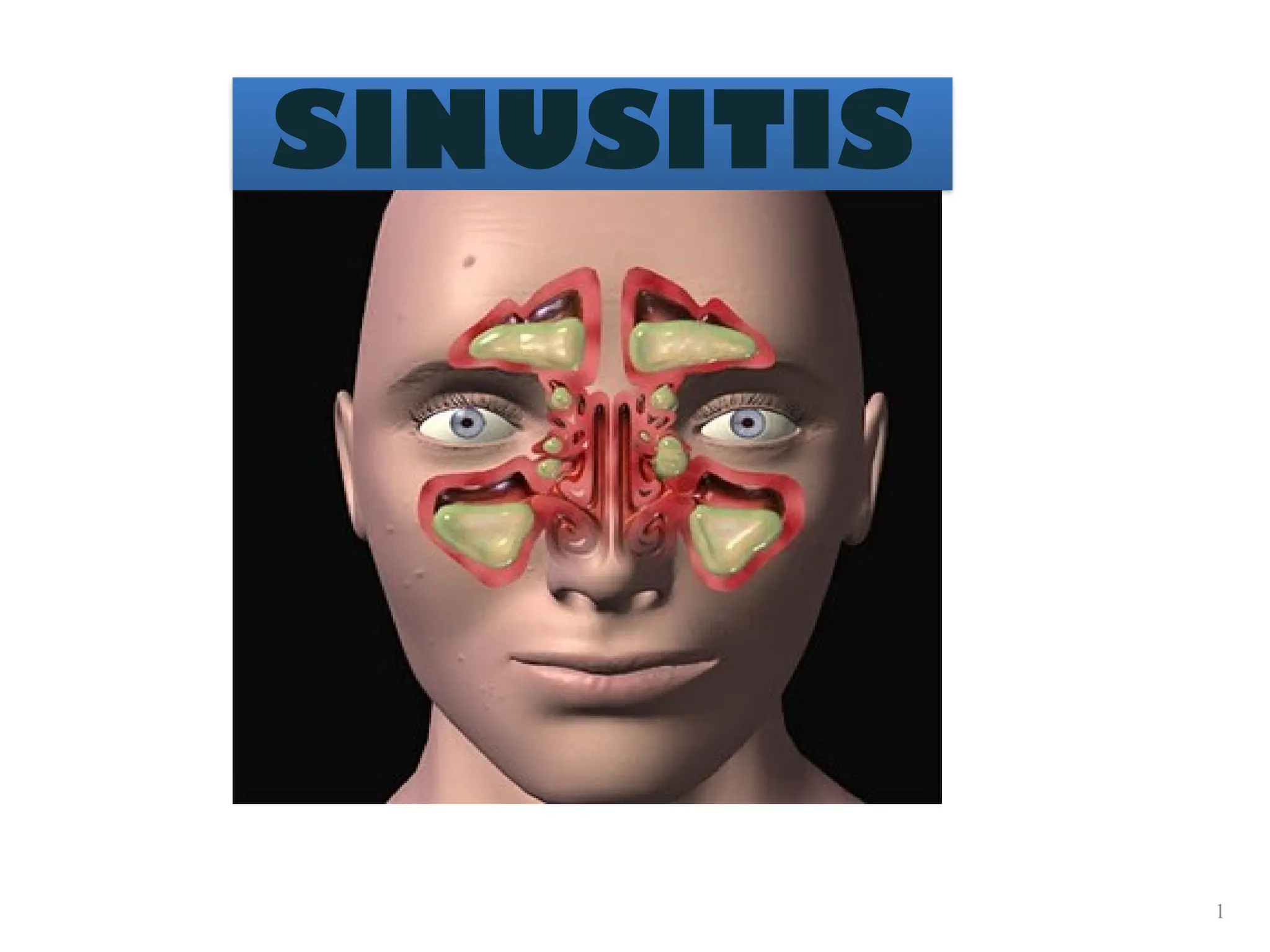
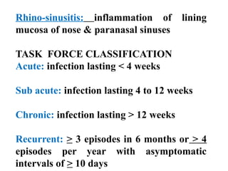
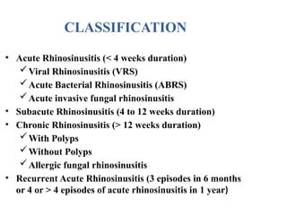
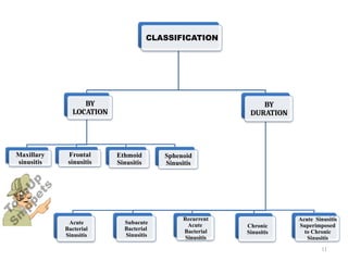
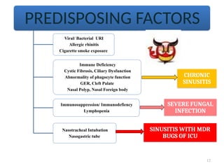
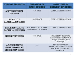
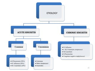
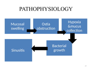
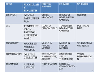
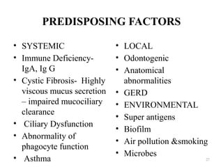
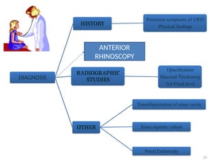
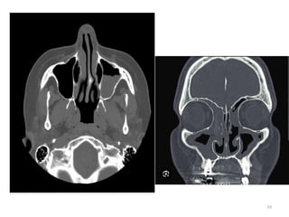
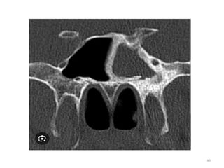
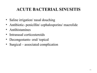
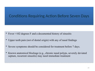
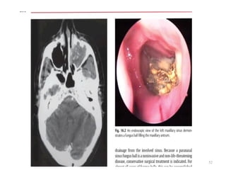
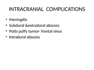
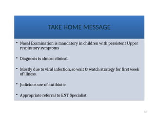
1
SINUSITIS
• hollow, air-filled cavities in skull that are lined by a mucous
membrane.
• known as the paranasal sinuses because of their formation from the
nasal mucosa and their continued communication with nasal fossas.
PARANASAL
SINUS
Anterior Sinus Anatomy Lateral Sinus Anatomy
3
Purpose of
sinuses
• Humidifying and warming
inspired air
• Regulation of intranasal pressure
• Increasing surface area for
olfaction
• Lightening the skull
• Resonance
• Absorbing shock
• Contribute to facial growth
Development
of sinuses
• Aerated at birth
• Maxillary sinuses
• Age 6-7
• Frontal /sphenoidal sinuses
• Puberty- approx 17-18 yrs
• Ethmoid
4
Maxillary Sinuses
• Largest sinuses
• 3.5 cm high
• 2.5 – 3 cm wide
• Within maxilla
• Above upper teeth
• Paired & symmetric
• Communicates with middle nasal
meatus
5
Frontal Sinuses
• Second largest sinuses
• 2 – 2.5 cm
• Normally:
• Between tables of vertical plate in frontal
bone
• Can extend beyond frontal bone inot the
orbital plates
• Rarely symmetrical & Number
varies (occassionally absent)
• Drain into middle nasal meatus
6
Sphenoid Sinuses
• Below sella turcica
• Extends between dorsum sellae
and post clinoid processes
• Can be single or paired
• Usually no more than two
• Drains into sphenoethmoidal recess
of nasal cavity
7
Ethmoid Sinuses
• Within lateral masses of ethmoid
bone
• Three groups:
• Anterior, middle & posterior
• Anterior & middle
• 2-8 cells
• Drains into middle nasal meatus
• Posterior
• 2-6 cells
• Drain into superior nasal meatus
8
Osteomeatal
complex – coronal
view
• Pathways of communication
• Frontal, ethmoid and maxillary
• 2 key passageways
• Infundibulum
• Middle nasal meatus
Rhino-sinusitis: inflammation of lining
mucosa of nose & paranasal sinuses
TASK FORCE CLASSIFICATION
Acute: infection lasting < 4 weeks
Sub acute: infection lasting 4 to 12 weeks
Chronic: infection lasting > 12 weeks
Recurrent: > 3 episodes in 6 months or > 4
episodes per year with asymptomatic
intervals of > 10 days
• Acute Rhinosinusitis (< 4 weeks duration)
Viral Rhinosinusitis (VRS)
Acute Bacterial Rhinosinusitis (ABRS)
Acute invasive fungal rhinosinusitis
• Subacute Rhinosinusitis (4 to 12 weeks duration)
• Chronic Rhinosinusitis (> 12 weeks duration)
With Polyps
Without Polyps
Allergic fungal rhinosinusitis
• Recurrent Acute Rhinosinusitis (3 episodes in 6 months
or 4 or > 4 episodes of acute rhinosinusitis in 1 year)
CLASSIFICATION
11
CLASSIFICATION
BY
LOCATION
Maxillary
sinusitis
Frontal
sinusitis
Ethmoid
Sinusitis
Sphenoid
Sinusitis
BY
DURATION
Acute
Bacterial
Sinusitis
Subacute
Bacterial
Sinusitis
Recurrent
Acute
Bacterial
Sinusitis
Chronic
Sinusitis
Acute Sinusitis
Superimposed
to Chronic
Sinusitis
12
PREDISPOSING FACTORS
Viral/ Bacterial URI
Allergic rhinitis
Cigarette smoke exposure
Immune Deficiency
Cystic Fibrosis, Ciliary Dysfunction
Abnormality of phagocyte function
GER, Cleft Palate
Nasal Polyp, Nasal Foreign body
Immunosuppression/ Immunodefiency
Lymphopenia
Nasotracheal Intubation
Nasogastric tube
CHRONIC
SINUSITIS
SEVERE FUNGAL
INFECTION
SINUSITIS WITH MDR
BUGS OF ICU
• Trauma
• Physical stigmata such as deviated nasal septum and
synechia.
• Swimming and diving: Infected water and high
content of chlorine gas (chemical inflammation)
enters the sinuses.
• Barotraumas.
• Dental infections and extraction of upper molars and
premolars:
Periapical dental abscess may burst into the sinus
Root of a tooth, during extraction, may be pushed
into maxillary sinus.
13
14
LOCAL FACTORS
LANZA &KENNEDY CRITERIA FOR SINUSITIS-
DIAGNOSIS
MAJOR MINOR
Purulent nasal discharge Headache
Purulent postnasal drip Ear pain/ fullness
Nasal congestion/ obstruction Halitosis
Facial congestion Dental Pain
Facial pressure/ pain Cough
Hyposmia / anosmia Fever(subacute)
Fever (acute) Fatigue
15
VIRAL
RHINOSINUSITIS
• Viruses:
Common viruses: Rhinovirus (most common in
adults) and parainfluenza viruses.
Other viruses: Adeno virus, picorna virus and its sub
groups such as rhinovirus, coxsackie, and ECHO
viruses.
Respiratory syncytial virus (RSV) and influenza
virus: More destructive to respiratory cilia.
• Transmission: Airborne droplets.
• Incubation period: 1- 4 days
• Duration: Lasts for 2-3 weeks.
COMMON COLD OR VIRAL
RHINOSINUSITIS (VRS)
18
VIRAL
RHINOSINUSITIS
Inflammation & Edema of nasal
mucosa block sinus drainage and
Impair mucociliary clearance
SINUSITIS
Nose blowing propel nasal
secretion & nasopharyngeal
bacteria into Sinus Cavity
• No approved treatment.
• Efficient in symptom control:
Antihistamines:
Oral decongestants:
Ipratropium bromide nasal spray: It reduces
rhinorrhea.
Topical decongestants
• Analgesics: Aspirin causes increased shedding of
viruses.
• Others: Chicken soup.
• Antibiotics have no role in VRS.
• General: Bed rest and plenty of fluids are encouraged.
Treatment
ACUTE
BACTERIAL
RHINOSINUSITIS
21
TYPE OF SINUSITIS DURATION OF
EACH EPISODE
SYMPTOMS IN
BETWEEN EPISODES
ACUTE BACTERIAL
SINUSITIS
< 30 DAYS COMPLETE RESOLUTION
SUB-ACUTE
BACTERIAL SINUSITIS
30- 90 DAYS COMPLETE RESOLUTION
RECURRENT ACUTE
BACTERIAL SINUSITIS
EACH EPISODE <30 DAYS
@ INTERVAL OF 10 DAYS
COMPLETE RESOLUTION
CHRONIC SINUSITIS > 90 DAYS PERSISTENT RESIDUAL
RESPIRATORY SYMPTOMS
(Cough, Rhinorrhea, Nasal
Blockage)
ACUTE SINUSITIS
SUPERIMPOSED TO
CHRONIC SINUSITIS
___ NEW RESPIRATORY
SYMPTOMS IN PATIENT WITH
RESIDUAL RESPIRATORY
SYMPTOMS
22
ETIOLOGY
ACUTE SINUSITIS
Common
● S.Pneumonia (30%)
● H. Influenza (20%)
● M. Catarrhalis (20%)
Uncommon
● S.Aureaus
● Other streptococci
● Anaerobes
CHRONIC SINUSITIS
● H. Influenza
● α & β hemolytic streptococci
● M.Catarrhalis
● S.Pneumoniae
● Coagulase negative staphylococci
PATHOPHYSIOLOGY
Mucosal
swelling
Ostia
obstruction
Hypoxia
&mucus
collection
Bacterial
growth
Sinusitis
23
SINUS MAXILLAR
Y
SINUSITIS
FRONTAL
SINUSTIS
ETHMOID SPHENOID
SYMPTOM DENTAL
PAIN UPPER
JAW
OFFICE
HEADACHE
BRIDGE OF
NOSE, MEDIAL
CANTHAL
OCCIPUT
O/E TENDERNE
SS ON
TAPPING
ANTERIOR
WALL
FLOOR OF
FRONTAL SINUS
TENDERNESS
NEAR MEDIAL
CANTHUS
POSTNASAL
DRIP
ENDOSCOPY MUCUS IN
MIDDLE
MEATUS
MUCUS IN
MIDDLE
MEATUS
MUCUS IN
MIDDLE
MEATUS
SPHENOETHM
OID RECESS
COMPLICATION ORBITAL
CELLLITIS
OSTEOMYELITI
S, MENINGITIS,
ABSCESS
CAVERNOUS
SINUS
THROMBOSIS
VISUAL
DETEORIRATIO
N
TREATMENT ANTRAL
LAVAGE
TREPHINATION EXTERNAL
ETHMOIDECTO
MY
24
CHRONIC
RHINOSINUSITIS
• S. aureus,
• Coagulase-negative staphylococcus,
• Anaerobic, and
• Gram-negative bacteria
CHRONIC RHINOSINUSITIS
PREDISPOSING FACTORS
• SYSTEMIC
• Immune Deficiency-
IgA, Ig G
• Cystic Fibrosis- Highly
viscous mucus secretion
– impaired mucociliary
clearance
• Ciliary Dysfunction
• Abnormality of
phagocyte function
• Asthma
• LOCAL
• Odontogenic
• Anatomical
abnormalities
• GERD
• ENVIRONMENTAL
• Super antigens
• Biofilm
• Air pollution &smoking
• Microbes
27
PATHOPHYSIOLOGY
28
29
DIAGNOSIS
HISTORY
Persistent symptoms of URTI
Physical findings
RADIOGRAPHIC
STUDIES
Opacification
Mucosal Thickening
Air-Fluid level
OTHER
Transillumination of sinus cavity
Sinus aspirate culture
Nasal Endoscopy
ANTERIOR
RHINOSCOPY
30
Technical Considerations
• Radiographic density is critical
• Overpenetration diminishes or obliterates pathology
• Underpenetration can simulate pathology
• Small focal spot
• Clean screens
• Perfect film/screen contact
• No high contrast
31
Lateral Projection
• All 4 sinuses
• Sphenoid of primary interest
• No rotation
• SI orbital roofs, mandibular rami
• Close beam restriction
• Clear air-fluid levels
32
CALDWELL
• Occipitofrontal -15-20
caudally
• Frontal &ethmoid air
cells
• Air fluid levels
• Close beam restriction
33
WATER’S
• OCCIPITOMENTAL
VIEW
• Maxilla & orbit
• Frontal
• Sphenoid
• Zygoma
• Nasal bone
• Clear air-fluid levels
34
35
Open Mouth Waters for Maxillary /
sphenoid sinuses
• Petrous ridges below maxillary
sinuses
• No rotation
• Maxillary sinuses
• Close beam restriction
• Clear air-fluid levels
• Sphenoid sinuses through open
mouth
36
Basillar View for ethmoidal
and sphenoidal sinuses
• No tilt or rotation
• Anterior frontal bone SI over
mental protuberance
• Mandibular condyles anterior to
petrous pyramids
• Clear air-fluid levels
37
ACUTE SINUSITIS CHRONIC SINUSITIS
The use of CT should be reserved for patients who (1) Present with severe unilateral maxillary
pain, facial swelling, and fever (2) Have not responded to antibiotic therapy
RADIOLOGICAL INVESTIGATIONS
• CT Scan
• MRI
38
39
40
41
42
43
TREATMENT
ACUTE BACTERIAL SINUSITIS
• Saline irrigation/ nasal douching
• Antibiotic- penicillin/ cephalosporins/ macrolide
• Antihistamines
• Intranasal corticosteroids
• Decongestants- oral/ topical
• Surgical – associated complication
44
45
Conditions Requiring Action Before Seven Days
• Fever >102 degrees F and a documented history of sinusitis
• Upper teeth pain (not of dental origin) with any of nasal findings
• Severe symptoms should be considered for treatment before 7 days.
• Known anatomical blockage (e.g., chronic nasal polyps, severely deviated
septum, recurrent sinusitis) may need immediate treatment
46
• Amoxicillin (45 mg/Kg/d)
• TMP-SMX in child with penicillin allergy
• Amoxi-Clav (80-90 mg/kg/d) in patient not
responding to amoxicillin in <72 hrs
1st
Line
• Macrolide (Azithromycin, Clarithromycin)
• Fluoroquinolone (Levofloxacin, Ofloxacin,
Moxyfloxacin)
• cefdinir, cefuroxime, and cefpodoxime
2nd
Line
ANTIBIOTICS
Recommended Course of antibiotic is 7 days after resolution of
symptoms
Parenteral antibiotic is recommended for (1)
Severe Sinusitis (2) Sinusitis with complication
47
Failure or No Response in 3 to 4 Weeks
• Consider referral to ENT or Allergy for further workup
Referral to ENT
• Need for microbiology
diagnosis
• Complications such
osteomyelitis, periorbital
infections or facial cellulites
• Polyps
• Treatment failures
Nasal steroid spray selected cases of recurrent sinusitis especially in the presence of an allergy or
inflammation
Referral to Allergy
• Recurrent episodes, may be
allergic greater than 30% of
the cases
• Polyps
• Treatment failures
• Seasonal pattern to the
occurrence of sinus
symptoms
48
CHRONIC OR
RECURRENT SINUSITIS
Referral to ENT
Surgical intervention
Balloon Sinuplasty
Functional Endoscopic Sinus Surgery
(FESS)
Caldwell-Luc Radical Antrostomy
FUNGAL SINUSITIS
• NON INVASIVE
• FUNGAL BALL
• ALLERGIC FUNGAL
• INVASIVE
• ACUTE
• CHRONIC
• GRANULOMATOUS
49
ASPERGILLUS,
RHIZOPUS, MUCOR,
ALTENARIA
50
FUNGAL BALL
• Dense accumulation of fungus and the
associated debris presenting as a noninvasive
mass in the sinuses.
• Nasal obstruction , postnasal drip
• O/E - 40% - purulent discharge from the
involved sinus and 10% have polyps.
• CT SCAN
51
CT SCAN
• Single opacified sinus with a central area of
high attenuation is highly suggestive of fungus
ball
• Centrally within the involved sinus are areas of
hyperattenuation that correspond to fungal
debris and punctate calcifications.
• There is usually minimal or no sinus
expansion, but there may be a thick osteitic
bone reaction from the chronic state.
52
53
AFRS
• Most common
• Symptoms- blowing dark chunks or rubbery
mucus, visual disturbance / diplopia
• O/E – proptosis or telecanthus due to sinus
expansion and mucocele formation.20
Intranasal examination will reveal polyposis
that is unilateral in up to 50% of cases.
• Inspissated yellowish mucus (allergic mucin)
may be seen within the nasal cavity.
54
• Diagnostic Criteria for Allergic Fungal
Rhinosinusitis Bent and Kuhn
• Type 1 hypersensitivity confirmed by history,
skin test, or serology
• Nasal polyposis
• Characteristic CT scan findings
• Positive fungal stain of sinus contents
Eosinophilic mucus without fungal invasion of
sinus mucosa
55
Fungus
enters nose
Triggers coombs type I & III
Reaction
Allergic
mucin, stasis
of secretion,
sinus ostia
obstruction
56
57
Acute invasive fungal sinusitis
• Aspergillus and mucor
• Immunocompromised or have diabetic
ketoacidosis.
• Risk factor - prolonged neutropenia.
prolonged treatment with systemic
corticosteroids.
58
• fever, headache, facial pain, and swelling are
rapidly followed by decreased visual acuity,
other cranial nerve deficits, and facial or
palatal necrosis.
• Extension into the cranial vault may cause
seizures, altered mental status, and then
death in a matter of days.
• O/E – middle turbinate most common site
59
• Reveal areas of pallor and nonbleeding,
insensate mucosa.
• As the disease progresses, the nasal tissues
will take on a gray to black appearance with
areas of ulceration
• Perineural or vascular invasion leads to areas
anesthesia, mycotic thrombosis, and ischemic
coagulative necrosis of tissue.
60
Chronic invasive fungal sinusitis
• Cranial nerve deficits, cavernous sinus
syndrome, orbital apex syndrome, seizures, or
mental status changes may develop before the
diagnosis.
• Diagnosis of CIFS is made by a combination of
the clinical presentation and histopathologic
examination.
61
Treatment
• FESS
• Amphotericin B
• Itraconazole
• Voriconazole
62
63
COMPLICATIONS
ORBITAL
Periorbital & Orbital
Cellulitis (Most often
secondary to Acute
bacterial Ethmoiditis)
- IV Antibiotic
- Surgical drainage of
ethmoidal sinus
INTRACRANIAL
1. Meningitis
2. Brain/ Epidural Abscess
3. Subdural Empyema
4. Cavernous sinus
thrombosis
- IV Antibiotic
- Surgical drainage of
Abscesses
OTHER
1. Pott Puffy Tumor
(Osteomyelitis of frontal
bone)
2. Mucocele
Surgical Drainage
64
ORBITAL COMPLICATIONS
• Most commonly following ethmoid sinusitis
• Lamina papyracea
• Osteitis or as thrombophlebitic process of
ethmoidal veins.
65
• Superior orbital fissure syndrome- 6, 3,4 palsy
• Orbital apex – optic nervand maxillary division
of trigeminal
66
67
68
CAVERNOUS SINUS THROMBOSIS
69
• Fever, chills and rigors
• Ophthalmoplegia
• V 1 anaesthetia
• CT, MRI
• Antibiotics
• Blood thinners
• Find out source of infection and surgical
management
70
INTRACRANIAL COMPLICATIONS
• Meningitis
• Subdural &extradural abscess
• Potts puffy tumor- frontal sinus
• Intradural abscess
71
Local
• Mucocele
• Osteomyelitis
72
TAKE HOME MESSAGE
• Nasal Examination is mandatory in children with persistent Upper
respiratory symptoms
• Diagnosis is almost clinical.
• Mostly due to viral infection, so wait & watch strategy for first week
of illness.
• Judicious use of antibiotic.
• Appropriate referral to ENT Specialist

sinusitis advanced.powerpoint presentations

  • 1.
  • 2.
    • hollow, air-filledcavities in skull that are lined by a mucous membrane. • known as the paranasal sinuses because of their formation from the nasal mucosa and their continued communication with nasal fossas. PARANASAL SINUS Anterior Sinus Anatomy Lateral Sinus Anatomy
  • 3.
    3 Purpose of sinuses • Humidifyingand warming inspired air • Regulation of intranasal pressure • Increasing surface area for olfaction • Lightening the skull • Resonance • Absorbing shock • Contribute to facial growth Development of sinuses • Aerated at birth • Maxillary sinuses • Age 6-7 • Frontal /sphenoidal sinuses • Puberty- approx 17-18 yrs • Ethmoid
  • 4.
    4 Maxillary Sinuses • Largestsinuses • 3.5 cm high • 2.5 – 3 cm wide • Within maxilla • Above upper teeth • Paired & symmetric • Communicates with middle nasal meatus
  • 5.
    5 Frontal Sinuses • Secondlargest sinuses • 2 – 2.5 cm • Normally: • Between tables of vertical plate in frontal bone • Can extend beyond frontal bone inot the orbital plates • Rarely symmetrical & Number varies (occassionally absent) • Drain into middle nasal meatus
  • 6.
    6 Sphenoid Sinuses • Belowsella turcica • Extends between dorsum sellae and post clinoid processes • Can be single or paired • Usually no more than two • Drains into sphenoethmoidal recess of nasal cavity
  • 7.
    7 Ethmoid Sinuses • Withinlateral masses of ethmoid bone • Three groups: • Anterior, middle & posterior • Anterior & middle • 2-8 cells • Drains into middle nasal meatus • Posterior • 2-6 cells • Drain into superior nasal meatus
  • 8.
    8 Osteomeatal complex – coronal view •Pathways of communication • Frontal, ethmoid and maxillary • 2 key passageways • Infundibulum • Middle nasal meatus
  • 9.
    Rhino-sinusitis: inflammation oflining mucosa of nose & paranasal sinuses TASK FORCE CLASSIFICATION Acute: infection lasting < 4 weeks Sub acute: infection lasting 4 to 12 weeks Chronic: infection lasting > 12 weeks Recurrent: > 3 episodes in 6 months or > 4 episodes per year with asymptomatic intervals of > 10 days
  • 10.
    • Acute Rhinosinusitis(< 4 weeks duration) Viral Rhinosinusitis (VRS) Acute Bacterial Rhinosinusitis (ABRS) Acute invasive fungal rhinosinusitis • Subacute Rhinosinusitis (4 to 12 weeks duration) • Chronic Rhinosinusitis (> 12 weeks duration) With Polyps Without Polyps Allergic fungal rhinosinusitis • Recurrent Acute Rhinosinusitis (3 episodes in 6 months or 4 or > 4 episodes of acute rhinosinusitis in 1 year) CLASSIFICATION
  • 11.
  • 12.
    12 PREDISPOSING FACTORS Viral/ BacterialURI Allergic rhinitis Cigarette smoke exposure Immune Deficiency Cystic Fibrosis, Ciliary Dysfunction Abnormality of phagocyte function GER, Cleft Palate Nasal Polyp, Nasal Foreign body Immunosuppression/ Immunodefiency Lymphopenia Nasotracheal Intubation Nasogastric tube CHRONIC SINUSITIS SEVERE FUNGAL INFECTION SINUSITIS WITH MDR BUGS OF ICU
  • 13.
    • Trauma • Physicalstigmata such as deviated nasal septum and synechia. • Swimming and diving: Infected water and high content of chlorine gas (chemical inflammation) enters the sinuses. • Barotraumas. • Dental infections and extraction of upper molars and premolars: Periapical dental abscess may burst into the sinus Root of a tooth, during extraction, may be pushed into maxillary sinus. 13
  • 14.
  • 15.
    LANZA &KENNEDY CRITERIAFOR SINUSITIS- DIAGNOSIS MAJOR MINOR Purulent nasal discharge Headache Purulent postnasal drip Ear pain/ fullness Nasal congestion/ obstruction Halitosis Facial congestion Dental Pain Facial pressure/ pain Cough Hyposmia / anosmia Fever(subacute) Fever (acute) Fatigue 15
  • 16.
  • 17.
    • Viruses: Common viruses:Rhinovirus (most common in adults) and parainfluenza viruses. Other viruses: Adeno virus, picorna virus and its sub groups such as rhinovirus, coxsackie, and ECHO viruses. Respiratory syncytial virus (RSV) and influenza virus: More destructive to respiratory cilia. • Transmission: Airborne droplets. • Incubation period: 1- 4 days • Duration: Lasts for 2-3 weeks. COMMON COLD OR VIRAL RHINOSINUSITIS (VRS)
  • 18.
    18 VIRAL RHINOSINUSITIS Inflammation & Edemaof nasal mucosa block sinus drainage and Impair mucociliary clearance SINUSITIS Nose blowing propel nasal secretion & nasopharyngeal bacteria into Sinus Cavity
  • 19.
    • No approvedtreatment. • Efficient in symptom control: Antihistamines: Oral decongestants: Ipratropium bromide nasal spray: It reduces rhinorrhea. Topical decongestants • Analgesics: Aspirin causes increased shedding of viruses. • Others: Chicken soup. • Antibiotics have no role in VRS. • General: Bed rest and plenty of fluids are encouraged. Treatment
  • 20.
  • 21.
    21 TYPE OF SINUSITISDURATION OF EACH EPISODE SYMPTOMS IN BETWEEN EPISODES ACUTE BACTERIAL SINUSITIS < 30 DAYS COMPLETE RESOLUTION SUB-ACUTE BACTERIAL SINUSITIS 30- 90 DAYS COMPLETE RESOLUTION RECURRENT ACUTE BACTERIAL SINUSITIS EACH EPISODE <30 DAYS @ INTERVAL OF 10 DAYS COMPLETE RESOLUTION CHRONIC SINUSITIS > 90 DAYS PERSISTENT RESIDUAL RESPIRATORY SYMPTOMS (Cough, Rhinorrhea, Nasal Blockage) ACUTE SINUSITIS SUPERIMPOSED TO CHRONIC SINUSITIS ___ NEW RESPIRATORY SYMPTOMS IN PATIENT WITH RESIDUAL RESPIRATORY SYMPTOMS
  • 22.
    22 ETIOLOGY ACUTE SINUSITIS Common ● S.Pneumonia(30%) ● H. Influenza (20%) ● M. Catarrhalis (20%) Uncommon ● S.Aureaus ● Other streptococci ● Anaerobes CHRONIC SINUSITIS ● H. Influenza ● α & β hemolytic streptococci ● M.Catarrhalis ● S.Pneumoniae ● Coagulase negative staphylococci
  • 23.
  • 24.
    SINUS MAXILLAR Y SINUSITIS FRONTAL SINUSTIS ETHMOID SPHENOID SYMPTOMDENTAL PAIN UPPER JAW OFFICE HEADACHE BRIDGE OF NOSE, MEDIAL CANTHAL OCCIPUT O/E TENDERNE SS ON TAPPING ANTERIOR WALL FLOOR OF FRONTAL SINUS TENDERNESS NEAR MEDIAL CANTHUS POSTNASAL DRIP ENDOSCOPY MUCUS IN MIDDLE MEATUS MUCUS IN MIDDLE MEATUS MUCUS IN MIDDLE MEATUS SPHENOETHM OID RECESS COMPLICATION ORBITAL CELLLITIS OSTEOMYELITI S, MENINGITIS, ABSCESS CAVERNOUS SINUS THROMBOSIS VISUAL DETEORIRATIO N TREATMENT ANTRAL LAVAGE TREPHINATION EXTERNAL ETHMOIDECTO MY 24
  • 25.
  • 26.
    • S. aureus, •Coagulase-negative staphylococcus, • Anaerobic, and • Gram-negative bacteria CHRONIC RHINOSINUSITIS
  • 27.
    PREDISPOSING FACTORS • SYSTEMIC •Immune Deficiency- IgA, Ig G • Cystic Fibrosis- Highly viscous mucus secretion – impaired mucociliary clearance • Ciliary Dysfunction • Abnormality of phagocyte function • Asthma • LOCAL • Odontogenic • Anatomical abnormalities • GERD • ENVIRONMENTAL • Super antigens • Biofilm • Air pollution &smoking • Microbes 27
  • 28.
  • 29.
    29 DIAGNOSIS HISTORY Persistent symptoms ofURTI Physical findings RADIOGRAPHIC STUDIES Opacification Mucosal Thickening Air-Fluid level OTHER Transillumination of sinus cavity Sinus aspirate culture Nasal Endoscopy ANTERIOR RHINOSCOPY
  • 30.
    30 Technical Considerations • Radiographicdensity is critical • Overpenetration diminishes or obliterates pathology • Underpenetration can simulate pathology • Small focal spot • Clean screens • Perfect film/screen contact • No high contrast
  • 31.
    31 Lateral Projection • All4 sinuses • Sphenoid of primary interest • No rotation • SI orbital roofs, mandibular rami • Close beam restriction • Clear air-fluid levels
  • 32.
    32 CALDWELL • Occipitofrontal -15-20 caudally •Frontal &ethmoid air cells • Air fluid levels • Close beam restriction
  • 33.
    33 WATER’S • OCCIPITOMENTAL VIEW • Maxilla& orbit • Frontal • Sphenoid • Zygoma • Nasal bone • Clear air-fluid levels
  • 34.
  • 35.
    35 Open Mouth Watersfor Maxillary / sphenoid sinuses • Petrous ridges below maxillary sinuses • No rotation • Maxillary sinuses • Close beam restriction • Clear air-fluid levels • Sphenoid sinuses through open mouth
  • 36.
    36 Basillar View forethmoidal and sphenoidal sinuses • No tilt or rotation • Anterior frontal bone SI over mental protuberance • Mandibular condyles anterior to petrous pyramids • Clear air-fluid levels
  • 37.
    37 ACUTE SINUSITIS CHRONICSINUSITIS The use of CT should be reserved for patients who (1) Present with severe unilateral maxillary pain, facial swelling, and fever (2) Have not responded to antibiotic therapy
  • 38.
  • 39.
  • 40.
  • 41.
  • 42.
  • 43.
  • 44.
    ACUTE BACTERIAL SINUSITIS •Saline irrigation/ nasal douching • Antibiotic- penicillin/ cephalosporins/ macrolide • Antihistamines • Intranasal corticosteroids • Decongestants- oral/ topical • Surgical – associated complication 44
  • 45.
    45 Conditions Requiring ActionBefore Seven Days • Fever >102 degrees F and a documented history of sinusitis • Upper teeth pain (not of dental origin) with any of nasal findings • Severe symptoms should be considered for treatment before 7 days. • Known anatomical blockage (e.g., chronic nasal polyps, severely deviated septum, recurrent sinusitis) may need immediate treatment
  • 46.
    46 • Amoxicillin (45mg/Kg/d) • TMP-SMX in child with penicillin allergy • Amoxi-Clav (80-90 mg/kg/d) in patient not responding to amoxicillin in <72 hrs 1st Line • Macrolide (Azithromycin, Clarithromycin) • Fluoroquinolone (Levofloxacin, Ofloxacin, Moxyfloxacin) • cefdinir, cefuroxime, and cefpodoxime 2nd Line ANTIBIOTICS Recommended Course of antibiotic is 7 days after resolution of symptoms Parenteral antibiotic is recommended for (1) Severe Sinusitis (2) Sinusitis with complication
  • 47.
    47 Failure or NoResponse in 3 to 4 Weeks • Consider referral to ENT or Allergy for further workup Referral to ENT • Need for microbiology diagnosis • Complications such osteomyelitis, periorbital infections or facial cellulites • Polyps • Treatment failures Nasal steroid spray selected cases of recurrent sinusitis especially in the presence of an allergy or inflammation Referral to Allergy • Recurrent episodes, may be allergic greater than 30% of the cases • Polyps • Treatment failures • Seasonal pattern to the occurrence of sinus symptoms
  • 48.
    48 CHRONIC OR RECURRENT SINUSITIS Referralto ENT Surgical intervention Balloon Sinuplasty Functional Endoscopic Sinus Surgery (FESS) Caldwell-Luc Radical Antrostomy
  • 49.
    FUNGAL SINUSITIS • NONINVASIVE • FUNGAL BALL • ALLERGIC FUNGAL • INVASIVE • ACUTE • CHRONIC • GRANULOMATOUS 49 ASPERGILLUS, RHIZOPUS, MUCOR, ALTENARIA
  • 50.
    50 FUNGAL BALL • Denseaccumulation of fungus and the associated debris presenting as a noninvasive mass in the sinuses. • Nasal obstruction , postnasal drip • O/E - 40% - purulent discharge from the involved sinus and 10% have polyps. • CT SCAN
  • 51.
    51 CT SCAN • Singleopacified sinus with a central area of high attenuation is highly suggestive of fungus ball • Centrally within the involved sinus are areas of hyperattenuation that correspond to fungal debris and punctate calcifications. • There is usually minimal or no sinus expansion, but there may be a thick osteitic bone reaction from the chronic state.
  • 52.
  • 53.
    53 AFRS • Most common •Symptoms- blowing dark chunks or rubbery mucus, visual disturbance / diplopia • O/E – proptosis or telecanthus due to sinus expansion and mucocele formation.20 Intranasal examination will reveal polyposis that is unilateral in up to 50% of cases. • Inspissated yellowish mucus (allergic mucin) may be seen within the nasal cavity.
  • 54.
    54 • Diagnostic Criteriafor Allergic Fungal Rhinosinusitis Bent and Kuhn • Type 1 hypersensitivity confirmed by history, skin test, or serology • Nasal polyposis • Characteristic CT scan findings • Positive fungal stain of sinus contents Eosinophilic mucus without fungal invasion of sinus mucosa
  • 55.
    55 Fungus enters nose Triggers coombstype I & III Reaction Allergic mucin, stasis of secretion, sinus ostia obstruction
  • 56.
  • 57.
    57 Acute invasive fungalsinusitis • Aspergillus and mucor • Immunocompromised or have diabetic ketoacidosis. • Risk factor - prolonged neutropenia. prolonged treatment with systemic corticosteroids.
  • 58.
    58 • fever, headache,facial pain, and swelling are rapidly followed by decreased visual acuity, other cranial nerve deficits, and facial or palatal necrosis. • Extension into the cranial vault may cause seizures, altered mental status, and then death in a matter of days. • O/E – middle turbinate most common site
  • 59.
    59 • Reveal areasof pallor and nonbleeding, insensate mucosa. • As the disease progresses, the nasal tissues will take on a gray to black appearance with areas of ulceration • Perineural or vascular invasion leads to areas anesthesia, mycotic thrombosis, and ischemic coagulative necrosis of tissue.
  • 60.
    60 Chronic invasive fungalsinusitis • Cranial nerve deficits, cavernous sinus syndrome, orbital apex syndrome, seizures, or mental status changes may develop before the diagnosis. • Diagnosis of CIFS is made by a combination of the clinical presentation and histopathologic examination.
  • 61.
    61 Treatment • FESS • AmphotericinB • Itraconazole • Voriconazole
  • 62.
  • 63.
    63 COMPLICATIONS ORBITAL Periorbital & Orbital Cellulitis(Most often secondary to Acute bacterial Ethmoiditis) - IV Antibiotic - Surgical drainage of ethmoidal sinus INTRACRANIAL 1. Meningitis 2. Brain/ Epidural Abscess 3. Subdural Empyema 4. Cavernous sinus thrombosis - IV Antibiotic - Surgical drainage of Abscesses OTHER 1. Pott Puffy Tumor (Osteomyelitis of frontal bone) 2. Mucocele Surgical Drainage
  • 64.
    64 ORBITAL COMPLICATIONS • Mostcommonly following ethmoid sinusitis • Lamina papyracea • Osteitis or as thrombophlebitic process of ethmoidal veins.
  • 65.
    65 • Superior orbitalfissure syndrome- 6, 3,4 palsy • Orbital apex – optic nervand maxillary division of trigeminal
  • 66.
  • 67.
  • 68.
  • 69.
    69 • Fever, chillsand rigors • Ophthalmoplegia • V 1 anaesthetia • CT, MRI • Antibiotics • Blood thinners • Find out source of infection and surgical management
  • 70.
    70 INTRACRANIAL COMPLICATIONS • Meningitis •Subdural &extradural abscess • Potts puffy tumor- frontal sinus • Intradural abscess
  • 71.
  • 72.
    72 TAKE HOME MESSAGE •Nasal Examination is mandatory in children with persistent Upper respiratory symptoms • Diagnosis is almost clinical. • Mostly due to viral infection, so wait & watch strategy for first week of illness. • Judicious use of antibiotic. • Appropriate referral to ENT Specialist