SHOCK,
Types & Management
Moderator: Dr. Mubasher Ahmad
Asst. professor dept. of anaesthesiology & critical care
GMC & associated hospitals, srinagar
Speaker: Dr.Faisal Rasool
Post graduate scholar (1st year)
anaesthesiology & critical care
• Definition
• Review basic physiologic aspects of shock
• Different categories with Etiology &Clinical features
• Management aspects
Objectives:
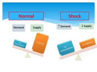
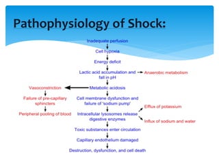
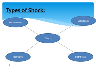
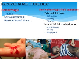
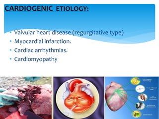
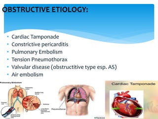
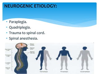
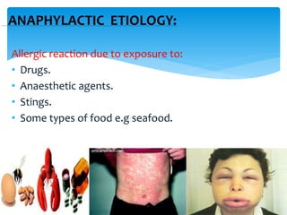
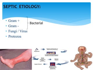
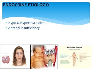
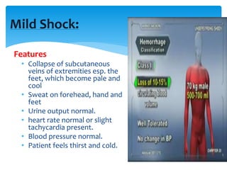
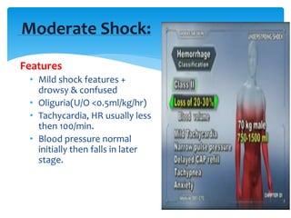
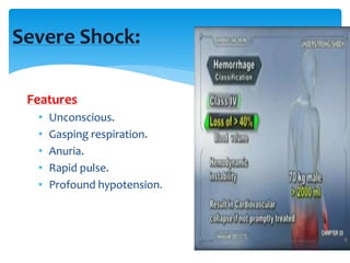
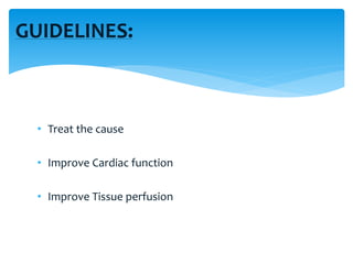
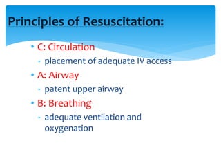
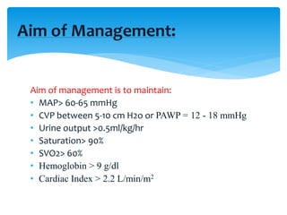
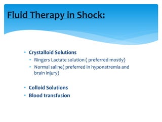
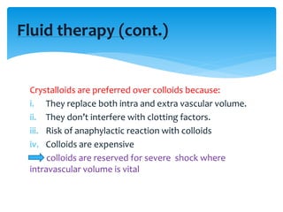
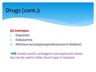
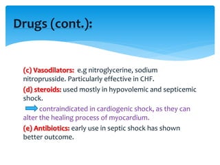
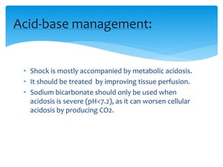
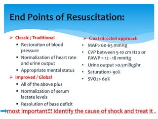
Shock is a state of poor tissue perfusion
with impaired cellular metabolism leading
to serious pathophysiological
abnormality.
Definition:
In other words;
• It’s a condition, in which circulation fails to meet the
metabolic need of the tissue & at the same time fails
to remove the metabolic waste products.
• Usually result of inadequate blood flow and/or
oxygen delivery
• Inadequate peripheral perfusion leading to failure of
tissue oxygenation
Definition (cont.):
aNormal Shock
Pathophysiology of Shock:
• Heart – ↓ CO / hypotension / myocardial depression
• Lung - ↓gas exchange / tachypnoea / pulmonary
edema
• Endocrine – ADH → ↑ reabsorption of water
• CNS – perfusion ↓ – drowsiness
• Blood - Coagulation abnormalities – DIC
• Renal - ↓ GFR - ↓ urine output
• GIT – mucosal ischemia – bleeding & ↑ hepatic
enzyme levels
Effects of Shock on Organs:
Types of Shock:
Shock
obstructive
Cardiogenic
Distributive
Hypovolemic
HYPOVOLAEMIC ETIOLOGY:
Hemorrhagic
Trauma
Gastrointestinal bl.
Retroperitoneal bl. Etc.
Non-Hemorrhagic(Fluid depletion)
 External fluid loss
- Dehydration
- Vomiting
- Diarrhea
Interstitial fluid redistribution
- Thermal injury
- Trauma
- Anaphylaxis
• Valvular heart disease (regurgitative type)
• Myocardial infarction.
• Cardiac arrhythmias.
• Cardiomyopathy
CARDIOGENIC ETIOLOGY:
OBSTRUCTIVE ETIOLOGY:
• Cardiac Tamponade
• Constrictive pericarditis
• Pulmonary Embolism
• Tension Pneumothorax
• Valvular disease (obstructitive type esp. AS)
• Air embolism
NEUROGENIC ETIOLOGY:
• Paraplegia.
• Quadriplegia.
• Trauma to spinal cord.
• Spinal anesthesia.
ANAPHYLACTIC ETIOLOGY:
Allergic reaction due to exposure to:
• Drugs.
• Anaesthetic agents.
• Stings.
• Some types of food e.g seafood.
• Gram +
• Gram -
• Fungi / Virus
• Protozoa
SEPTIC ETIOLOGY:
Bacterial
ENDOCRINE ETIOLOGY:
• Hypo & Hyperthyroidism.
• Adrenal insufficiency.
Clinical Features:
Features of shock depend on the degree of loss
of volume & on duration of shock.
Types
• Mild shock.
• Moderate shock.
• Severe shock.
Mild Shock:
Features
• Collapse of subcutaneous
veins of extremities esp. the
feet, which become pale and
cool
• Sweat on forehead, hand and
feet
• Urine output normal.
• heart rate normal or slight
tachycardia present.
• Blood pressure normal.
• Patient feels thirst and cold.
Moderate Shock:
Features
• Mild shock features +
drowsy & confused
• Oliguria(U/O <0.5ml/kg/hr)
• Tachycardia, HR usually less
then 100/min.
• Blood pressure normal
initially then falls in later
stage.
Severe Shock:
Features
• Unconscious.
• Gasping respiration.
• Anuria.
• Rapid pulse.
• Profound hypotension.
Stages of shock:
• Initial : The cells become leaky and switch to anaerobic
metabolism.
• Non-progressive(compensated stage): Attempt to correct
the metabolic upset of shock.
• Progressive (decompensated stage ): Eventually the
compensation will begin to fail.
• Refractory : Organs fail and the shock can no longer be reversed.
MANAGEMENT
OF
SHOCK
• Blood pressure
• Heart rate
• Pulse- oximetry
• Respiratory rate
• Urine output
• ECG
GENERAL MONITORING:
• PCWP/CVP ( PCWP is considered better guide for fluid
resuscitation than CVP but due to cost and tech.
feasibility generally CVP is preferred)
• Blood gas analysis (metabolic acidosis usu. Present)
• Mixed venous oxygen saturation (SVO2): best guide
for tissue perfusion.
SPECIFIC MONITORING:
GUIDELINES:
• Treat the cause
• Improve Cardiac function
• Improve Tissue perfusion
Principles of Resuscitation:
• C: Circulation
• placement of adequate IV access
• A: Airway
• patent upper airway
• B: Breathing
• adequate ventilation and
oxygenation
Aim of management is to maintain:
• MAP> 60-65 mmHg
• CVP between 5-10 cm H2o or PAWP = 12 - 18 mmHg
• Urine output >0.5ml/kg/hr
• Saturation> 90%
• SVO2> 60%
• Hemoglobin > 9 g/dl
• Cardiac Index > 2.2 L/min/m2
Aim of Management:
Fluid Therapy in Shock:
• Crystalloid Solutions
• Ringers Lactate solution ( preferred mostly)
• Normal saline( preferred in hyponatremia and
brain injury)
• Colloid Solutions
• Blood transfusion
Crystalloids are preferred over colloids because:
i. They replace both intra and extra vascular volume.
ii. They don’t interfere with clotting factors.
iii. Risk of anaphylactic reaction with colloids
iv. Colloids are expensive
colloids are reserved for severe shock where
intravascular volume is vital
Fluid therapy (cont.)
Dynamic Fluid Response:
Infusing 250-500ml of Fluid rapidly in 5 - 10 mts.
• Responders – Improvement
• Transient responders – revert back
• Non – responders
Fluid therapy (cont.):
• Oxygen Carrying Capacity:
• Only RBC contribute to oxygen carrying capacity
(hemoglobin)
• Replacement with all other solutions will
• support volume
• Improve end organ perfusion
• Will Not provide additional oxygen carrying capacity
(a) Vasopressors:
• Phenylephrine
• Ephedrine
• Nor epinephrine
• Mephenteramine
• Vasopressin
• adrenaline
Drugs:
Vasopressors are more effective if
sympathetic blockade is present
therefore they are useful in:
1.Neurogenic shock: Phenylephrine is
preferred
2.Spinal hypotension: Ephedrine is
preferred
3.Septicemic shock: Nor epinephrine is
preferred
(b) Inotropes:
i. Dopamine
ii. Dobutamine
iii. Milrinone lactate(phosphodiesterase iii inhibitor)
mostly used in cardiogenic and septicemic shock,
but can be used in other shock types if required.
Drugs (cont.):
(c) Vasodilators: e.g nitroglycerine, sodium
nitroprusside. Particularly effective in CHF.
(d) steroids: used mostly in hypovolemic and septicemic
shock.
contraindicated in cardiogenic shock, as they can
alter the healing process of myocardium.
(e) Antibiotics: early use in septic shock has shown
better outcome.
Drugs (cont.):
• Shock is mostly accompanied by metabolic acidosis.
• It should be treated by improving tissue perfusion.
• Sodium bicarbonate should only be used when
acidosis is severe (pH<7.2), as it can worsen cellular
acidosis by producing CO2.
Acid-base management:
End Points of Resuscitation:
 Classic / Traditional
 Restoration of blood
pressure
 Normalization of heart rate
and urine output
 Appropriate mental status
 Improved / Global
 All of the above plus
 Normalization of serum
lactate levels
 Resolution of base deficit
 Goal directed approach
• MAP> 60-65 mmHg
• CVP between 5-10 cm H2o or
PAWP = 12 - 18 mmHg
• Urine output >0.5ml/kg/hr
• Saturation> 90%
• SVO2> 60%
most important!!! Identify the cause of shock and treat it .
THANK
YOU

Shock its types and management

  • 1.
    SHOCK, Types & Management Moderator:Dr. Mubasher Ahmad Asst. professor dept. of anaesthesiology & critical care GMC & associated hospitals, srinagar Speaker: Dr.Faisal Rasool Post graduate scholar (1st year) anaesthesiology & critical care
  • 2.
    • Definition • Reviewbasic physiologic aspects of shock • Different categories with Etiology &Clinical features • Management aspects Objectives:
  • 3.
    Shock is astate of poor tissue perfusion with impaired cellular metabolism leading to serious pathophysiological abnormality. Definition:
  • 4.
    In other words; •It’s a condition, in which circulation fails to meet the metabolic need of the tissue & at the same time fails to remove the metabolic waste products. • Usually result of inadequate blood flow and/or oxygen delivery • Inadequate peripheral perfusion leading to failure of tissue oxygenation Definition (cont.):
  • 5.
  • 6.
  • 7.
    • Heart –↓ CO / hypotension / myocardial depression • Lung - ↓gas exchange / tachypnoea / pulmonary edema • Endocrine – ADH → ↑ reabsorption of water • CNS – perfusion ↓ – drowsiness • Blood - Coagulation abnormalities – DIC • Renal - ↓ GFR - ↓ urine output • GIT – mucosal ischemia – bleeding & ↑ hepatic enzyme levels Effects of Shock on Organs:
  • 9.
  • 10.
    HYPOVOLAEMIC ETIOLOGY: Hemorrhagic Trauma Gastrointestinal bl. Retroperitonealbl. Etc. Non-Hemorrhagic(Fluid depletion)  External fluid loss - Dehydration - Vomiting - Diarrhea Interstitial fluid redistribution - Thermal injury - Trauma - Anaphylaxis
  • 11.
    • Valvular heartdisease (regurgitative type) • Myocardial infarction. • Cardiac arrhythmias. • Cardiomyopathy CARDIOGENIC ETIOLOGY:
  • 12.
    OBSTRUCTIVE ETIOLOGY: • CardiacTamponade • Constrictive pericarditis • Pulmonary Embolism • Tension Pneumothorax • Valvular disease (obstructitive type esp. AS) • Air embolism
  • 13.
    NEUROGENIC ETIOLOGY: • Paraplegia. •Quadriplegia. • Trauma to spinal cord. • Spinal anesthesia.
  • 14.
    ANAPHYLACTIC ETIOLOGY: Allergic reactiondue to exposure to: • Drugs. • Anaesthetic agents. • Stings. • Some types of food e.g seafood.
  • 15.
    • Gram + •Gram - • Fungi / Virus • Protozoa SEPTIC ETIOLOGY: Bacterial
  • 16.
    ENDOCRINE ETIOLOGY: • Hypo& Hyperthyroidism. • Adrenal insufficiency.
  • 17.
    Clinical Features: Features ofshock depend on the degree of loss of volume & on duration of shock. Types • Mild shock. • Moderate shock. • Severe shock.
  • 18.
    Mild Shock: Features • Collapseof subcutaneous veins of extremities esp. the feet, which become pale and cool • Sweat on forehead, hand and feet • Urine output normal. • heart rate normal or slight tachycardia present. • Blood pressure normal. • Patient feels thirst and cold.
  • 19.
    Moderate Shock: Features • Mildshock features + drowsy & confused • Oliguria(U/O <0.5ml/kg/hr) • Tachycardia, HR usually less then 100/min. • Blood pressure normal initially then falls in later stage.
  • 20.
    Severe Shock: Features • Unconscious. •Gasping respiration. • Anuria. • Rapid pulse. • Profound hypotension.
  • 21.
    Stages of shock: •Initial : The cells become leaky and switch to anaerobic metabolism. • Non-progressive(compensated stage): Attempt to correct the metabolic upset of shock. • Progressive (decompensated stage ): Eventually the compensation will begin to fail. • Refractory : Organs fail and the shock can no longer be reversed.
  • 22.
  • 23.
    • Blood pressure •Heart rate • Pulse- oximetry • Respiratory rate • Urine output • ECG GENERAL MONITORING:
  • 24.
    • PCWP/CVP (PCWP is considered better guide for fluid resuscitation than CVP but due to cost and tech. feasibility generally CVP is preferred) • Blood gas analysis (metabolic acidosis usu. Present) • Mixed venous oxygen saturation (SVO2): best guide for tissue perfusion. SPECIFIC MONITORING:
  • 25.
    GUIDELINES: • Treat thecause • Improve Cardiac function • Improve Tissue perfusion
  • 26.
    Principles of Resuscitation: •C: Circulation • placement of adequate IV access • A: Airway • patent upper airway • B: Breathing • adequate ventilation and oxygenation
  • 27.
    Aim of managementis to maintain: • MAP> 60-65 mmHg • CVP between 5-10 cm H2o or PAWP = 12 - 18 mmHg • Urine output >0.5ml/kg/hr • Saturation> 90% • SVO2> 60% • Hemoglobin > 9 g/dl • Cardiac Index > 2.2 L/min/m2 Aim of Management:
  • 28.
    Fluid Therapy inShock: • Crystalloid Solutions • Ringers Lactate solution ( preferred mostly) • Normal saline( preferred in hyponatremia and brain injury) • Colloid Solutions • Blood transfusion
  • 29.
    Crystalloids are preferredover colloids because: i. They replace both intra and extra vascular volume. ii. They don’t interfere with clotting factors. iii. Risk of anaphylactic reaction with colloids iv. Colloids are expensive colloids are reserved for severe shock where intravascular volume is vital Fluid therapy (cont.)
  • 30.
    Dynamic Fluid Response: Infusing250-500ml of Fluid rapidly in 5 - 10 mts. • Responders – Improvement • Transient responders – revert back • Non – responders Fluid therapy (cont.):
  • 31.
    Fluid therapy (cont.): •Oxygen Carrying Capacity: • Only RBC contribute to oxygen carrying capacity (hemoglobin) • Replacement with all other solutions will • support volume • Improve end organ perfusion • Will Not provide additional oxygen carrying capacity
  • 32.
    (a) Vasopressors: • Phenylephrine •Ephedrine • Nor epinephrine • Mephenteramine • Vasopressin • adrenaline Drugs: Vasopressors are more effective if sympathetic blockade is present therefore they are useful in: 1.Neurogenic shock: Phenylephrine is preferred 2.Spinal hypotension: Ephedrine is preferred 3.Septicemic shock: Nor epinephrine is preferred
  • 33.
    (b) Inotropes: i. Dopamine ii.Dobutamine iii. Milrinone lactate(phosphodiesterase iii inhibitor) mostly used in cardiogenic and septicemic shock, but can be used in other shock types if required. Drugs (cont.):
  • 34.
    (c) Vasodilators: e.gnitroglycerine, sodium nitroprusside. Particularly effective in CHF. (d) steroids: used mostly in hypovolemic and septicemic shock. contraindicated in cardiogenic shock, as they can alter the healing process of myocardium. (e) Antibiotics: early use in septic shock has shown better outcome. Drugs (cont.):
  • 35.
    • Shock ismostly accompanied by metabolic acidosis. • It should be treated by improving tissue perfusion. • Sodium bicarbonate should only be used when acidosis is severe (pH<7.2), as it can worsen cellular acidosis by producing CO2. Acid-base management:
  • 36.
    End Points ofResuscitation:  Classic / Traditional  Restoration of blood pressure  Normalization of heart rate and urine output  Appropriate mental status  Improved / Global  All of the above plus  Normalization of serum lactate levels  Resolution of base deficit  Goal directed approach • MAP> 60-65 mmHg • CVP between 5-10 cm H2o or PAWP = 12 - 18 mmHg • Urine output >0.5ml/kg/hr • Saturation> 90% • SVO2> 60% most important!!! Identify the cause of shock and treat it .
  • 37.