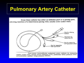
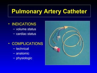
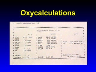
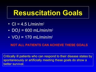
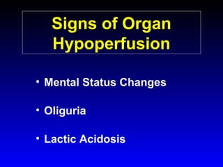
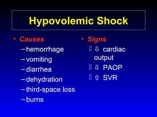
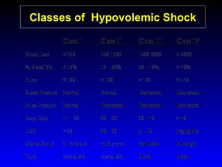
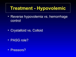
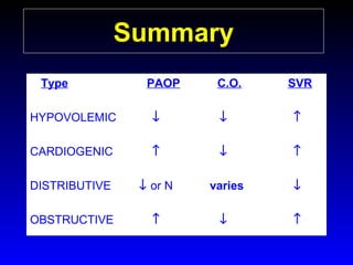
This document provides an overview of shock and hemodynamic monitoring using a pulmonary artery catheter. It defines shock as inadequate organ perfusion and outlines the main categories of shock: hypovolemic, cardiogenic, distributive, and obstructive. It discusses goals of fluid resuscitation in shock and reviews hemodynamic parameters measured by a pulmonary artery catheter such as cardiac output, vascular resistances, and oxygen transport variables. The document uses case studies to demonstrate how these parameters can be utilized to diagnose and guide treatment in different shock states.
This document discusses the basic cardiac physiology used to manage critically ill patients, including determinants of mean arterial pressure, cardiac output, and oxygen delivery. It describes how these concepts are used to diagnose different types of shock. Monitoring of central venous pressure is discussed as well as equations relating mean arterial pressure, stroke volume, heart rate, and systemic vascular resistance. The importance of oxygen delivery to tissues and maintaining an optimal venous oxygen saturation is emphasized.
Haemodynamic data can be acquired in many ways. However we obtain the raw data we still have a big problem…
What do all these figures mean? How can we put it all together to help our patients?
Associate Professor Brendan E. Smith.
School of Biomedical Science, Charles Sturt University,
Specialist in Anaesthesia and Intensive Care, Bathurst Base Hospital, Bathurst, NSW, Australia
This document discusses the use of hemodynamic monitoring to guide fluid administration and optimize stroke volume. It notes that physical exam signs are often inaccurate indicators of a patient's clinical status. Various methods for measuring stroke volume are presented, with esophageal Doppler monitoring found to be accurate, easy to use, and supported by evidence from randomized controlled trials showing it can reduce length of hospital stay. The document advocates using changes in stroke volume in response to fluids or medications to guide treatment to optimize preload and cardiac function.
The USCOM 1A is a non-invasive device for accurate measurement and monitoring of circulation. USCOM allows simple measurement of preload, contractility and afterload for goal direction of therapy, and accurate monitoring of post intervention changes, taking advanced haemodynamics beyond the ICU.
USCOM The measure of life!
This document discusses cardiac resynchronization therapy (CRT) for heart failure patients. Some key points:
- CRT improves symptoms, exercise tolerance, quality of life and reduces mortality for selected heart failure patients.
- Non-response to CRT remains a problem, occurring in 30-45% of patients.
- Patient selection factors like QRS duration, bundle branch block pattern and degree of ventricular dyssynchrony impact response.
- Optimal lead placement and device programming are important for response. Follow-up optimization of atrioventricular and interventricular delays can improve outcomes.
Physiology of hemodynamics & PiCCO parameters in detailmeducationdotnet
The document discusses physiology of hemodynamics and PiCCO parameters. It begins by stating the goal of intensive care medicine is ensuring adequate oxygenation of organs and tissues. It then provides details on cardiac output, factors that influence it like preload and contractility, and how cardiac output relates to oxygen delivery. It emphasizes the importance of monitoring parameters like cardiac output, hemoglobin, arterial oxygen saturation, and mixed or central venous oxygen saturation to assess oxygen delivery and consumption.
1) Pressure tracing of the left ventricle involves using fluid-filled catheters connected to pressure transducers to record intracardiac pressures. Factors such as catheter size, damping, and natural frequency determine recording quality.
2) Sources of error in pressure measurements include catheter whip, end-pressure artifacts, and deterioration of frequency response. Invasive monitoring is useful for evaluating conditions like aortic stenosis and mitral stenosis when noninvasive data is discrepant or unclear.
3) Distinguishing constrictive pericarditis from restrictive cardiomyopathy involves assessing ventricular interdependence, pulmonary pressures, and left ventricular pressure tracings. Hemodynamic data aids in diagnosis and management of many conditions.
1) The document discusses choosing cardiac output monitoring devices for peri-operative and ICU settings. It considers devices' reliability with changing vascular resistance and ability to provide useful clinical information.
2) For peri-operative monitoring of high-risk surgical patients, less invasive devices using uncalibrated pulse contour analysis like Vigileo and Clearsight may be suitable when vascular resistance does not change significantly.
3) For ICU patients receiving vasopressors where resistance changes greatly, more reliable thermodilution methods like PiCCO, EV1000 and pulmonary artery catheter are recommended to measure cardiac output and assess ventricular function.
The document discusses acute right ventricular (RV) failure, including:
1) The RV's main job is to maintain low right atrial pressure to optimize venous return to the heart. RV dysfunction can lead to reduced cardiac output.
2) Treatment for RV failure differs from left ventricular failure - RV failure may require fluid administration while left sided failure uses diuretics.
3) RV infarction is associated with worse outcomes than left ventricular infarction such as higher mortality, and requires a tailored treatment approach including fluid administration in some cases rather than diuretics. Early revascularization can help recovery.
Monitoring of physiologic variables is an integral part of caring for critically ill patients. Debate exists regarding the usefulness and safety of invasive hemodynamic monitoring in the intensive care unit. Several studies have shown improved outcomes with hemodynamic monitoring in high-risk surgical patients, but evidence is conflicting for medical patients. PiCCO monitoring combines transpulmonary thermodilution and pulse contour analysis to continuously measure cardiac output and other hemodynamic parameters noninvasively. It provides useful information to guide fluid management and assess fluid responsiveness.
I. Dr. Badamali discusses hemodynamic monitoring of critically ill patients to improve outcomes. He outlines key equations related to cardiac output, oxygen delivery, and blood pressure.
II. Four goals of hemodynamic optimization are discussed: improving blood pressure, cardiac output, regional blood flow, and microcirculation. Dynamic parameters like stroke volume variation and passive leg raising can predict fluid responsiveness.
III. Evidence from studies like Rivers et al. show that early goal-directed therapy integrating hemodynamic monitoring into a treatment protocol can reduce mortality from sepsis. Goals must be part of an organized resuscitation and care program to successfully impact outcomes.
1. Clinical examination alone is not sufficient to assess hemodynamic status in critically ill patients as individual vital signs do not reflect overall status.
2. Arterial lines can be used to monitor blood pressure, heart rate, and derive parameters like cardiac output but waveforms require interpretation and may be affected by various artifacts.
3. Pulmonary artery catheters can measure central venous and pulmonary artery pressures as well as cardiac output but have potential complications and their use remains controversial with no proven benefits shown in large trials.
This document discusses hemodynamic monitoring techniques to guide treatment for patients with hemodynamic failure. It describes using the PiCCO device to noninvasively monitor parameters such as cardiac output, lung water content, contractility, and fluid responsiveness to determine whether a patient needs vasopressors, volume expansion, inotropes, and when to stop fluid administration. Tests like pulse pressure variation, end-expiratory occlusion, and passive leg raising used with continuous cardiac output monitoring can predict fluid responsiveness, while extravascular lung water and pulmonary vascular permeability index measurements help avoid excessive fluid loading and lung injury.
The document discusses various techniques for hemodynamic monitoring, including both conventional and advanced methods. It provides an overview of the history of hemodynamic monitoring and outlines some of the goals of different monitoring devices. The document then reviews several specific monitoring techniques, such as arterial lines, central venous catheters, pulmonary artery catheters, echocardiography, pulse contour analysis, and electrical bioimpedance. Both advantages and disadvantages of each method are discussed.
This document provides an overview of hemodynamic monitoring principles in the ICU. It discusses the steps for assessing global and regional perfusion, including initial clinical assessment and basic monitoring of vital signs and lactate levels. It then covers monitoring of preload and fluid responsiveness, methods for measuring cardiac output, assessing cardiac contractility and tissue perfusion. A variety of invasive and non-invasive monitoring techniques are explained, from pulmonary artery catheters and arterial waveform analysis to echocardiography, near-infrared spectroscopy and analysis of the microcirculation. Key principles emphasized are that no single monitor determines outcomes, and monitoring needs may change over time based on equipment and integrating multiple variables.
This document discusses hemodynamic monitoring of patients during acute respiratory distress syndrome (ARDS). It covers hemodynamic interactions between the lungs and heart during spontaneous breathing and mechanical ventilation. Positive pressure ventilation decreases venous return and right ventricular stroke volume while increasing pressures. It also discusses ventricular interdependence and the effects of PEEP and prone positioning. Tools for hemodynamic monitoring discussed include assessing afterload, contractility, the right ventricle, perfusion, preload, and preload responsiveness. Considerations for different monitoring tools like echocardiography, pulmonary artery catheters, and arterial pulse-wave analysis are provided.
This document provides an introduction to hemodynamic monitoring, which involves measuring factors that influence blood flow and pressure. It defines hemodynamic monitoring and outlines its purposes, which include diagnosing and managing shock states, determining fluid status, and measuring cardiac output. The document discusses indications for hemodynamic monitoring as well as contraindications for invasive pulmonary artery catheters. It also reviews important hemodynamic values and concepts, pulmonary artery catheter insertion and positioning, waveform analysis, and removal of pulmonary artery catheters.
Hemodynamic monitoring involves measuring and monitoring the factors that influence blood flow and pressure in the body. It is concerned with five main areas: the right heart, lungs, left heart, fluid status, and blood pressures. Normal hemodynamic values include a central venous pressure of 2-6 mmHg, pulmonary artery pressure of 6-15 mmHg, pulmonary capillary wedge pressure of 6-12 mmHg, and a cardiac output of 4-8 L/min. Treatment for different types of shock depends on the underlying cause but may include inotropes, vasopressors, antibiotics, and fluid resuscitation.
Inotropy Index accurately predicts fluid responsiveness in volume resuscitation.
Brendan E. Smith and Veronica M. Madigan
School of Biomedical Science,
Charles Sturt University, Bathurst, NSW, Australia.
Specialist in Anaesthesia and Intensive Care,
Bathurst Base Hospital, Bathurst, NSW, Australia.
This document discusses various hemodynamic parameters and waveforms seen in different cardiac conditions:
- It compares pressures, waveforms and compliance in conditions like constrictive pericarditis, right ventricular failure, cardiac tamponade and pulmonary hypertension.
- Key waveforms and pressures are described for valvular lesions like mitral stenosis, aortic stenosis, mitral regurgitation and aortic regurgitation.
- Equations for calculating cardiac output, shunt fractions and valvular areas are provided.
- Diagnostic criteria for pseudosevere versus true severe aortic stenosis on stress echocardiography are outlined.
- A case example is presented of a patient evaluated for pulmonary
Critically Appraised Topic: Fluid Loading in Right Ventricular InfarctionMoneer Basalyous
Three key points:
1) Volume loading has variable effects on patients with right ventricular myocardial infarction (RVMI) and hypotension based on 7 clinical studies. It had no effect in 5 studies, a modest effect in 1 study, and an effect within limits in 1 study.
2) Reviews recommend an initial trial of volume loading for RVMI patients with low output and no pulmonary congestion, targeting a CVP below 15 mmHg, but avoiding excessive volume that could reduce preload.
3) The conclusion is that RVMI patients are sensitive to volume changes. An initial fluid challenge is reasonable for hypotensive patients with low CVP and no congestion, but invasive monitoring may be needed if no response.
1) Hemodynamic monitoring involves measuring the cardiovascular system to describe its performance, with the goal of oxygen delivery. While monitoring provides data, it does not constitute therapy on its own.
2) There are various methods of hemodynamic monitoring, including non-invasive and invasive techniques. Invasive methods such as arterial lines, central venous catheters, and pulmonary artery catheters allow for more continuous monitoring but carry greater risks.
3) Shock is a state of end-organ dysfunction due to inadequate tissue perfusion and oxygen delivery. It can be classified as hypovolemic, cardiogenic, distributive, or obstructive. Goal-directed resuscitation aims to first restore intravascular volume, support
Cardiac catheterization is useful for assessing left-to-right shunts through three main techniques: oximetry runs to detect oxygen saturation step-ups, indicator dye dilution to detect early recirculation of dye injected into the proximal chamber, and angiocardiography to directly visualize the anatomic site of the shunt. While oximetry is best to localize the shunt, dye dilution can detect smaller shunts and angiography confirms anatomy. Together these techniques allow diagnosis and quantification of left-to-right intracardiac shunts.
Basic hemodynamic principles viewed through pressure volume relationsInsideScientific
The goal of this webinar is to provide an overview of the fundamental principles of preload, afterload, contractility and lusitropy (diastolic properties), how these are quantified on the pressure-volume diagram, and how they are affected in heart failure. Links are made to underlying properties of cardiac muscle and ventricular structure. After establishing basic concepts, it will be demonstrated how pressure-volume analysis can lead to a quantitative understanding of how heart and vasculature interact to determine stroke volume, cardiac output and blood pressure. The implications for understanding therapeutic effects will also be discussed.
Key Topics:
- Preload, Afterload, Contractility and Lusitropy
- Cardiac Muscle and Ventricular Structure
- Understanding Heart-Vasculature Interactions
- PV Loops in Heart Failure
- Understanding Therapies and Their Effects on Cardiac Pump Performance
Robert Koch identified Mycobacterium tuberculosis in 1882 and received the Nobel Prize for this discovery. In 1906, Calmette and Guerin developed the BCG vaccine for tuberculosis. The vaccine was first used in humans in 1921 in France but did not become widely used in places like the US, UK, and Germany until after World War II. Tuberculosis is caused by the bacterium M. tuberculosis and spreads through airborne droplets when infected people cough, sneeze, or talk. It affects mostly the lungs but can spread to other organs, and if left untreated it can be fatal.
Hodgkin's lymphoma is characterized by the presence of Reed-Sternberg cells. It is classified into five subtypes based on cell morphology. Hodgkin's lymphoma most often presents as painless enlargement of single or adjacent lymph nodes and commonly spreads in an orderly fashion. Investigations include CBC, ESR, LDH, imaging and bone marrow biopsy. Treatment involves chemotherapy such as ABVD or MOPP regimens depending on disease stage and prognosis. Late effects of treatment can include leukemia, second malignancies, and organ damage.
Meningitis is an inflammation of the membranes covering the brain and spinal cord caused by bacterial or viral infections. The two most common types of bacterial meningitis are caused by Neisseria meningitidis (meningococcal meningitis) and Streptococcus pneumoniae (pneumococcal meningitis). Viruses from the enterovirus group are the most common cause of viral meningitis. Symptoms of meningitis include fever, headache, stiff neck, and sensitivity to light. While bacterial meningitis requires intravenous antibiotics, viral meningitis is usually treated with supportive care. Handwashing is the best way to prevent transmission between people through contact with nose and throat secretions.
This document provides information on normal pressure hydrocephalus (NPH), including its causes, symptoms, diagnosis, treatment with shunt surgery, and patient outcomes. It discusses how NPH presents with a triad of gait disturbance, urinary incontinence, and dementia. Diagnostic tests include brain imaging and CSF flow studies. Patients with mild symptoms and gait issues are most likely to benefit from shunt surgery, while those with significant dementia tend to show little improvement. However, long-term outcomes of shunt surgery are variable, with benefits often declining over time.
This document summarizes several presentations on recent developments in Hodgkin lymphoma. It begins with a discussion of a study comparing the predictive value of interim PET scans done after one or two cycles of therapy. The study found PET scans after one cycle had a higher negative predictive value. Another section summarizes a trial finding patients with low stage disease and a negative PET scan after 3 cycles of ABVD had excellent prognosis without further treatment. Later sections discuss outcomes of combining brentuximab vedotin with standard therapies for relapsed/refractory and advanced stage disease. Results showed improved survival correlated with response to brentuximab vedotin.
Iron deficiency anemia is caused by a lack of iron in the body. Common symptoms include fatigue, palpitations, tinnitus, and headaches. Diagnosis involves blood tests showing low iron levels and microcytic, hypochromic red blood cells. Treatment depends on the severity, and involves oral or intravenous iron supplements to replenish iron stores over 6-12 months. Parenteral iron is used for severe cases or those unable to tolerate oral iron.
This document discusses osteomyelitis, an infection of bone tissue. It defines osteomyelitis and classifies it as either acute or chronic, and by mechanism and host response. For acute hematogenous osteomyelitis, it describes the typical microbial patterns, diagnosis process using tests, imaging and scans, and treatment approach involving antibiotics and sometimes surgery. For chronic osteomyelitis, it discusses the compromised soft tissue and bone involvement, classification, diagnosis through clinical and imaging evaluation, and focus on surgical treatment including debridement along with antibiotic therapy.
This document summarizes key concepts relating to cell injury, adaptation, and death. It discusses how cells respond to stress through reversible or irreversible injury, adaptation, or death. It also describes various types of cellular adaptation like hypertrophy, hyperplasia, atrophy, and metaplasia that cells undergo in response to stress. Additionally, it covers the mechanisms and morphological changes associated with different types of cell injury like necrosis and apoptosis.
A patient named Mary Jacob Chiyedath, a first year MSc Nursing student, underwent a CT and MRI scan that showed a meningioma with mass effect. She then had a craniotomy procedure to excise the meningioma, and the excised tissue was sent for histopathological examination.
1) Tuberculosis remains a major global health problem, infecting around 1/3 of the world's population and causing millions of deaths each year, especially in developing countries.
2) Treatment involves a combination of antibiotics over a long period of time to prevent drug resistance, with first-line drugs like isoniazid and rifampin being most effective but also posing toxicity risks.
3) Control efforts face challenges from factors like poverty, HIV co-infection, and the emergence of drug-resistant strains, but expanded treatment programs could prevent over 200 million infections and 35 million deaths by 2020.
Meningioma es un tumor benigno del cerebro que se origina en las células meningoteliales de la aracnoides. Crecen lentamente y se unen a la duramadre, comprimiendo el cerebro y causando síntomas como cefalea. Son más comunes en mujeres y se asocian con otros tumores como schwannomas. La mayoría tienen mutaciones en el gen NF2 en el cromosoma 22.
This document provides an introduction to pathology. It defines pathology as the scientific study of disease and discusses its main branches of general pathology and systemic pathology. It also outlines several key techniques used in pathology like microbiologic, molecular, immunologic, and morphologic analysis. The document then discusses key aspects of the disease process including etiology, pathogenesis, clinical manifestations, and molecular and morphologic changes. It provides examples of different types of etiologies and explains key pathology concepts such as reversible and irreversible cell injury, necrosis, apoptosis, and intracellular accumulations.
This document defines massive transfusion as replacing one blood volume or more within 24 hours, which corresponds to approximately 10 units of blood for a 70 kg adult. Massive transfusion can cause numerous complications including dilution coagulopathies, hypothermia, acidosis, and tissue hypoxia. The overall mortality for patients requiring massive transfusion is around 40% but increases to over 75% for those who develop hemostatic disorders. Proper use of massive transfusion protocols which rapidly provide blood products can help minimize complications and reduce mortality rates.
This document discusses viral oncogenesis, or how viruses can cause cancer. It defines oncogenes as viral genes that can cause tumors and proto-oncogenes as normal cellular genes that viruses can modify to become oncogenes. It describes the two main types of oncogenic viruses as DNA viruses and RNA viruses, listing some examples of each. For RNA viruses, it explains that all oncogenic viruses are retroviruses and describes the general life cycle of retroviruses, from entering the cell to replicating and exiting. The document concludes that while not all retroviruses contain viral oncogenes, they can integrate near proto-oncogenes and modulate host cell growth to cause cancer.
Benign tumors called meningiomas originate from cells of the arachnoid layer of the meninges and are attached to the dura mater. They commonly present as well-defined masses compressing the brain but can be easily separated. Multiple meningiomas, acoustic schwannomas, and gliomas may indicate neurofibromatosis type 2, while around 50% of solitary meningiomas have a mutation in the 22q gene. Meningiomas demonstrate distinct histological patterns including syncytial, fibroblastic, transitional, psammomatous, and secretory variants.
This document discusses hemodynamic disorders and edema. It begins by defining the normal composition of body water and the three body compartments it is contained in. It then defines edema as excess fluid in the interstitial tissue space and describes different types of edema based on location. The pathophysiology of edema involves either increased hydrostatic pressure or reduced plasma osmotic pressure. Specific causes of edema are discussed like congestive heart failure, liver disease, and malnutrition. The document also covers morphology, hemorrhage, congestion, hemostasis, and hemorrhagic disorders.
1. Hemodynamic disorders involve changes in intravascular volume, pressure, or protein content that affect fluid movement across vessel walls and can cause edema, hyperemia, congestion, hemorrhage, thrombosis, embolism, infarction, or shock.
2. Edema is increased fluid in tissues, caused by increased hydrostatic pressure, reduced plasma proteins, lymphatic obstruction, sodium retention, or inflammation.
3. Thrombosis is inappropriate blood clot formation from endothelial injury, blood stasis, or hypercoagulability per Virchow's triad, and thrombi can embolize or organize.
4. Embolism occurs when a detached mass is
meningioma tumors presentation include definition, causes, symptoms, and treatment options
prepared by Abbas Wael Abbas
supervised by Dr Jawad Ziyadah ( neurosurgeon)
1. Programmed cell death (PCD) refers to regulated cell death processes that eliminate cells when they are no longer useful or potentially harmful.
2. Apoptosis is a form of PCD characterized by nuclear fragmentation, chromatin condensation, cell shrinkage and blebbing, and formation of apoptotic bodies that are quickly phagocytosed, avoiding inflammation. It relies on caspase activation through the mitochondrial or death receptor pathways.
3. Alternative cell death pathways include necroptosis, pyroptosis and autophagy. Necroptosis resembles necrosis morphologically but is caspase-independent and regulated. Pyroptosis involves caspase-1 and occurs during microbial infection, triggering inflammation. Autophagy
This document provides an overview of shock and hemodynamic monitoring. It defines shock as inadequate organ perfusion and classifies shock into four categories: hypovolemic, cardiogenic, distributive, and obstructive. For each category, it describes the pathophysiology, signs, and treatment approach. It also discusses goals of fluid resuscitation and introduces concepts of oxygen delivery, consumption, and tools like pulmonary artery catheters that are used to monitor hemodynamics and guide shock management. Vasopressor and inotropic agents commonly used to treat shock like dopamine, norepinephrine, and epinephrine are also outlined.
This document provides information on different types of shock, including definitions, pathophysiology, stages, types, treatments, and case examples. It defines shock as a state of inadequate tissue perfusion and oxygen delivery. The main types discussed are hypovolemic, cardiogenic, distributive (septic), and obstructive shock. It outlines the stages of shock from compensated to decompensated to irreversible. Case examples are provided to demonstrate how to identify the type of shock based on presenting signs and symptoms. Initial treatment approaches focus on oxygenation, ventilation, fluid resuscitation and vasoactive drugs. Prognosis depends on the cause, with septic shock having higher mortality.
This document provides an overview of shock and its pathophysiology. It defines shock as a clinical syndrome resulting from inadequate tissue perfusion due to alterations in circulation. The stages of shock are described as compensatory, progressive, and irreversible. Compensatory mechanisms aimed at maintaining homeostasis in response to shock are discussed for various body systems. Nursing interventions for shock focus on treating its underlying cause, restoring circulating volume and hemodynamics through fluid resuscitation and vasoactive drugs, and minimizing oxygen consumption.
1. Shock is defined as a systemic state of low tissue perfusion that is inadequate for normal cellular respiration. It occurs when there is insufficient delivery of oxygen and glucose to cells, causing cells to switch from aerobic to anaerobic metabolism. If perfusion is not restored, cell death ensues.
2. The main types of shock are hypovolemic, cardiogenic, obstructive, distributive, and endocrine shock. Hypovolemic shock, the most common type, is caused by blood or fluid loss. Cardiogenic shock results from cardiac dysfunction that reduces cardiac output.
3. The goals of shock resuscitation are to increase oxygen delivery, decrease oxygen demand, improve cardiac
This document discusses shock, including its definition, pathophysiology, stages, types (hypovolemic, distributive, cardiogenic), and management. Shock is defined as inadequate tissue perfusion with oxygenated blood. It outlines the initial, compensatory, progressive, and irreversible stages of shock. Hypovolemic shock is the most common type in trauma patients and results from blood or fluid loss. Initial fluid resuscitation for trauma patients in hemorrhagic shock consists of 2 L of isotonic saline as rapidly as possible. Ongoing fluid resuscitation is guided by monitoring the patient's response and signs of end organ perfusion. Blood transfusion may be needed for patients who are transient or non
This document defines and discusses the pathophysiology of different types of shock: cardiogenic, obstructive, hypovolemic, and distributive. It notes that shock occurs when there is inadequate perfusion and oxygenation of cells, leading to cellular and organ dysfunction. The key signs of shock include tachycardia, hypotension, altered mental status, and decreased urine output. Early goal-directed resuscitation is important to prevent end organ damage and death, and should focus on airway management, oxygenation, fluid resuscitation, and treating the underlying cause.
This document discusses shock, sepsis management, and fluid resuscitation. It addresses:
1) Types of shock including hypovolemic, distributive, obstructive, cardiogenic, and neurogenic.
2) Principles of fluid resuscitation including increasing preload to improve stroke volume and cardiac output. However, fluid boluses only improve cardiac output in about 50% of ICU patients.
3) Dynamic measures like pulse pressure variation, stroke volume variation, passive leg raise, and echocardiography changes after fluid bolus are better than static measures at predicting fluid responsiveness.
Pediatric shock can be septic, cardiogenic, hypovolemic, distributive, or obstructive based on the underlying pathophysiology. Septic shock can present as warm shock with increased cardiac output and decreased systemic vascular resistance or cold shock with decreased cardiac output. Early recognition and treatment is key to managing shock. Treatment involves fluid resuscitation, inotropic support, and addressing the underlying cause of shock according to institutional guidelines. Differentiating between septic and cardiogenic shock involves clinical assessment of signs and symptoms as well as generating a therapeutic plan.
Cardiogenic shock is a low cardiac output state resulting from inadequate tissue perfusion despite adequate left ventricular filling pressures. It is usually caused by acute myocardial infarction which accounts for about 80% of cases. Clinically, it is defined by sustained hypotension with signs of hypoperfusion and a systolic blood pressure less than 90 mmHg for at least 30 minutes or the need for vasopressor/inotropic support. The mortality rate for cardiogenic shock remains high at over 80% despite advances in management. Early diagnosis and aggressive treatment including revascularization, inotropic support, and mechanical circulatory support are aimed at improving outcomes.
This document provides an overview of shock, including its definition, types, physiology, and management. It discusses the key features and immediate treatment of hemorrhagic, neurogenic, septic, anaphylactic, cardiogenic, and obstructive shock through case examples. The main points are that shock results from inadequate tissue perfusion, early recognition and aggressive fluid/vasopressor resuscitation are critical to improving outcomes across different shock types.
Shock is characterized by a systemic reduction in tissue perfusion resulting in decreased oxygen delivery. There are four main types of shock: hypovolemic, cardiogenic, obstructive, and distributive. The goals of resuscitation are to increase oxygen delivery and decrease demand. Treatment involves establishing IV access, fluid resuscitation, vasopressors, inotropes, antibiotics for infection, and treating the underlying cause. Endpoints of resuscitation include restoration of blood pressure, normalization of heart rate, urine output, lactate levels, and mental status.
Shock its pathopysiology and managementSHAKIL JAWED
This document discusses shock, including definitions, types, causes, pathophysiology, diagnosis, and treatment. It defines shock as a state of low tissue perfusion from inadequate oxygen and glucose delivery. The main types of shock discussed are hypovolemic, cardiogenic, obstructive, distributive, and septic shock. Treatment involves identifying and treating the underlying cause, restoring circulating blood volume and tissue perfusion through fluid resuscitation, and providing vasopressor support if needed to maintain blood pressure. Goals of resuscitation are optimizing oxygen delivery while avoiding fluid overload.
The document discusses the three fluid compartments in the body, the mechanisms of osmosis, different types of shock, water and electrolyte disorders including hyponatremia, and case examples to illustrate concepts such as hypotonic hyponatremia and hyponatremic encephalopathy. It provides an overview of fluid, electrolyte, and shock physiology for medical students and residents.
This document provides an overview of fluids, electrolytes and shock. It discusses the three fluid compartments in the body, the three membranes that regulate fluid movement, and the principles of osmosis. It describes the causes and types of shock and explains how fluids are used to treat shock. It also covers water and electrolyte balance, discussing the homeostatic systems that regulate these. Specifically, it details the causes and treatment of various hyponatremia conditions like isotonic, hypertonic, and hypotonic hyponatremia.
Shock
what is shock
stages of shock
types of shock, their presentation and management
presentation is made for medical students using kumar and clark and guyton.
Assessment of haemodynamics a critically ill patient and its management has always been a matter if debate. Over time a lot of studies and therapeutic interventions have been carried out. This presentation is a review of such interventions and their impact on the outcome.
This document discusses various methods for hemodynamic monitoring including invasive and non-invasive techniques. It begins with an overview of initial clinical assessment steps like vital signs and urine output monitoring. It then covers basic global perfusion monitoring using upstream markers like blood pressure and downstream markers like lactate levels. Advanced monitoring techniques discussed include methods for assessing preload like central venous pressure and fluid responsiveness. Cardiac output monitoring methods covered are thermodilution, Fick method, and newer minimally invasive techniques using arterial waveform analysis. The document provides details on the principles, clinical applications, and limitations of these various hemodynamic monitoring measures.
Shock is defined as a life-threatening condition where blood flow to organs is low, decreasing oxygen and nutrient delivery and waste removal. There are four main types of shock: hypovolemic from low blood volume, cardiogenic from low cardiac output despite adequate volume, distributive from low vascular resistance usually due to sepsis, and obstructive from outflow obstruction. Hypovolemic shock is caused by blood loss, fluid loss, or decreased intake and presents with tachycardia, hypotension, and decreased urine output. Initial management focuses on restoring circulating volume through fluid resuscitation and controlling any bleeding. Cardiogenic shock presents with cool skin, tachypnea, hypotension, and altered mental status and
The Swan-Ganz catheter, also known as a pulmonary artery catheter, is a specialized catheter used to monitor a patient's hemodynamics. It is inserted into the internal jugular or subclavian vein and threaded through the heart into the pulmonary artery. This allows direct measurement of pressures in the right atrium, right ventricle, pulmonary artery, and indirect measurement of left-sided pressures. The catheter is useful for diagnosis and management of conditions affecting heart function or pulmonary circulation. However, randomized controlled trials found no improvement in outcomes with its use and increased risks, so the catheter's benefits must be weighed against risks for each individual patient.
Does Over-Masturbation Contribute to Chronic Prostatitis.pptxwalterHu5
In some case, your chronic prostatitis may be related to over-masturbation. Generally, natural medicine Diuretic and Anti-inflammatory Pill can help mee get a cure.
Histololgy of Female Reproductive System.pptxAyeshaZaid1
Dive into an in-depth exploration of the histological structure of female reproductive system with this comprehensive lecture. Presented by Dr. Ayesha Irfan, Assistant Professor of Anatomy, this presentation covers the Gross anatomy and functional histology of the female reproductive organs. Ideal for students, educators, and anyone interested in medical science, this lecture provides clear explanations, detailed diagrams, and valuable insights into female reproductive system. Enhance your knowledge and understanding of this essential aspect of human biology.
- Video recording of this lecture in English language: https://youtu.be/kqbnxVAZs-0
- Video recording of this lecture in Arabic language: https://youtu.be/SINlygW1Mpc
- Link to download the book free: https://nephrotube.blogspot.com/p/nephrotube-nephrology-books.html
- Link to NephroTube website: www.NephroTube.com
- Link to NephroTube social media accounts: https://nephrotube.blogspot.com/p/join-nephrotube-on-social-media.html
TEST BANK For Community Health Nursing A Canadian Perspective, 5th Edition by...Donc Test
TEST BANK For Community Health Nursing A Canadian Perspective, 5th Edition by Stamler, Verified Chapters 1 - 33, Complete Newest Version Community Health Nursing A Canadian Perspective, 5th Edition by Stamler, Verified Chapters 1 - 33, Complete Newest Version Community Health Nursing A Canadian Perspective, 5th Edition by Stamler Community Health Nursing A Canadian Perspective, 5th Edition TEST BANK by Stamler Test Bank For Community Health Nursing A Canadian Perspective, 5th Edition Pdf Chapters Download Test Bank For Community Health Nursing A Canadian Perspective, 5th Edition Pdf Download Stuvia Test Bank For Community Health Nursing A Canadian Perspective, 5th Edition Study Guide Test Bank For Community Health Nursing A Canadian Perspective, 5th Edition Ebook Download Stuvia Test Bank For Community Health Nursing A Canadian Perspective, 5th Edition Questions and Answers Quizlet Test Bank For Community Health Nursing A Canadian Perspective, 5th Edition Studocu Test Bank For Community Health Nursing A Canadian Perspective, 5th Edition Quizlet Test Bank For Community Health Nursing A Canadian Perspective, 5th Edition Stuvia Community Health Nursing A Canadian Perspective, 5th Edition Pdf Chapters Download Community Health Nursing A Canadian Perspective, 5th Edition Pdf Download Course Hero Community Health Nursing A Canadian Perspective, 5th Edition Answers Quizlet Community Health Nursing A Canadian Perspective, 5th Edition Ebook Download Course hero Community Health Nursing A Canadian Perspective, 5th Edition Questions and Answers Community Health Nursing A Canadian Perspective, 5th Edition Studocu Community Health Nursing A Canadian Perspective, 5th Edition Quizlet Community Health Nursing A Canadian Perspective, 5th Edition Stuvia Community Health Nursing A Canadian Perspective, 5th Edition Test Bank Pdf Chapters Download Community Health Nursing A Canadian Perspective, 5th Edition Test Bank Pdf Download Stuvia Community Health Nursing A Canadian Perspective, 5th Edition Test Bank Study Guide Questions and Answers Community Health Nursing A Canadian Perspective, 5th Edition Test Bank Ebook Download Stuvia Community Health Nursing A Canadian Perspective, 5th Edition Test Bank Questions Quizlet Community Health Nursing A Canadian Perspective, 5th Edition Test Bank Studocu Community Health Nursing A Canadian Perspective, 5th Edition Test Bank Quizlet Community Health Nursing A Canadian Perspective, 5th Edition Test Bank Stuvia
Basavarajeeyam is a Sreshta Sangraha grantha (Compiled book ), written by Neelkanta kotturu Basavaraja Virachita. It contains 25 Prakaranas, First 24 Chapters related to Rogas& 25th to Rasadravyas.
These lecture slides, by Dr Sidra Arshad, offer a quick overview of the physiological basis of a normal electrocardiogram.
Learning objectives:
1. Define an electrocardiogram (ECG) and electrocardiography
2. Describe how dipoles generated by the heart produce the waveforms of the ECG
3. Describe the components of a normal electrocardiogram of a typical bipolar lead (limb II)
4. Differentiate between intervals and segments
5. Enlist some common indications for obtaining an ECG
6. Describe the flow of current around the heart during the cardiac cycle
7. Discuss the placement and polarity of the leads of electrocardiograph
8. Describe the normal electrocardiograms recorded from the limb leads and explain the physiological basis of the different records that are obtained
9. Define mean electrical vector (axis) of the heart and give the normal range
10. Define the mean QRS vector
11. Describe the axes of leads (hexagonal reference system)
12. Comprehend the vectorial analysis of the normal ECG
13. Determine the mean electrical axis of the ventricular QRS and appreciate the mean axis deviation
14. Explain the concepts of current of injury, J point, and their significance
Study Resources:
1. Chapter 11, Guyton and Hall Textbook of Medical Physiology, 14th edition
2. Chapter 9, Human Physiology - From Cells to Systems, Lauralee Sherwood, 9th edition
3. Chapter 29, Ganong’s Review of Medical Physiology, 26th edition
4. Electrocardiogram, StatPearls - https://www.ncbi.nlm.nih.gov/books/NBK549803/
5. ECG in Medical Practice by ABM Abdullah, 4th edition
6. Chapter 3, Cardiology Explained, https://www.ncbi.nlm.nih.gov/books/NBK2214/
7. ECG Basics, http://www.nataliescasebook.com/tag/e-c-g-basics
Local Advanced Lung Cancer: Artificial Intelligence, Synergetics, Complex Sys...Oleg Kshivets
Overall life span (LS) was 1671.7±1721.6 days and cumulative 5YS reached 62.4%, 10 years – 50.4%, 20 years – 44.6%. 94 LCP lived more than 5 years without cancer (LS=2958.6±1723.6 days), 22 – more than 10 years (LS=5571±1841.8 days). 67 LCP died because of LC (LS=471.9±344 days). AT significantly improved 5YS (68% vs. 53.7%) (P=0.028 by log-rank test). Cox modeling displayed that 5YS of LCP significantly depended on: N0-N12, T3-4, blood cell circuit, cell ratio factors (ratio between cancer cells-CC and blood cells subpopulations), LC cell dynamics, recalcification time, heparin tolerance, prothrombin index, protein, AT, procedure type (P=0.000-0.031). Neural networks, genetic algorithm selection and bootstrap simulation revealed relationships between 5YS and N0-12 (rank=1), thrombocytes/CC (rank=2), segmented neutrophils/CC (3), eosinophils/CC (4), erythrocytes/CC (5), healthy cells/CC (6), lymphocytes/CC (7), stick neutrophils/CC (8), leucocytes/CC (9), monocytes/CC (10). Correct prediction of 5YS was 100% by neural networks computing (error=0.000; area under ROC curve=1.0).
Cell Therapy Expansion and Challenges in Autoimmune DiseaseHealth Advances
There is increasing confidence that cell therapies will soon play a role in the treatment of autoimmune disorders, but the extent of this impact remains to be seen. Early readouts on autologous CAR-Ts in lupus are encouraging, but manufacturing and cost limitations are likely to restrict access to highly refractory patients. Allogeneic CAR-Ts have the potential to broaden access to earlier lines of treatment due to their inherent cost benefits, however they will need to demonstrate comparable or improved efficacy to established modalities.
In addition to infrastructure and capacity constraints, CAR-Ts face a very different risk-benefit dynamic in autoimmune compared to oncology, highlighting the need for tolerable therapies with low adverse event risk. CAR-NK and Treg-based therapies are also being developed in certain autoimmune disorders and may demonstrate favorable safety profiles. Several novel non-cell therapies such as bispecific antibodies, nanobodies, and RNAi drugs, may also offer future alternative competitive solutions with variable value propositions.
Widespread adoption of cell therapies will not only require strong efficacy and safety data, but also adapted pricing and access strategies. At oncology-based price points, CAR-Ts are unlikely to achieve broad market access in autoimmune disorders, with eligible patient populations that are potentially orders of magnitude greater than the number of currently addressable cancer patients. Developers have made strides towards reducing cell therapy COGS while improving manufacturing efficiency, but payors will inevitably restrict access until more sustainable pricing is achieved.
Despite these headwinds, industry leaders and investors remain confident that cell therapies are poised to address significant unmet need in patients suffering from autoimmune disorders. However, the extent of this impact on the treatment landscape remains to be seen, as the industry rapidly approaches an inflection point.
Cell Therapy Expansion and Challenges in Autoimmune Disease
Shock
1. Shock
Scott G. Sagraves, MD, FACS
Assistant Professor
Trauma & Surgical Critical Care
Associate Director of Trauma
UHS of Eastern Carolina
2. Objectives
•
•
•
•
Define & classify shock
Outline management principles
Discuss goals of fluid resuscitation
Understand the concepts of oxygen
supply and demand in managing shock.
• Describe the physiologic effects of
vasopressors and inotropic agents
3. Goals
• Review hemodynamic techniques in the ICU
• Introduce the concept of the cardiac cycle
• Review of the pulmonary artery catheter
parameters
• Utilize the presentation to analyze clinical
cases and to feel comfortable with pa-c
parameters.
5. Hypotension
• In Adults:
– systolic BP ≤ 90 mm Hg
– mean arterial pressure ≤ 60 mm Hg
systolic BP > 40 mm Hg from the
patient’s baseline pressure
8. Pathophysiology
ATP + H2O ⇒ ADP + Pi + H+ + Energy
Acidosis results from the accumulation of acid
when during anaerobic metabolism the
creation of ATP from ADP is slowed.
H+ shift extracellularly and a metabolic acidosis
develops
9. Pathophysiology
• ATP production fails, the Na+/K+ pump
fails resulting in the inability to correct
the cell electronic potential.
• Cell swelling occurs leading to rupture
and death.
• Oxidative Phosphorylation stops &
anaerobic metabolism begins leading to
lactic acid production.
10. Why Monitor?
• Essential to understanding their disease
• Describe the patient’s physiologic
status
• Facilitates diagnosis and treatment of
shock
11. History
• 1960’s
– low BP = shock; MSOF resulted after BP
restored
• 1970’s
– Swan & Ganz - flow-directed catheter
– thermistor → cardiac output
• 1980’s
– resuscitation based on oxygen delivery,
consumption & oxygen transport balance.
17. Standard Parameters
• Measured
– Blood pressure
– Pulmonary A.
pressure
– Heart rate
– Cardiac Output
– Stroke volume
– Wedge pressure
– CVP
• Calculated
–
–
–
–
Mean BP
Mean PAP
Cardiac Index
Stroke volume
index
– SVRI
– LVSWI
– BSA
18. Why Index?
• Body habitus and size is individual
• Inter-patient variability does not allow
“normal” ranges
• “Indexing” to patient with BSA allows for
reproducible standard
19. Index Example
PATIENT A
•
•
•
•
60 yo male
50 kg
CO = 4.0 L/min
BSA = 1.86
CI = 2.4 L/min/m2
PATIENT B
•
•
•
•
60 yo male
150 kg
CO = 4.0 L/min
BSA = 2.64
CI = 1.5 L/min/m2
25. Hemodynamic Calculations
Parameter
Cardiac Index (CI)
Normal
2.8 - 4.2
Stroke Volume Index (SVI)
30 - 65
Sys Vasc Resistance Index (SVRI)
1600 - 2400
Left Vent Stroke Work Index (LVSWI) 43 - 62
26. Cardiac Index
C.I. = HR x SVI
SVI measures the amount of blood ejected by the
ventricle with each cardiac contraction.
Total blood flow = beats per minute x blood volume ejected per beat
27. Vascular Resistance Index
SYSTEMIC (SVRI)
MAP - CVP
CI
x 80
↑ SVR = vasoconstriction
↓ SVR = vasodilation
PULMONARY (PVRI)
MPAP - PAOP
CI
x 80
↑PVR = constriction
PE, hypoxia
Vascular resistance = change in pressure/blood flow
28. Stroke Work
LVSWI = (MAP-PAOP) x SVI x 0.0136
normal = 43 - 62
VSWI describe how well the ventricles
are contracting and can be used to
identify patients who have poor
cardiac function.
ventricular stroke work = ∆ pressure x vol. ejected
30. Definitions
• O2 Delivery - volume of gaseous O2
delivered to the LV/min.
• O2 Consumption - volume of gaseous
O2 which is actually used by the
tissue/min.
• O2 Demand - volume of O2 actually
needed by the tissues to function in an
aerobic manner
Demand > consumption = anaerobic metabolism
31. Rationale for Improving
O2 Delivery
Insult
Tissue Hypoxia
Demands are met
Increased Delivery
Increased Consumption
32. VO2I
Critical O2 Delivery
The critical value is
variable
& is dependent upon the
patient, disease, and the
metabolic demands of the
patient.
DO2I
33. Oxygen Calculations
• Arterial Oxygen Content
(CaO2)
• Venous Oxygen Content
(CvO2)
• Arteriovenous Oxygen
Difference (avDO2)
• Delivery (O2AVI)
• Consumption (VO2I)
Efficiency of
the
oxygenation
of blood and
the rates of
oxygen
delivery and
consumption
34. Arterial Oxygen Content
CaO2 = (1.34 x Hgb x SaO2) + (PaO2 x 0.0031)
If low, check hemoglobin or pulmonary gas
exchange
36. Oxygen Delivery (DO2I)
O2AVI = CI x CaO2 x 10
Normal values suggests that the heart
& lungs are working efficiently to
provide oxygen to the tissues.
< 400 is bad sign
37. Oxygen Consumption
VO2I = CI x (CaO2 - CvO2)
If VO2I < 100 suggest tissues are not
getting enough oxygen
40. Resuscitation Goals
• CI = 4.5 L/min/m2
• DO2I = 600 mL/min/m2
• VO2I = 170 mL/min/m2
NOT ALL PATIENTS CAN ACHIEVE THESE GOALS
Critically ill patients who can respond to their disease states by
spontaneously or artificially meeting these goals do show a
better survival.
50. Treatment - Hypovolemic
• Reverse hypovolemia vs. hemorrhage
control
• Crystalloid vs. Colloid
• PASG role?
• Pressors?
51. Resuscitation
• Transport times < 15 minutes showed
pre-hospital fluids were ineffective,
however, if transport time > 100 minutes
fluid was beneficial.
• Penetrating torso trauma benefited from
limited resuscitation prior to bleeding
control. Not applicable to BLUNT
victims.
52. Fluid Administration
•
•
•
•
1 L crystalloid ≈ 250 ml colloid
crystalloids are cheaper
blood must supplement either
FFP for coagulopathy, NOT volume
• Watch for hyperchloremic metabolic acidosis
when large volumes of NaCl are infused
• NO survival benefit with colloids
53. Role of PASG?
• Houston - Higher mortality rate in penetrating
thoracic, cardiac trauma
• No benefit in penetrating cardiac trauma
• Role undefined in rural, blunt trauma
• Splinting role
54. Cardiogenic Shock
• Cause
– defect in cardiac function
• Signs
cardiac output
PAOP
SVR
left ventricular stroke work (LVSW)
58. SIRS - Distributive Shock
• Prompt volume replacement - fill the tank
• Early antibiotic administration - treat the cause
• Inotropes - first try Dopamine
• If MAP < 60
– Dopamine = 2 - 3 µg/kg/min
– Norepinephrine = titrate (1-100 µg/min)
• R/O missed injury
59. Adrenal Crisis
Distributive Shock
• Causes
– Autoimmune adrenalitis
– Adrenal apoplexy = B hemorrhage or infarct
– heparin may predispose
• Steroids may be lifesaving in the patient
who is unresponsive to fluids, inotropic,
and vasopressor support. Which one?
66. Dobutamine
∀ β-agonist
• 5 - 20 µg/kg/min
• potent inotrope, variable chronotrope
• caution in hypotension (inadequate volume)
may precipitate tachycardia or worsen
hypotension
67. Norepinephrine
• Potent α-adrenergic vasopressor
• Some β-adrenergic, inotropic, chronotropic
• Dose 1 - 100 µg/min
• Unproven effect with low-dose dopamine to
protect renal and mesenteric flow.
68. Epinephrine
∀ α- and β-adrenergic effects
• potent inotrope and chronotrope
• dose 1 - 10 µg/min
• increases myocardial oxygen consumption
particularly in coronary heart disease
69. Amrinone
• Phosphodiesterase inhibitor, positive inotropic
and vasodilatory effects
• increased cardiac stroke output without an
increase in cardiac stroke work
• most often added with dobutamine as a second
agent
• load dose = 0.75 -1.5 mg/kg → 5 - 10 µg/kg/min
drip
• main side-effect - thrombocytopenia
73. GSW
• 24 year old male victim of a
shotgun blast to his right lower
quadrant/groin at close range.
• Hemodynamically unstable in the
field and his right lower extremity
was cool and pulseless upon
arrival to the trauma resuscitation
area.
75. Post-op
• Patient received 12 L crystalloid, 15
units of blood, 6 units of FFP, and 2 6
packs of platelets.
• HR 130, BP 96/48, T 34.7° C
• PAWP 8, CVP 6, CI 4.2, SVRI 2700,
LVSWI 42.
Diagnosis? Treatment?
81. Auto-Pedestrian Crash
• Thrown from the
rear bed of pick up
truck during a MVC
at 60 mph.
• Hemodynamically
unstable
• Pain to palpation of
the pelvis
• Hematuria with
Foley® insertion
85. Sepsis
• Fluids
• Correct the cause
• Antibiotics
• Debridement
• Vasopressors
– Phenylephrine
– Levophed
86. Initial Resuscitation
•
•
•
•
•
CVP: 8- 12 mm Hg
MAP ≥ 65 mm Hg
UOP ≥ 0.5 cc/kg/hr
Mixed venous Oxygen Sat ≥ 70%
Consider:
– Transfusion to Hgb ≥ 10
– Dobutamine up to 20 µg/kg/min
87. Vasopressors
• Assure adequate fluid volume
• Administer via CVL
• Do not use dopamine for renal
protection
• Requires arterial line placement
• Vasopressin:
– Refractory shock
– Infusion rate 0.01 – 0.04 Units/min
88. Steroid Use in Sepsis
• Refractory shock 200-300 mg/day of
hydrocortisone in divided doses for
7 days
• ACTH test
• Once septic shock resolves, taper
dose
• Add fludrocortisone 50 µg po q day
89. Geriatric Trauma
• 70 year old female
• MVC while talking on
her cell phone
• ruptured diaphragm
and spleen s/p OR
• Intubated and PA-C