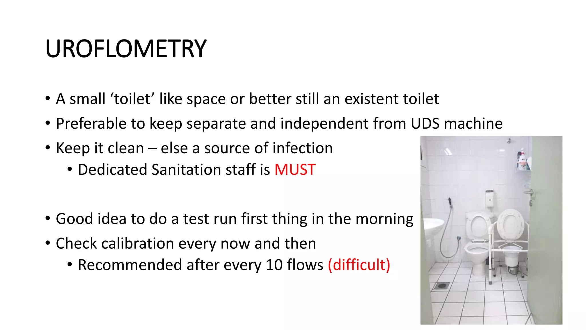
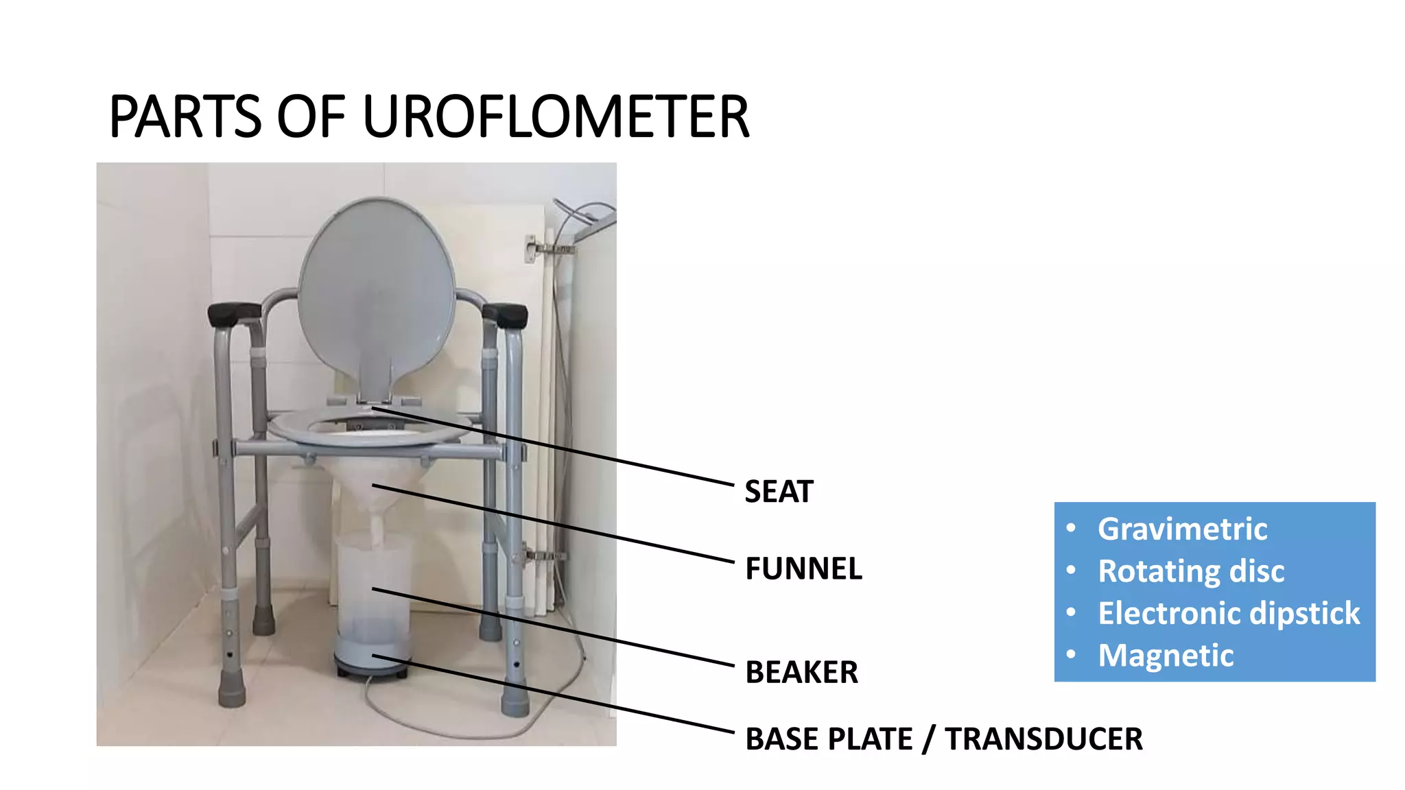
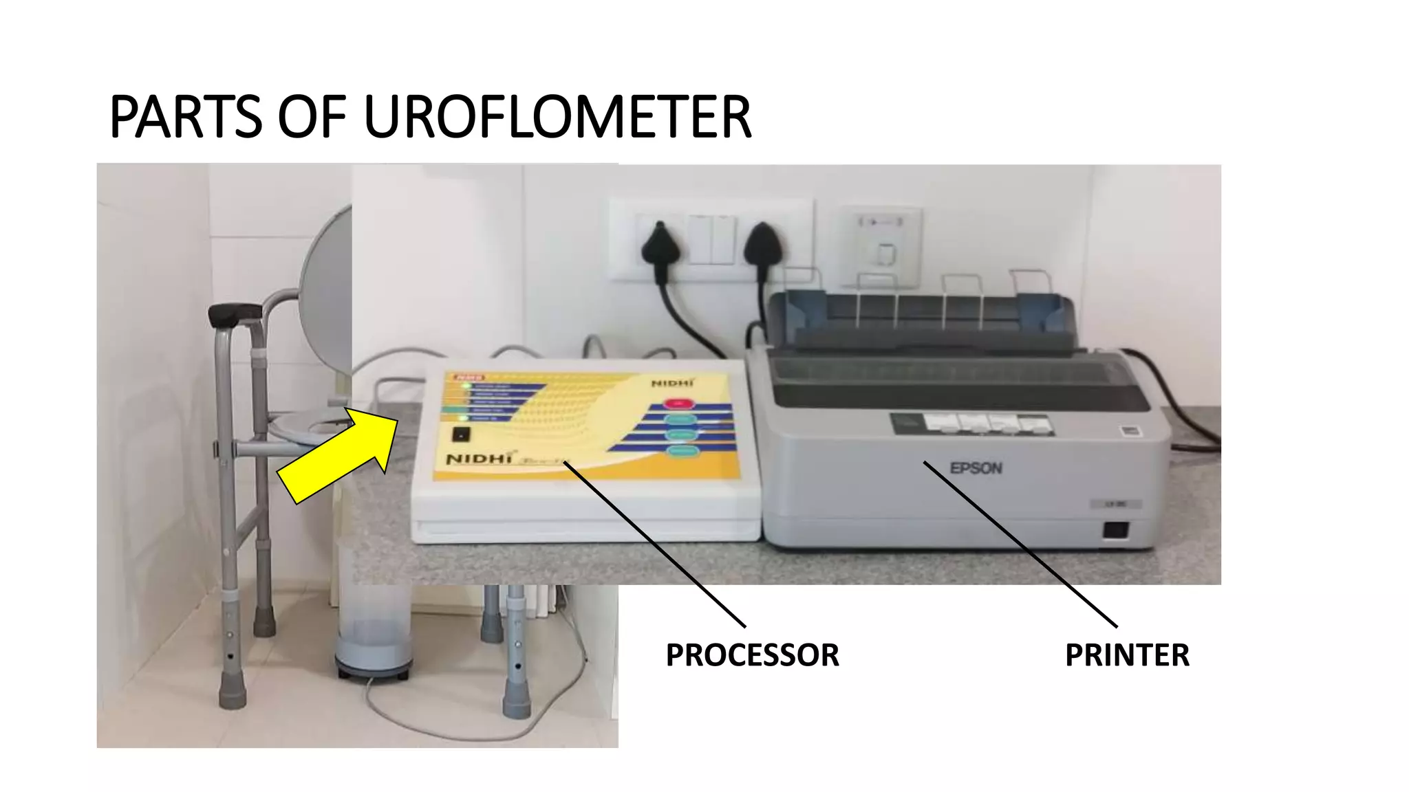
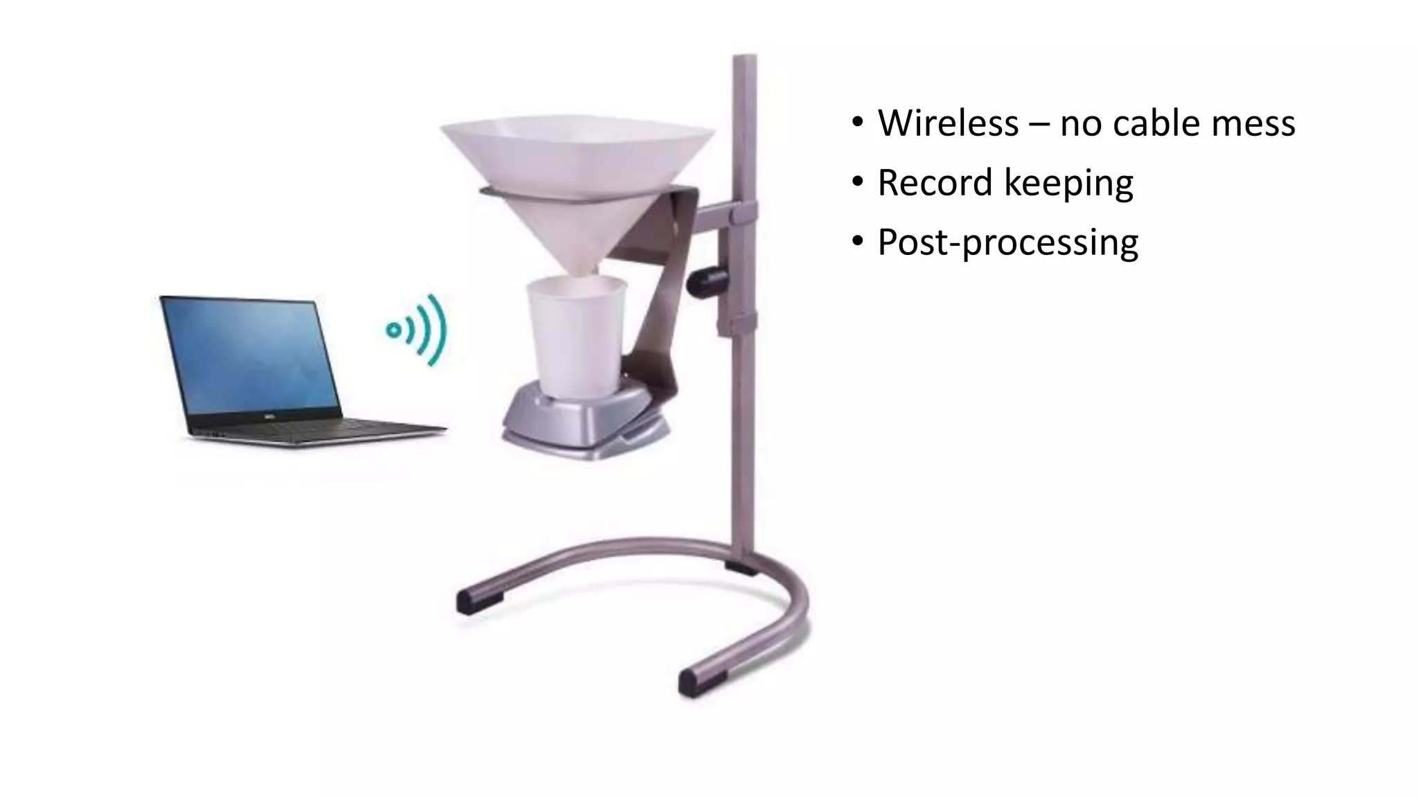
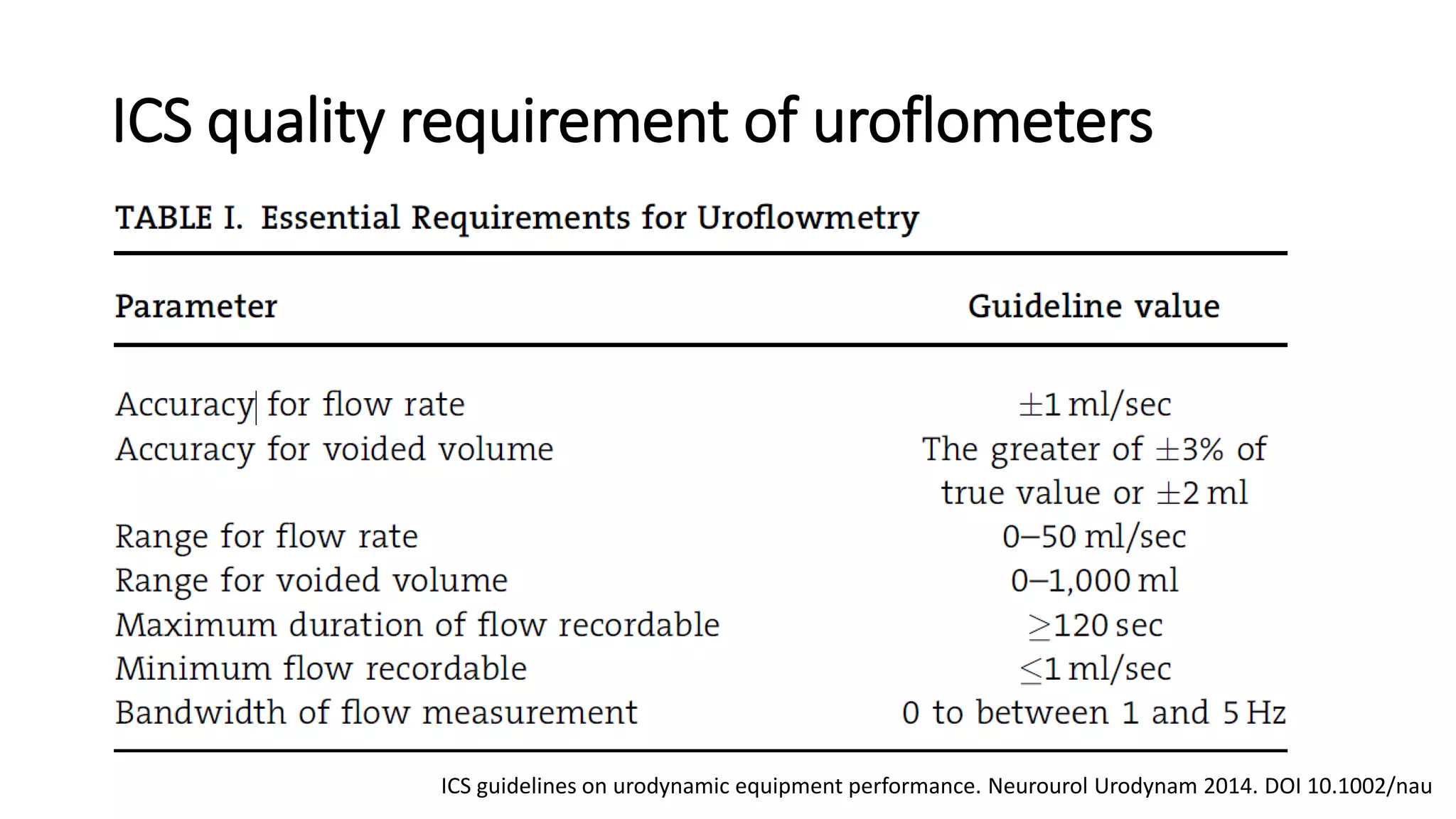
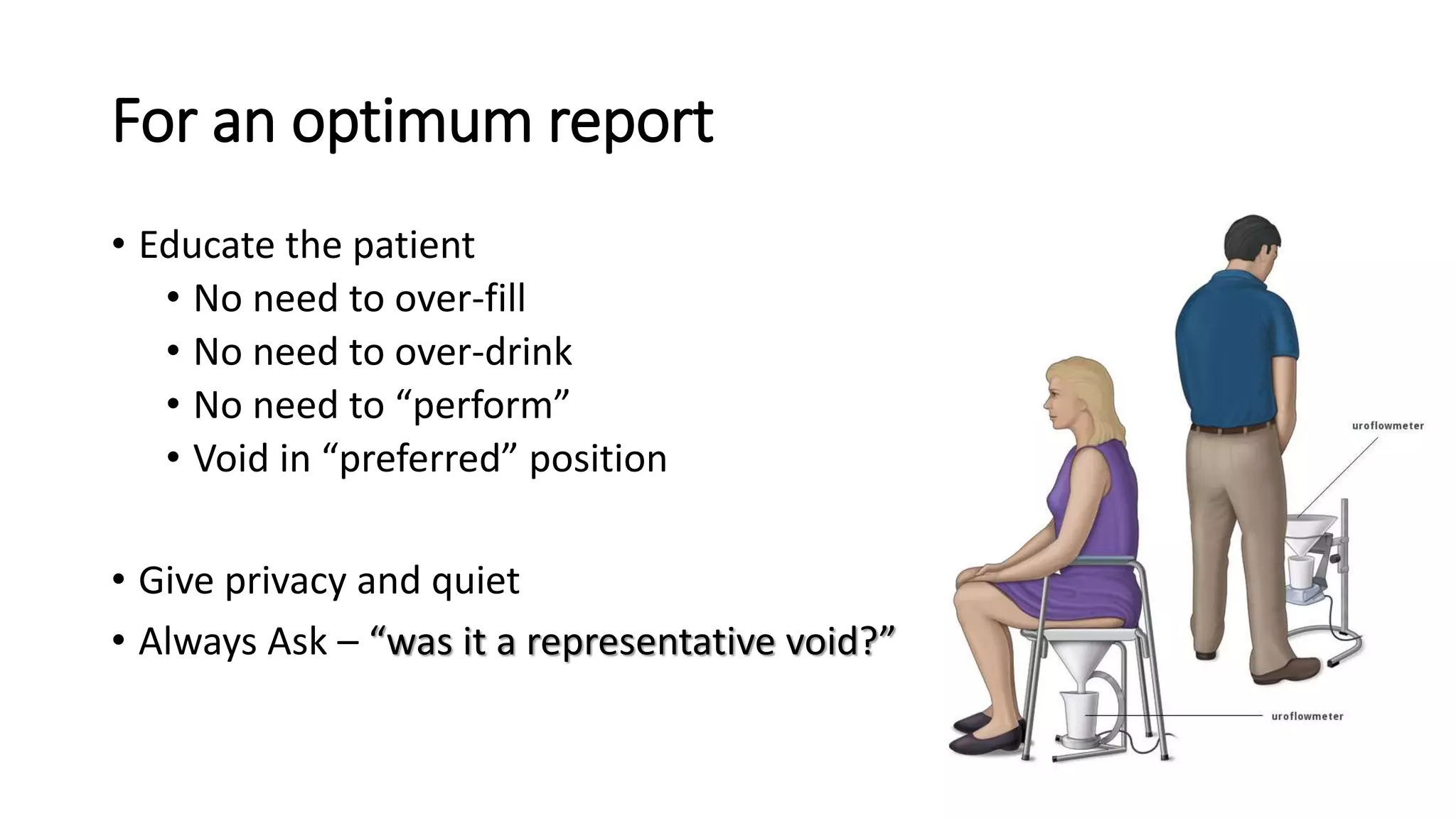
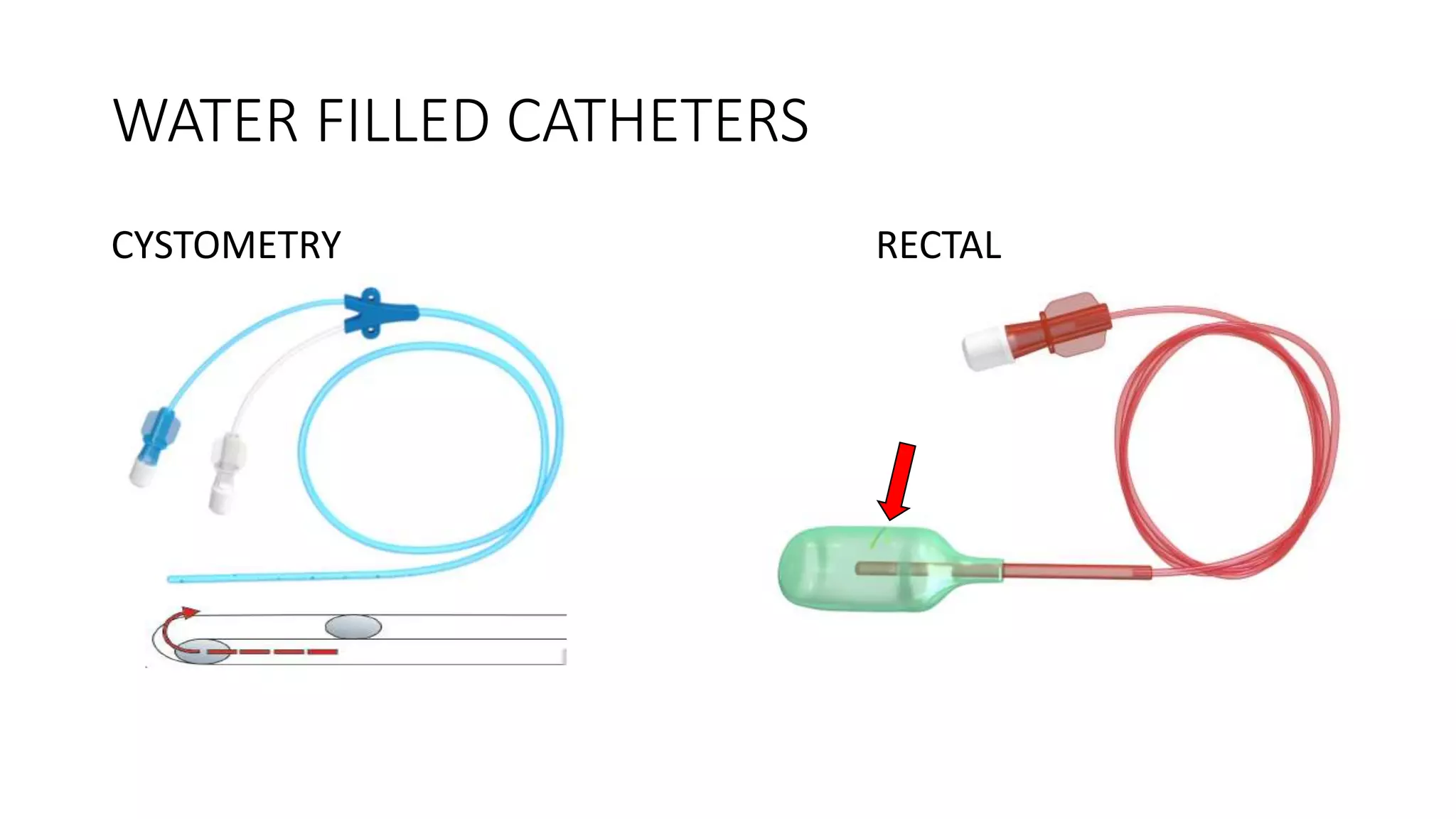
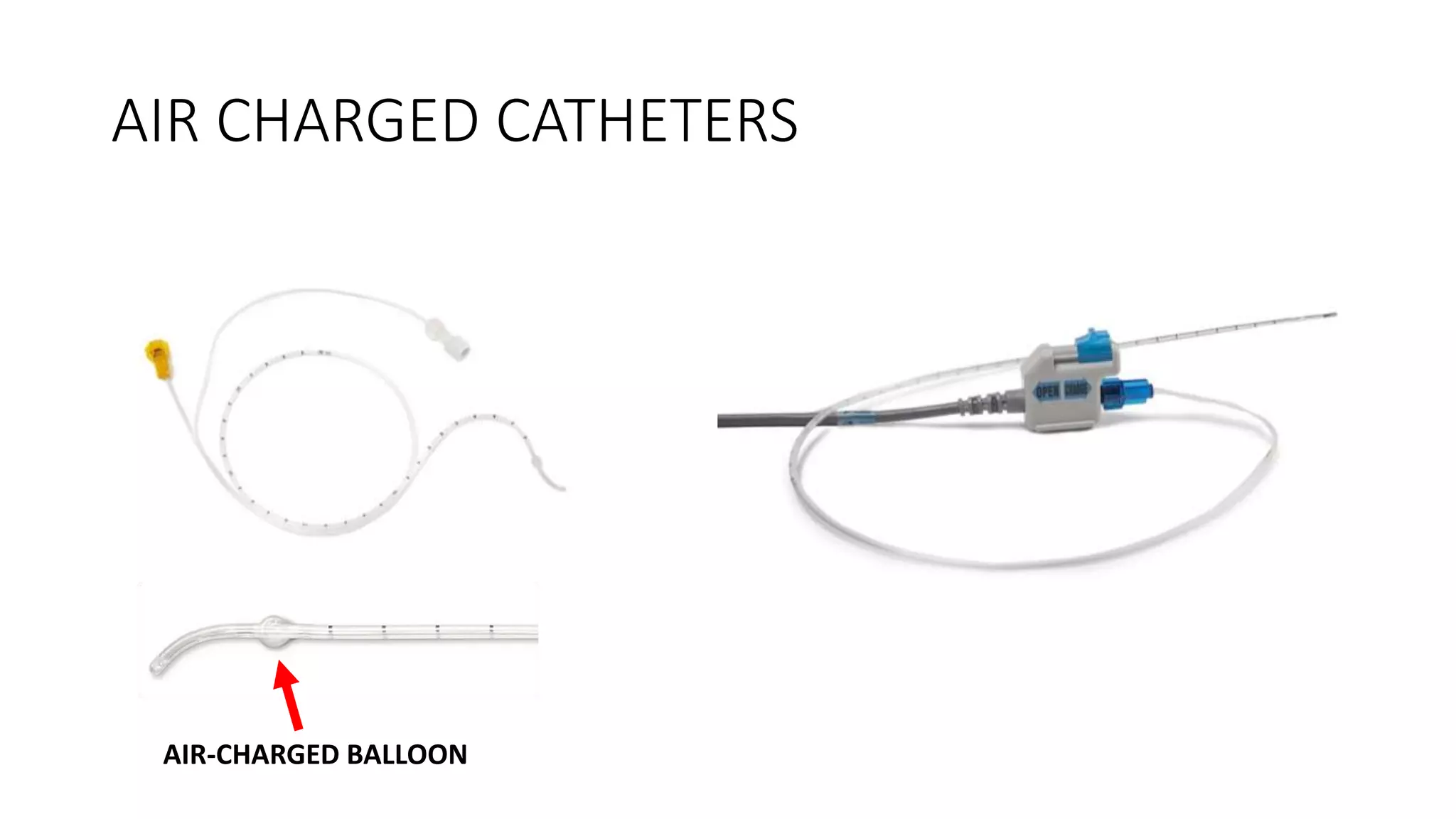
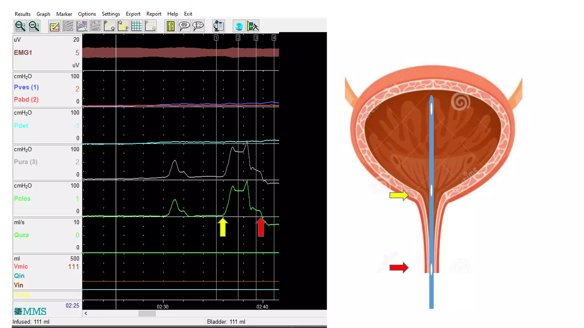
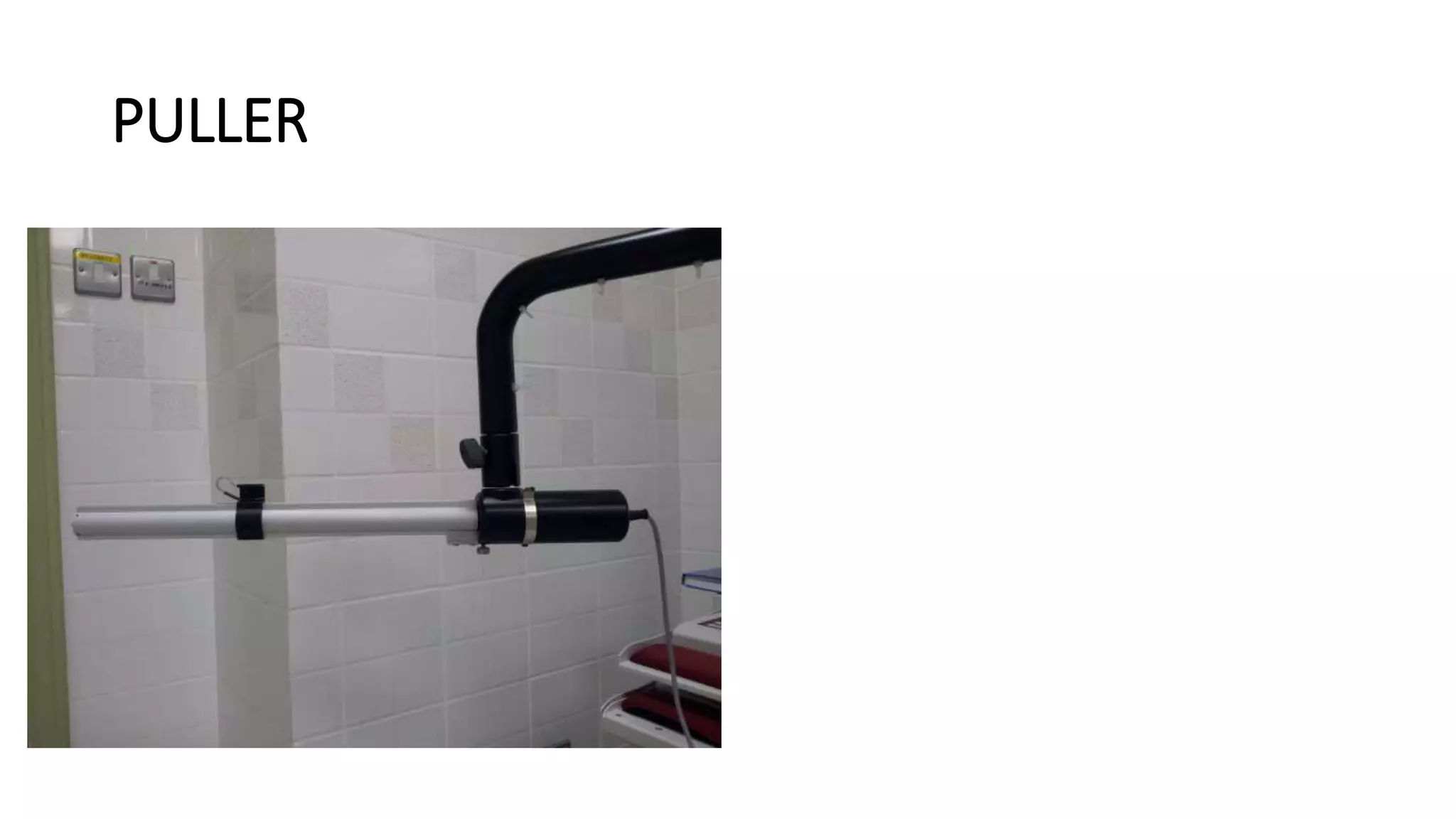
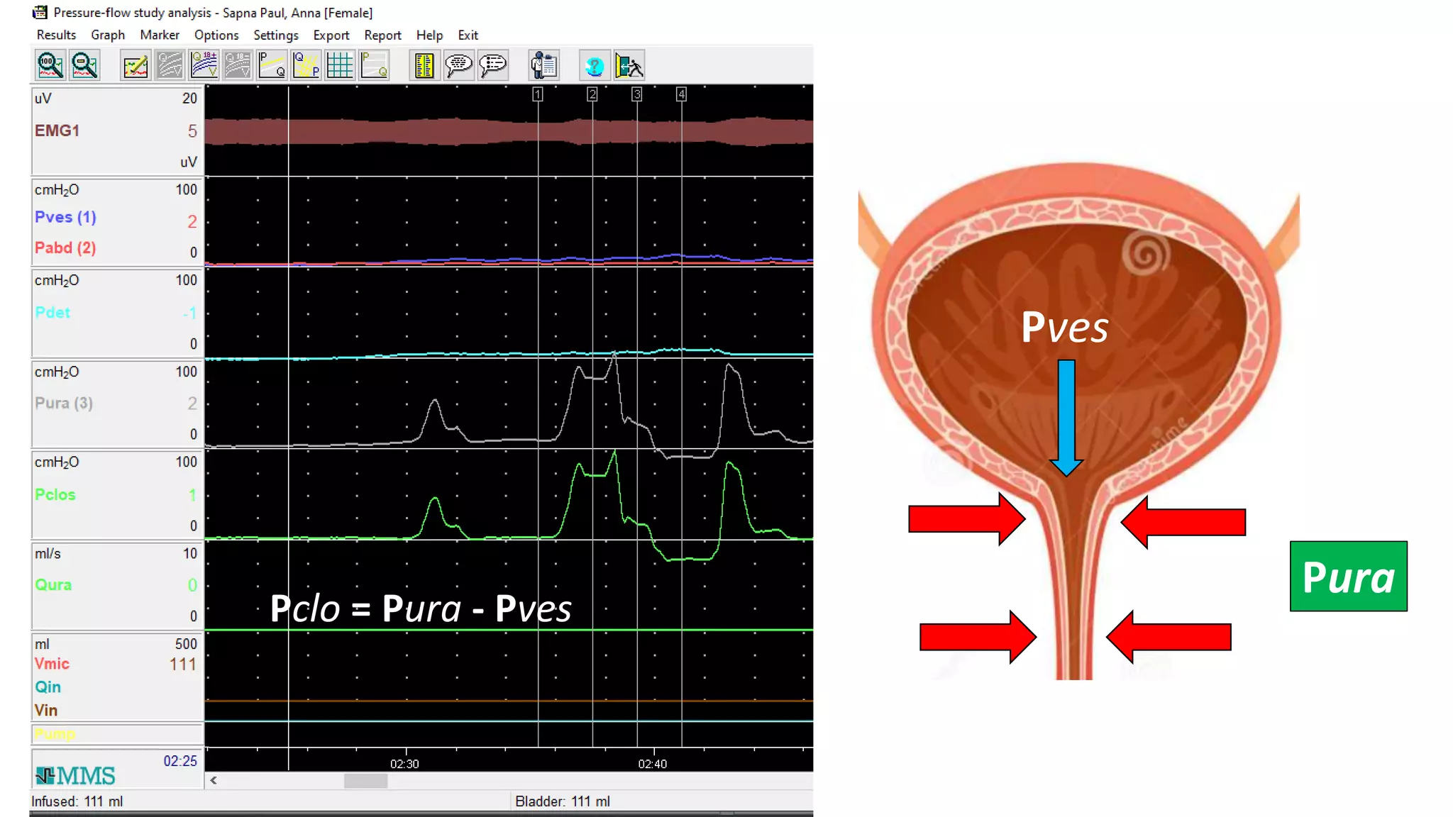
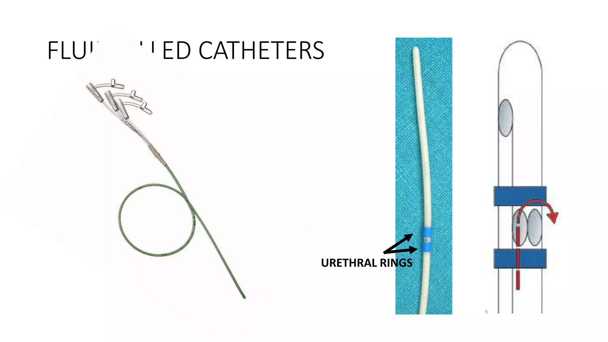
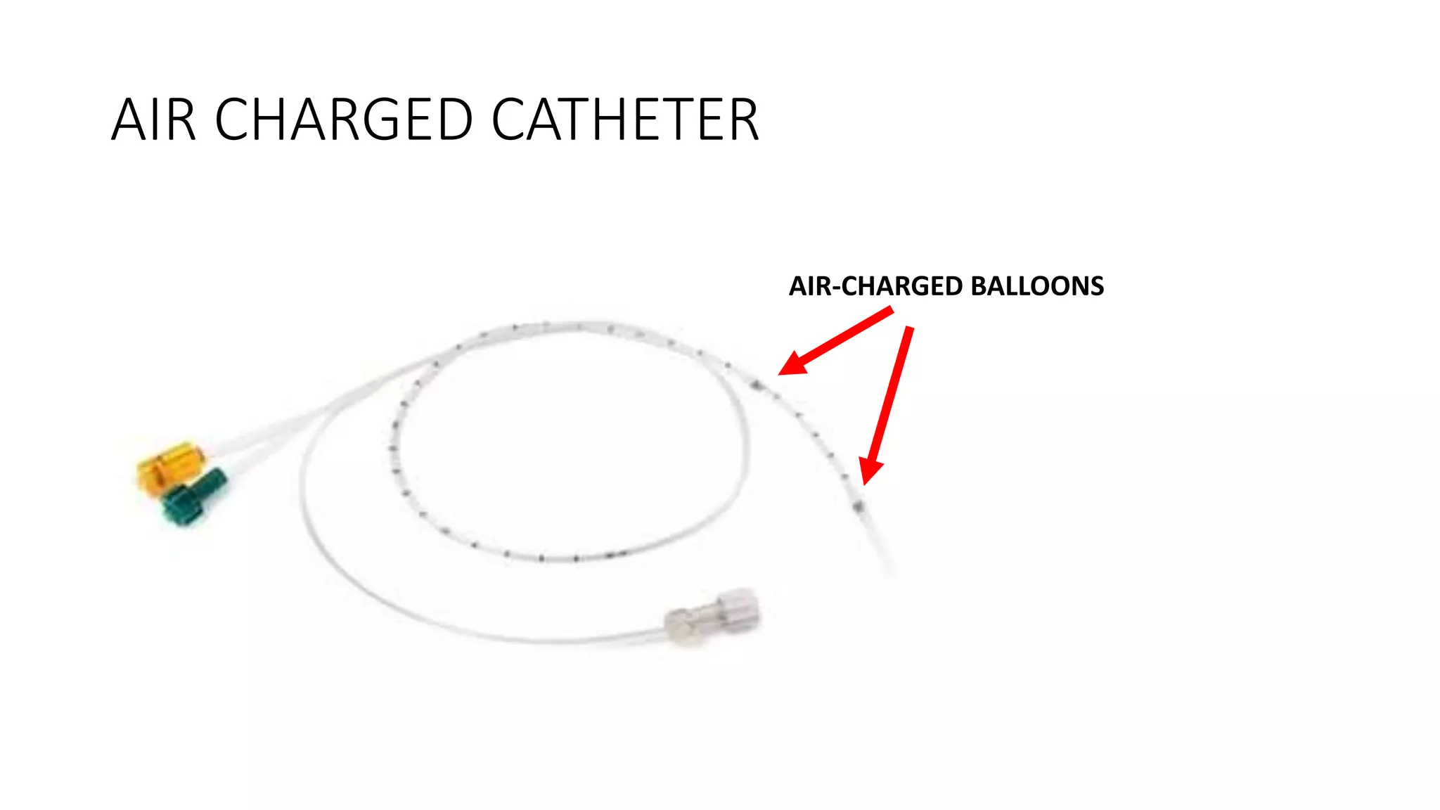
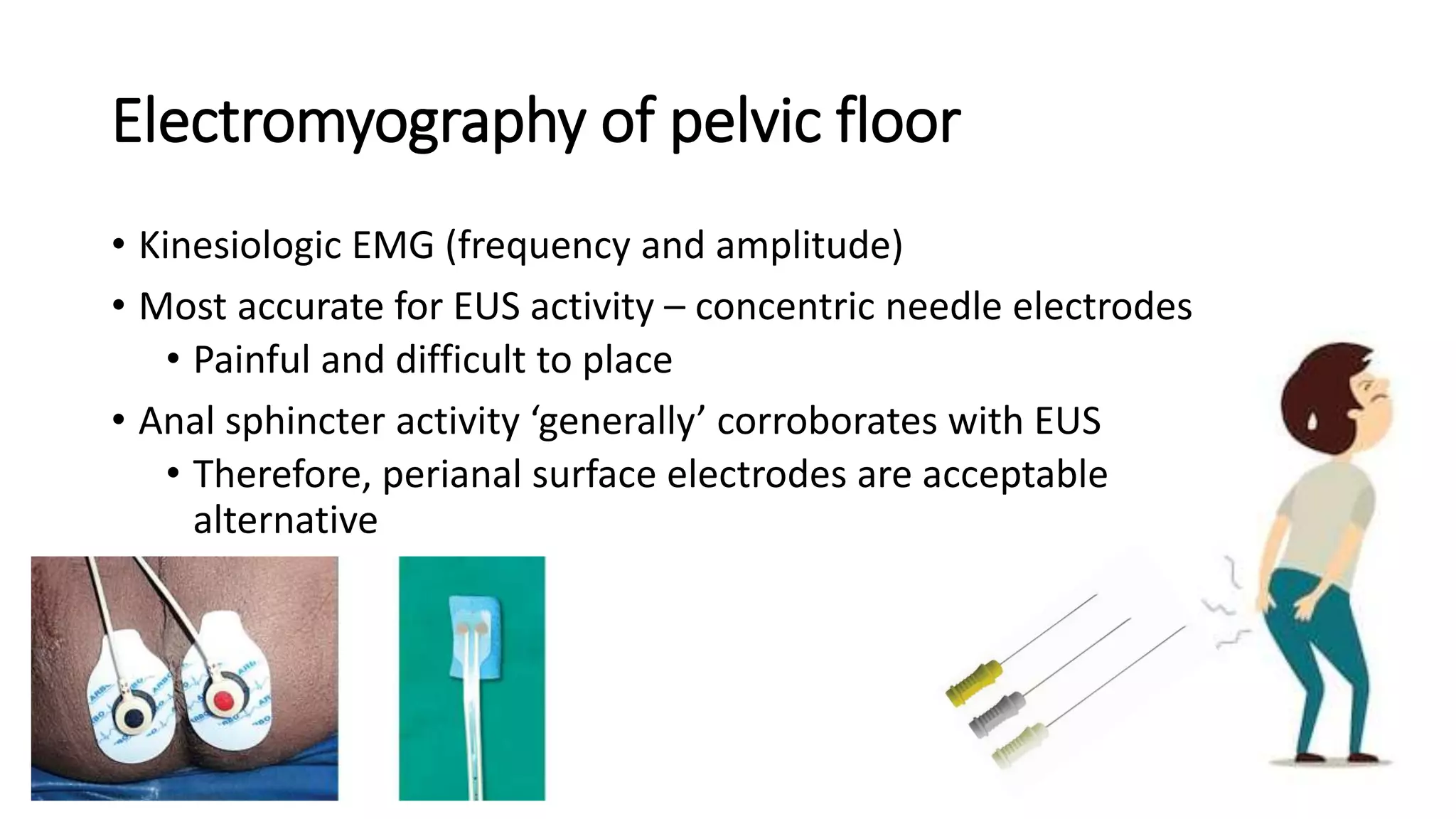
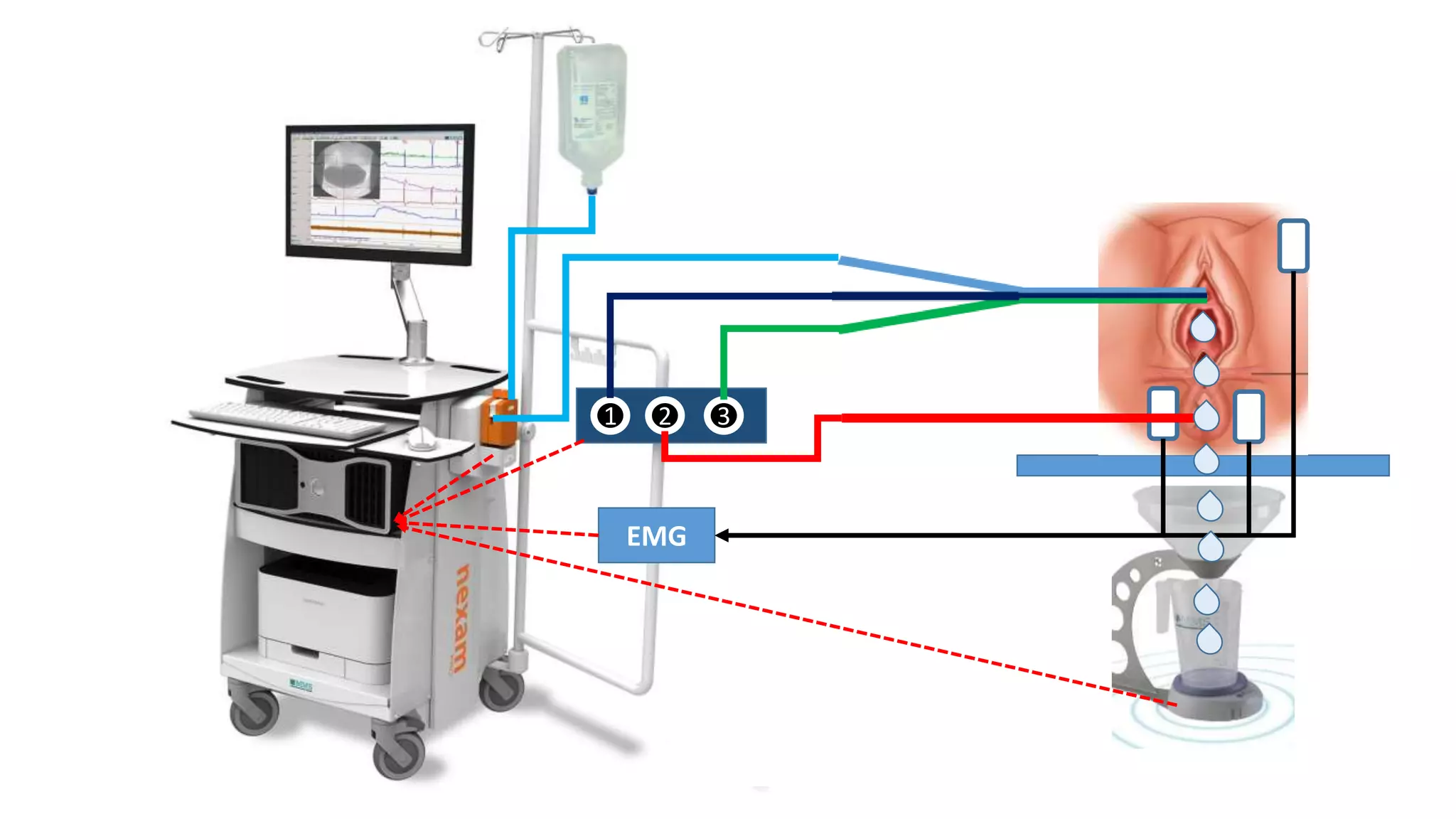
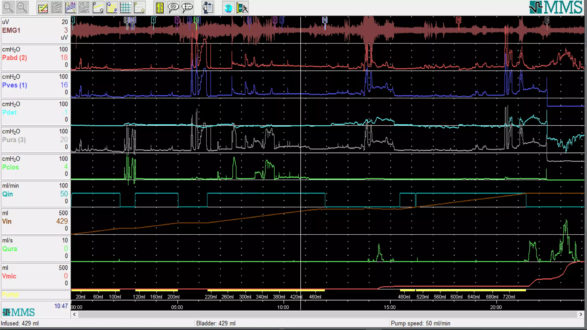
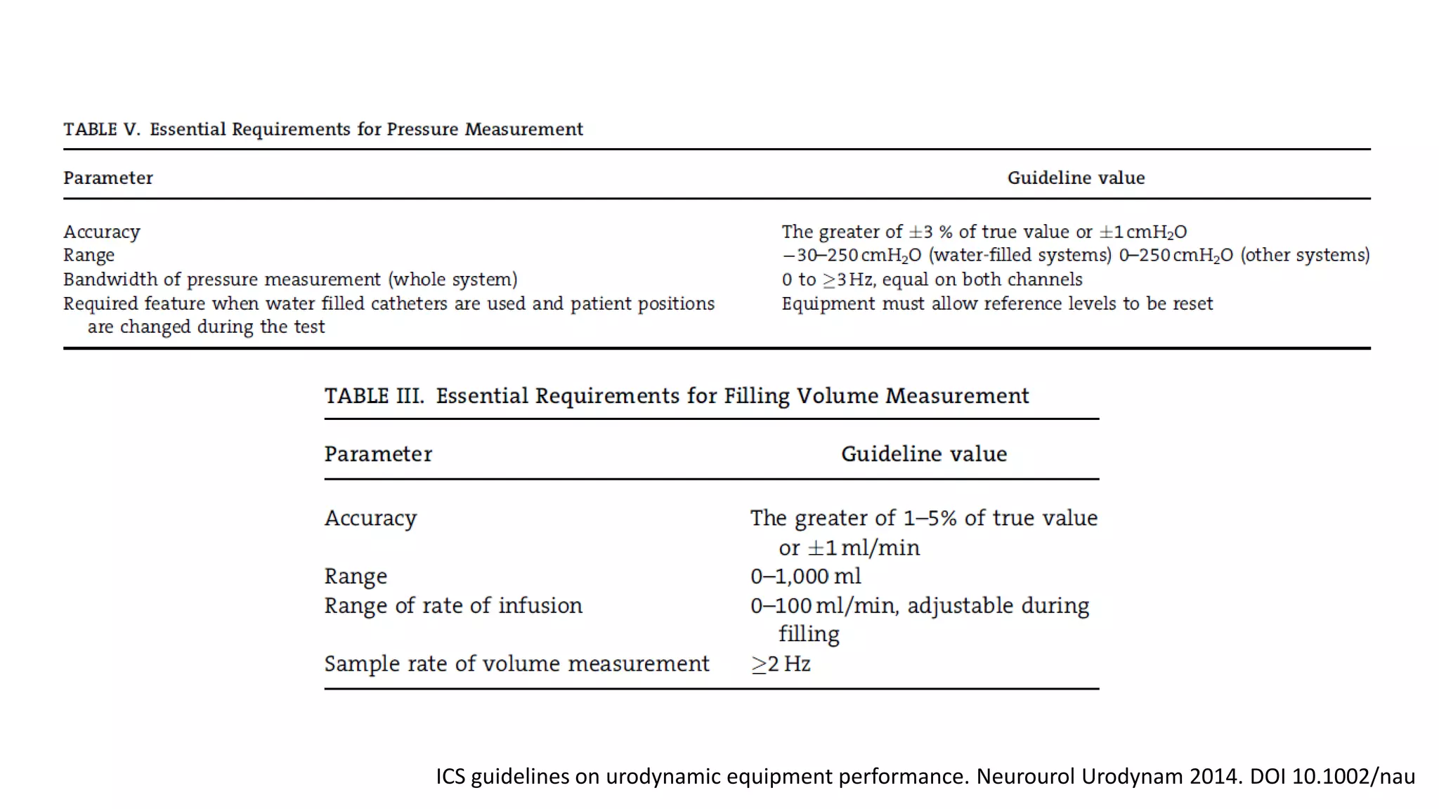
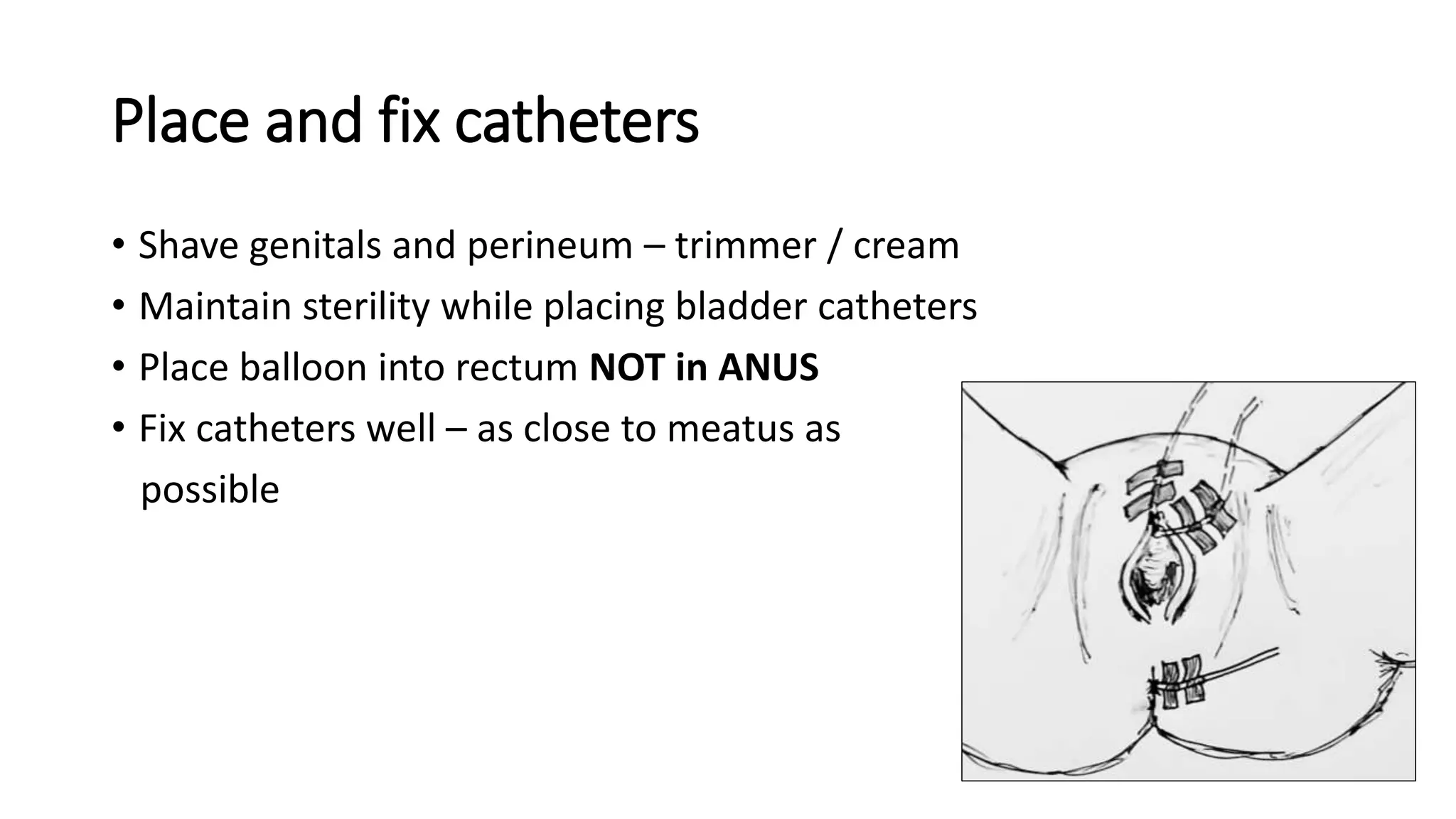
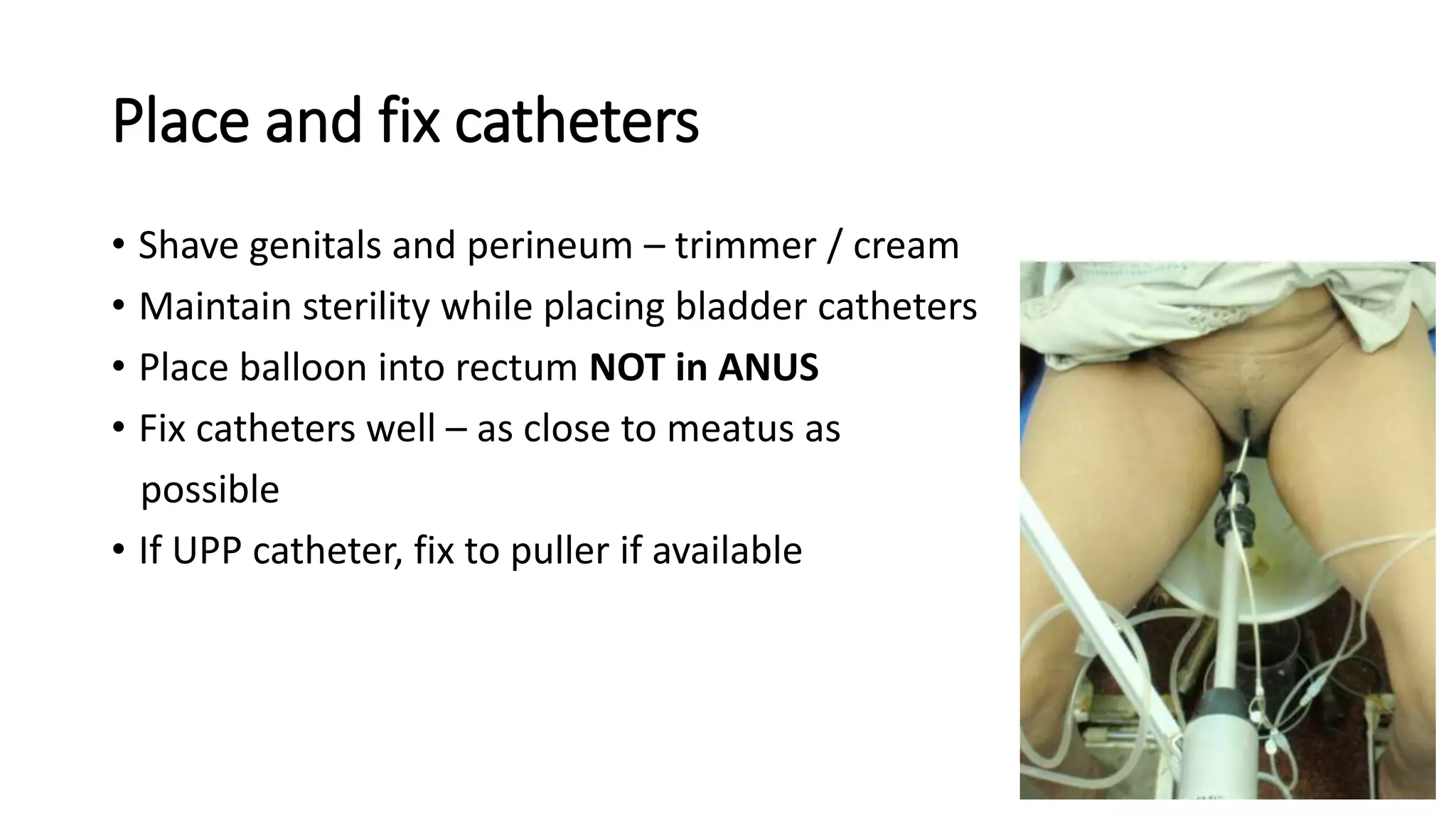
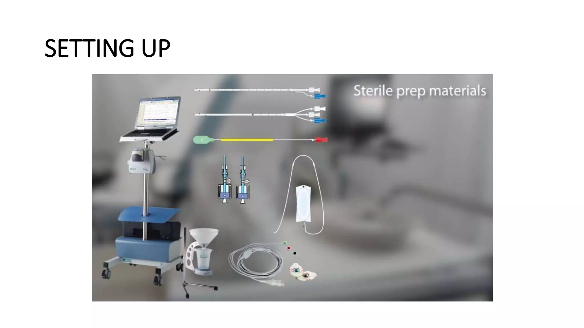
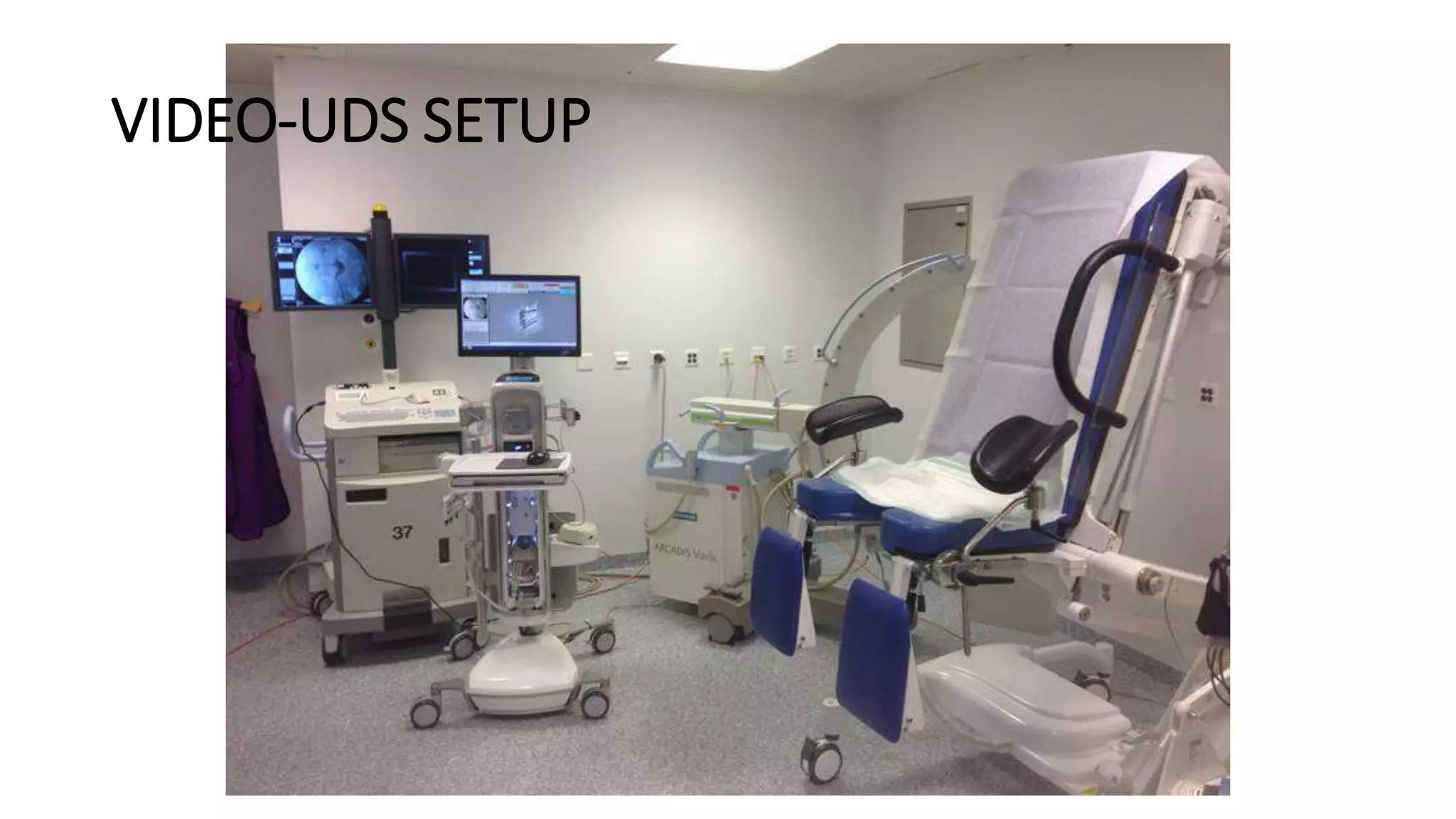
URODYNAMICS SETUP
​Dr Mayank Mohan Agarwal
​MS, MRCS(Ed), ​DNB, MCh (PGI, Chd)
​VMMF and IAUA Fellowships Uro-Oncology, Urogynecology
(MSKCC, NY; UCLA, LA; WFUBMC, NC)​
Associate Director
Urology and Kidney transplant surgery
Medanta Awadh Hospital
Lucknow
Introduction
• Setup of uroflometry
• Setup of urodynamics
• Setup of video
UROFLOMETRY
UROFLOMETRY
• A small ‘toilet’ like space or better still an existent toilet
• Preferable to keep separate and independent from UDS machine
• Keep it clean – else a source of infection
• Dedicated Sanitation staff is MUST
• Good idea to do a test run first thing in the morning
• Check calibration every now and then
• Recommended after every 10 flows (difficult)
POUR 300ML AT
‘PHYSIOLOGICAL SPEEDS’
AND CHECK READING OF
THE MACHINE
PARTS OF UROFLOMETER
BASE PLATE / TRANSDUCER
BEAKER
FUNNEL
SEAT
• Gravimetric
• Rotating disc
• Electronic dipstick
• Magnetic
PARTS OF UROFLOMETER
PROCESSOR PRINTER
• Wireless – no cable mess
• Record keeping
• Post-processing
ICS quality requirement of uroflometers
ICS guidelines on urodynamic equipment performance. Neurourol Urodynam 2014. DOI 10.1002/nau
For an optimum report
• Educate the patient
• No need to over-fill
• No need to over-drink
• No need to “perform”
• Void in “preferred” position
• Give privacy and quiet
• Always Ask – “was it a representative void?”
Post-void residual urine
• Very important adjunct to a uroflo report
• Almost always by ultrasound
• Must avoid catheterization as far as possible
• Dedicated “bladder scans” available
but prohibitively expensive
Multichannel urodynamics
WHAT DO WE WANT TO KNOW
• Pressures and sensations in bladder
• Pressure – flow relations
• Pressure – leak relations
• Pressures in urethra
• EMG activity
• Verify findings with fluoroscopy
WHAT DO WE WANT TO KNOW
• Pressures and sensations in bladder
• Pressure – flow relations
• Pressure – leak relations
Pves = Pdet + Pabd
• Pabd = Prectum
Pves
Pdet
Pabd
WATER FILLED CATHETERS
CYSTOMETRY RECTAL
SAVE COST
8-9FR INFANT FEEDING TUBE GLOVE FINGER FOR BALLOON 150CM ARTERIAL LINE
AIR CHARGED CATHETERS
AIR-CHARGED BALLOON
WHAT DO WE WANT TO KNOW
• Pressures and sensations in bladder
• Pressure – flow relations
• Pressure – leak relations
• Pressures in urethra
• EMG activity
• Verify findings with fluoroscopy
PULLER
MANUAL PULLER : SAVE COST
Pves
Pura
Pclo = Pura - Pves
FLUID FILLED CATHETERS
URETHRAL RINGS
AIR CHARGED CATHETER
AIR-CHARGED BALLOONS
WHAT DO WE WANT TO KNOW
• Pressures and sensations in bladder
• Pressure – flow relations
• Pressure – leak relations
• Pressures in urethra
• EMG activity
• Verify findings with fluoroscopy
Electromyography of pelvic floor
• Kinesiologic EMG (frequency and amplitude)
• Most accurate for EUS activity – concentric needle electrodes
• Painful and difficult to place
• Anal sphincter activity ‘generally’ corroborates with EUS
• Therefore, perianal surface electrodes are acceptable
alternative
21 3
EMG
ICS guidelines on urodynamic equipment performance. Neurourol Urodynam 2014. DOI 10.1002/nau
CALIBRATION
• Check calibration once every 10 measurements or so (difficult)
• For water filled system –
‘zero’ the transducer
 raise the catheter ≥ 50cm (by scale)
 look for pressure change in system
CALIBRATION
• For air-charged or catheter tip transducer system
‘zero’ the transducer
 submerge the tripod mounted catheter
system into a deep tube
 fill water to defined height and watch
corresponding change in computer
Set up for a study
• Maintain sterility
• Maintain privacy
• Talk-communicate-talk
• Unless the patient is mentally relaxed – the findings can drastically
changed
Place and fix catheters
• Shave genitals and perineum – trimmer / cream
• Maintain sterility while placing bladder catheters
• Place balloon into rectum NOT in ANUS
Place and fix catheters
• Shave genitals and perineum – trimmer / cream
• Maintain sterility while placing bladder catheters
• Place balloon into rectum NOT in ANUS
Place and fix catheters
• Shave genitals and perineum – trimmer / cream
• Maintain sterility while placing bladder catheters
• Place balloon into rectum NOT in ANUS
• Fix catheters well – as close to meatus as
possible
Place and fix catheters
• Shave genitals and perineum – trimmer / cream
• Maintain sterility while placing bladder catheters
• Place balloon into rectum NOT in ANUS
• Fix catheters well – as close to meatus as
possible
• If UPP catheter, fix to puller if available
SETTING UP
• Cough periodically
during the study
• Cough at the end of
the study
VIDEO-UDS SETUP
SEPARATE MCUG
CORRECT POSITIONING OF PATIENT RESPECTIVE TO X RAY BEAMS FOR GOOD VIEW OF OUTLET
A EXAMPLE OF THE SAME
THANK YOU

setting up for urodynamics

  • 1.
    URODYNAMICS SETUP ​Dr MayankMohan Agarwal ​MS, MRCS(Ed), ​DNB, MCh (PGI, Chd) ​VMMF and IAUA Fellowships Uro-Oncology, Urogynecology (MSKCC, NY; UCLA, LA; WFUBMC, NC)​ Associate Director Urology and Kidney transplant surgery Medanta Awadh Hospital Lucknow
  • 2.
    Introduction • Setup ofuroflometry • Setup of urodynamics • Setup of video
  • 3.
  • 4.
    UROFLOMETRY • A small‘toilet’ like space or better still an existent toilet • Preferable to keep separate and independent from UDS machine • Keep it clean – else a source of infection • Dedicated Sanitation staff is MUST • Good idea to do a test run first thing in the morning • Check calibration every now and then • Recommended after every 10 flows (difficult)
  • 5.
    UROFLOMETRY • A small‘toilet’ like space or better still an existent toilet • Preferable to keep separate and independent from UDS machine • Keep it clean – else a source of infection • Dedicated Sanitation staff is MUST • Good idea to do a test run first thing in the morning • Check calibration every now and then • Recommended after every 10 flows (difficult) POUR 300ML AT ‘PHYSIOLOGICAL SPEEDS’ AND CHECK READING OF THE MACHINE
  • 6.
    PARTS OF UROFLOMETER BASEPLATE / TRANSDUCER BEAKER FUNNEL SEAT • Gravimetric • Rotating disc • Electronic dipstick • Magnetic
  • 7.
  • 8.
    • Wireless –no cable mess • Record keeping • Post-processing
  • 9.
    ICS quality requirementof uroflometers ICS guidelines on urodynamic equipment performance. Neurourol Urodynam 2014. DOI 10.1002/nau
  • 10.
    For an optimumreport • Educate the patient • No need to over-fill • No need to over-drink • No need to “perform” • Void in “preferred” position • Give privacy and quiet • Always Ask – “was it a representative void?”
  • 11.
    Post-void residual urine •Very important adjunct to a uroflo report • Almost always by ultrasound • Must avoid catheterization as far as possible • Dedicated “bladder scans” available but prohibitively expensive
  • 12.
  • 13.
    WHAT DO WEWANT TO KNOW • Pressures and sensations in bladder • Pressure – flow relations • Pressure – leak relations • Pressures in urethra • EMG activity • Verify findings with fluoroscopy
  • 14.
    WHAT DO WEWANT TO KNOW • Pressures and sensations in bladder • Pressure – flow relations • Pressure – leak relations Pves = Pdet + Pabd • Pabd = Prectum Pves Pdet Pabd
  • 16.
  • 17.
    SAVE COST 8-9FR INFANTFEEDING TUBE GLOVE FINGER FOR BALLOON 150CM ARTERIAL LINE
  • 18.
  • 19.
    WHAT DO WEWANT TO KNOW • Pressures and sensations in bladder • Pressure – flow relations • Pressure – leak relations • Pressures in urethra • EMG activity • Verify findings with fluoroscopy
  • 21.
  • 22.
    MANUAL PULLER :SAVE COST
  • 23.
  • 24.
  • 25.
  • 26.
    WHAT DO WEWANT TO KNOW • Pressures and sensations in bladder • Pressure – flow relations • Pressure – leak relations • Pressures in urethra • EMG activity • Verify findings with fluoroscopy
  • 27.
    Electromyography of pelvicfloor • Kinesiologic EMG (frequency and amplitude) • Most accurate for EUS activity – concentric needle electrodes • Painful and difficult to place • Anal sphincter activity ‘generally’ corroborates with EUS • Therefore, perianal surface electrodes are acceptable alternative
  • 28.
  • 30.
    ICS guidelines onurodynamic equipment performance. Neurourol Urodynam 2014. DOI 10.1002/nau
  • 31.
    CALIBRATION • Check calibrationonce every 10 measurements or so (difficult) • For water filled system – ‘zero’ the transducer  raise the catheter ≥ 50cm (by scale)  look for pressure change in system
  • 32.
    CALIBRATION • For air-chargedor catheter tip transducer system ‘zero’ the transducer  submerge the tripod mounted catheter system into a deep tube  fill water to defined height and watch corresponding change in computer
  • 33.
    Set up fora study • Maintain sterility • Maintain privacy • Talk-communicate-talk • Unless the patient is mentally relaxed – the findings can drastically changed
  • 34.
    Place and fixcatheters • Shave genitals and perineum – trimmer / cream • Maintain sterility while placing bladder catheters • Place balloon into rectum NOT in ANUS
  • 35.
    Place and fixcatheters • Shave genitals and perineum – trimmer / cream • Maintain sterility while placing bladder catheters • Place balloon into rectum NOT in ANUS
  • 36.
    Place and fixcatheters • Shave genitals and perineum – trimmer / cream • Maintain sterility while placing bladder catheters • Place balloon into rectum NOT in ANUS • Fix catheters well – as close to meatus as possible
  • 37.
    Place and fixcatheters • Shave genitals and perineum – trimmer / cream • Maintain sterility while placing bladder catheters • Place balloon into rectum NOT in ANUS • Fix catheters well – as close to meatus as possible • If UPP catheter, fix to puller if available
  • 38.
  • 39.
    • Cough periodically duringthe study • Cough at the end of the study
  • 40.
  • 41.
  • 42.
    CORRECT POSITIONING OFPATIENT RESPECTIVE TO X RAY BEAMS FOR GOOD VIEW OF OUTLET
  • 43.
    A EXAMPLE OFTHE SAME
  • 44.