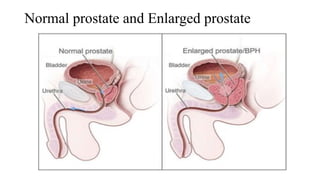
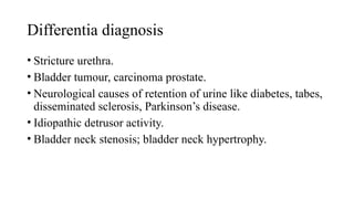
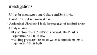
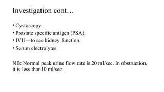
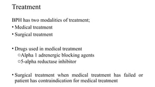
CMT 06207
Benign Prostatic Hyperplasia (BPH) and
Urethral Stricture
Session 14
By Dr. Stephen S. Kasaizi
Learning Objectives
By the end of this session, students are expected to be able to:
• Define Benign prostate hypertrophy (BPH) and urethral
stricture
• Describe clinical feature of BPH and urethral stricture
• Describe the causes of urethral stricture
• List the differential diagnosis of BPH and urethral stricture
• Describe the management of BPH and urethral stricture
Definition
• It is benign enlargement of prostate which occurs after 50
years, usually between 60 and 70 years.
• BPH affects both glandular epithelium and connective tissue
stroma.
Normal prostate and Enlarged prostate
Subdivision of the prostate
• The prostate can be
subdivided in to two ways
1. In lobes
2. In Zones
Division of Prostate in to lobes
• The "lobe" classification is more often used in anatomy. The
prostate is incompletely divided into five lobes:
oAnterior lobe (or isthmus)
oPosterior lobe
oRight lateral lobe
oLeft lateral lobe
oMedian lobe( or middle lobe)
Division of Prostate in to Zones
• The prostate has been described as consisting of three or four
zones.
• This "zone" classification is more often used in pathology.
The prostate gland has four distinct glandular regions, two of
which arise from different segments of the prostatic urethra:
oPeripheral zone
oCentral zone
oTransition zone
oAnterior fibro-muscular zone (or stroma
Pathology
• BPH usually involves median and lateral lobes or one of them.
• It involves adenomatous zone of prostate, i.e. submucosal
glands.
• Median lobe enlarges into the bladder.
• Lateral lobes narrow the urethra causing obstruction.
• Urethra above the verumontanum gets elongated and narrowed.
• Bladder initially takes the pressure burden causing
trabeculations, sacculations and later diverticula formation.
Activity: Brainstorming
• What are the clinical presentations of a patient with BPH?
Clinical features
• Frequency occurs due to introversion of sensitive urethral
mucosa into the bladder or due to cystitis and urethritis.
• Urgency, hesitancy, nocturia.
• Overflow and terminal dribbling.
• Difficulty in micturition with weak stream and dribble.
• Pain in suprapubic region and in loin due to cystitis and
hydronephrosis respectively.
• Acute retention of urine.
Summary of clinical features
Symptoms of voiding
• Hesitancy
• Poor flow not improving by
straining
• Dribbling even after micturition
• Intermittent stream—stops and
starts
• Poor bladder emptying
• Episodes of near retention
Symptoms of storage
• Frequency
• Nocturia
• Urgency
• Urge incontinence
• Nocturnal incontinence
Activity: Brainstorming
• Mention three (3) differential diagnosis of BPH.
Differentia diagnosis
• Stricture urethra.
• Bladder tumour, carcinoma prostate.
• Neurological causes of retention of urine like diabetes, tabes,
disseminated sclerosis, Parkinson’s disease.
• Idiopathic detrusor activity.
• Bladder neck stenosis; bladder neck hypertrophy.
Investigations
• Urine for microscopy and Culture and Sensitivity
• Blood urea and serum creatinine.
• Abdominal Ultrasound-look for presence of residual urine.
• Urodynamics.
oUrine flow rate >15 ml/sec is normal. 10–15 ml is
equivocal; <10 ml is low.
oVoiding pressure <60 cm of water is normal; 60–80 is
equivocal; >80 is high.
Investigation cont…
• Cystoscopy.
• Prostate specific antigen (PSA).
• IVU—to see kidney function.
• Serum electrolytes.
NB: Normal peak urine flow rate is 20 ml/sec. In obstruction,
it is less than10 ml/sec.
Treatment
BPH has two modalities of treatment;
• Medical treatment
• Surgical treatment
• Drugs used in medical treatment
oAlpha 1 adrenergic blocking agents
o5-alpha reductase inhibitor
• Surgical treatment when medical treatment has failed or
patient has contraindication for medical treatment
Indications for Surgery
• Prostatism (frequency, dysuria, urgency).
• Acute retention of urine.
• Chronic retention of urine with residual urine more than 200
ml.
• Complications like hydroureter, hydronephrosis, stone
formation, recurrent infection, bladder changes, bladder
stones
• Haematuria.
Urethral stricture
Definition
• Urethral stricture is narrowing of the urethral lumen causing
functional obstruction
oThis is due to formation of fibrous tissue following
damage to the urethral mucosa
• Most common cause of urethral stricture is ureteropelvic
junction (UPJ) obstruction which could be congenital or
acquired narrowing at the level of UPJ
Activity: Brainstorming
• What are the causes of urethral stricture?
Causes and risk factors
• It can follow treatment for another urological condition,
pelvic radiation therapy or urinary diversion surgery
• External traumatic injury like straddle injuries.
• Congenital anomalies may result in to stricture
• Most commonly caused by gonococal urethritis and
chlymidia, transmitted through sexual intercourse.
• Could be associated with pelvic fracture
• Can follow urological procedures like catheterization
Clinical features
• Poor urinary stream
• Forking and spraying of the stream
• Incomplete emptying
• Frequency, dysuria
• Retention and often with overflow
• Pain, burning micturition, suprapubic tenderness
• Thickening and button-like feeling in bulbar urethra (Bulbous
urethra is felt clinically by lifting the scrotum in midline in
the perineum)
Investigations
• Urine microscopy and culture.
• Blood urea and serum creatinine.
• IVU to see hydronephrosis and function of kidney.
• Ultrasound abdomen.
• X-ray of pelvis to see old fracture with history of trauma.
Investigation cont…
• Ascending urethrogram is an essential investigation to see the
site, type, extent and false passage.
• Urodynamic studies.
• Urethroscopy
Treatment
• Intermittent dilatation
• Surgery.
• Refer the patient to a higher center for further evaluation and
treatment.
Key points
• BPH is benign enlargement of prostate which occurs after 50
years, usually between 60 and 70 years.
• The prostate is subdivided in to five lobes or four zones.
• There are two treatment modalities for BPH; Medical and
surgical
• Urethral stricture is narrowing of the urethral lumen causing
functional obstruction
• Urethral stricture is most commonly caused by gonococal
urethritis transmitted through sexual intercourse.
Evaluation
• Mention lobes of prostate
• Mention for investigation for the patient with BPH
• Define urethral stricture
• Mention four (4) causes of urethral stricture
Reference
• Siram Bhat M (2015) SRB’s Manual of surgery( 5th
edition).
Prakash Rao Thangam venghese Joshua.
• K.Rajgopal Shenoy, Anitha shenoy (2016) Manipal Manual
of surgery (4th
edition). CBS New Delhi
• F.Charles Brunkardi (2019) Schwartz’s principles of surgery
(11th
edition). McGraw-Hill Education
• Russell R.C.G., Norman S.W. & Bulstrode C.J.K. (2006).
Bailey and Love’s Short Practice of Surgery (25th
edition.).
London: Hodder Arnold

Session 14- Benign prostate hyperplasia(BPH).pptx

  • 1.
    CMT 06207 Benign ProstaticHyperplasia (BPH) and Urethral Stricture Session 14 By Dr. Stephen S. Kasaizi
  • 2.
    Learning Objectives By theend of this session, students are expected to be able to: • Define Benign prostate hypertrophy (BPH) and urethral stricture • Describe clinical feature of BPH and urethral stricture • Describe the causes of urethral stricture • List the differential diagnosis of BPH and urethral stricture • Describe the management of BPH and urethral stricture
  • 3.
    Definition • It isbenign enlargement of prostate which occurs after 50 years, usually between 60 and 70 years. • BPH affects both glandular epithelium and connective tissue stroma.
  • 4.
    Normal prostate andEnlarged prostate
  • 5.
    Subdivision of theprostate • The prostate can be subdivided in to two ways 1. In lobes 2. In Zones
  • 6.
    Division of Prostatein to lobes • The "lobe" classification is more often used in anatomy. The prostate is incompletely divided into five lobes: oAnterior lobe (or isthmus) oPosterior lobe oRight lateral lobe oLeft lateral lobe oMedian lobe( or middle lobe)
  • 7.
    Division of Prostatein to Zones • The prostate has been described as consisting of three or four zones. • This "zone" classification is more often used in pathology. The prostate gland has four distinct glandular regions, two of which arise from different segments of the prostatic urethra: oPeripheral zone oCentral zone oTransition zone oAnterior fibro-muscular zone (or stroma
  • 8.
    Pathology • BPH usuallyinvolves median and lateral lobes or one of them. • It involves adenomatous zone of prostate, i.e. submucosal glands. • Median lobe enlarges into the bladder. • Lateral lobes narrow the urethra causing obstruction. • Urethra above the verumontanum gets elongated and narrowed. • Bladder initially takes the pressure burden causing trabeculations, sacculations and later diverticula formation.
  • 9.
    Activity: Brainstorming • Whatare the clinical presentations of a patient with BPH?
  • 10.
    Clinical features • Frequencyoccurs due to introversion of sensitive urethral mucosa into the bladder or due to cystitis and urethritis. • Urgency, hesitancy, nocturia. • Overflow and terminal dribbling. • Difficulty in micturition with weak stream and dribble. • Pain in suprapubic region and in loin due to cystitis and hydronephrosis respectively. • Acute retention of urine.
  • 11.
    Summary of clinicalfeatures Symptoms of voiding • Hesitancy • Poor flow not improving by straining • Dribbling even after micturition • Intermittent stream—stops and starts • Poor bladder emptying • Episodes of near retention Symptoms of storage • Frequency • Nocturia • Urgency • Urge incontinence • Nocturnal incontinence
  • 12.
    Activity: Brainstorming • Mentionthree (3) differential diagnosis of BPH.
  • 13.
    Differentia diagnosis • Strictureurethra. • Bladder tumour, carcinoma prostate. • Neurological causes of retention of urine like diabetes, tabes, disseminated sclerosis, Parkinson’s disease. • Idiopathic detrusor activity. • Bladder neck stenosis; bladder neck hypertrophy.
  • 14.
    Investigations • Urine formicroscopy and Culture and Sensitivity • Blood urea and serum creatinine. • Abdominal Ultrasound-look for presence of residual urine. • Urodynamics. oUrine flow rate >15 ml/sec is normal. 10–15 ml is equivocal; <10 ml is low. oVoiding pressure <60 cm of water is normal; 60–80 is equivocal; >80 is high.
  • 15.
    Investigation cont… • Cystoscopy. •Prostate specific antigen (PSA). • IVU—to see kidney function. • Serum electrolytes. NB: Normal peak urine flow rate is 20 ml/sec. In obstruction, it is less than10 ml/sec.
  • 16.
    Treatment BPH has twomodalities of treatment; • Medical treatment • Surgical treatment • Drugs used in medical treatment oAlpha 1 adrenergic blocking agents o5-alpha reductase inhibitor • Surgical treatment when medical treatment has failed or patient has contraindication for medical treatment
  • 17.
    Indications for Surgery •Prostatism (frequency, dysuria, urgency). • Acute retention of urine. • Chronic retention of urine with residual urine more than 200 ml. • Complications like hydroureter, hydronephrosis, stone formation, recurrent infection, bladder changes, bladder stones • Haematuria.
  • 18.
  • 19.
    Definition • Urethral strictureis narrowing of the urethral lumen causing functional obstruction oThis is due to formation of fibrous tissue following damage to the urethral mucosa • Most common cause of urethral stricture is ureteropelvic junction (UPJ) obstruction which could be congenital or acquired narrowing at the level of UPJ
  • 20.
    Activity: Brainstorming • Whatare the causes of urethral stricture?
  • 21.
    Causes and riskfactors • It can follow treatment for another urological condition, pelvic radiation therapy or urinary diversion surgery • External traumatic injury like straddle injuries. • Congenital anomalies may result in to stricture • Most commonly caused by gonococal urethritis and chlymidia, transmitted through sexual intercourse. • Could be associated with pelvic fracture • Can follow urological procedures like catheterization
  • 22.
    Clinical features • Poorurinary stream • Forking and spraying of the stream • Incomplete emptying • Frequency, dysuria • Retention and often with overflow • Pain, burning micturition, suprapubic tenderness • Thickening and button-like feeling in bulbar urethra (Bulbous urethra is felt clinically by lifting the scrotum in midline in the perineum)
  • 23.
    Investigations • Urine microscopyand culture. • Blood urea and serum creatinine. • IVU to see hydronephrosis and function of kidney. • Ultrasound abdomen. • X-ray of pelvis to see old fracture with history of trauma.
  • 24.
    Investigation cont… • Ascendingurethrogram is an essential investigation to see the site, type, extent and false passage. • Urodynamic studies. • Urethroscopy
  • 25.
    Treatment • Intermittent dilatation •Surgery. • Refer the patient to a higher center for further evaluation and treatment.
  • 26.
    Key points • BPHis benign enlargement of prostate which occurs after 50 years, usually between 60 and 70 years. • The prostate is subdivided in to five lobes or four zones. • There are two treatment modalities for BPH; Medical and surgical • Urethral stricture is narrowing of the urethral lumen causing functional obstruction • Urethral stricture is most commonly caused by gonococal urethritis transmitted through sexual intercourse.
  • 27.
    Evaluation • Mention lobesof prostate • Mention for investigation for the patient with BPH • Define urethral stricture • Mention four (4) causes of urethral stricture
  • 28.
    Reference • Siram BhatM (2015) SRB’s Manual of surgery( 5th edition). Prakash Rao Thangam venghese Joshua. • K.Rajgopal Shenoy, Anitha shenoy (2016) Manipal Manual of surgery (4th edition). CBS New Delhi • F.Charles Brunkardi (2019) Schwartz’s principles of surgery (11th edition). McGraw-Hill Education • Russell R.C.G., Norman S.W. & Bulstrode C.J.K. (2006). Bailey and Love’s Short Practice of Surgery (25th edition.). London: Hodder Arnold