1) Molecular dynamics simulations were used to explore the effects of sodium ions and the mechanism of their role as allosteric modulators of dopaminergic G protein coupled receptors.
2) The simulations showed sodium ions binding to the receptor in two steps: first interacting with negatively charged residues on the extracellular surface, then visiting three binding sites between transmembrane regions.
3) Sodium ions preferentially bound to an allosteric site between transmembrane regions 2 and 3, where they induced a conformational change in a "toggle switch" tryptophan residue to lock the receptor in an inactive state.
This document summarizes an ab initio study of the denaturation of the Small Ubiquitin-like Modifier (SUMO) protein using molecular dynamics simulations and NMR calculations. The study found that after denaturing, some residues in SUMO still showed propensities to form secondary structure rather than becoming fully random coils. Molecular dynamics simulations of different SUMO topologies under denaturing conditions were performed. NMR properties were then calculated and compared to experimental observations, showing some residues maintained beta sheet or alpha helical propensities when denatured. This suggests denatured proteins can become trapped in local energy minima rather than fully unfolding.
Insights into All-Atom Protein Structure Prediction via in silico Simulationsdwang953
The document discusses using in silico molecular dynamics simulations to predict the 3D structures of proteins from their amino acid sequences in order to better understand protein folding. It provides background on proteins and the protein folding problem, and describes current models of protein folding as well as molecular dynamics and replica exchange molecular dynamics simulation techniques for studying protein folding at an atomistic level in silico.
Molecular dynamics (MD) simulations allow atoms and molecules to interact over time, representing a virtual experiment. MD was used to give dynamics to SUMO proteins in solution. The SUMO protein was divided into fragments which were given random conformations using CYANA. These conformations were then converted to GROMACS format and molecular dynamics simulations were performed using GROMACS. The simulations involved energy minimization to relieve strain, followed by production runs. Various analysis tools were then used to analyze the results.
This document provides an overview of molecular dynamics (MD) simulations and their analysis. MD simulations calculate the time-dependent behavior of molecules and can be used to study conformational changes in proteins and nucleic acids. The document outlines various analyses that can be done on MD simulations including root mean square deviation (RMSD), root mean square fluctuation (RMSF), radius of gyration, hydrogen bonding, secondary structure analysis using Ramachandran plots, free energy surfaces, and principal component analysis. It also provides examples of running MD simulations using VMD and applications of MD simulations such as understanding allostery and molecular docking.
Monte Carlo Simulations & Membrane Simulation and DynamicsArindam Ghosh
Monte Carlo simulations and molecular dynamics simulations are common computational methods to study membrane proteins and lipid bilayers. Monte Carlo simulations use random sampling to explore the behavior of complex systems. Molecular dynamics simulations numerically simulate particle motions under internal and external forces based on empirical energy functions. There are different levels of molecular dynamics simulations including atomistic, united atom, and coarse grained simulations, each with varying degrees of atomic detail and accessible timescales. Parameterized force fields are used to model interactions in lipid and protein systems. These computational methods provide insights into membrane and protein dynamics that are difficult to obtain experimentally.
This presentation discusses protein structure prediction using Rosetta. It begins with an overview of the Critical Assessment of Protein Structure Prediction (CASP) experiments and notes that Rosetta is one of the top performing free-modeling servers. The presentation then describes the basic ab initio protocol used by Rosetta, which involves fragment insertion, scoring, and refinement. It also discusses limitations and success rates. Key aspects of the Rosetta energy functions and sampling algorithms are presented. Examples of specific Rosetta applications including low-resolution modeling and refinement are provided.
Homology modeling is a technique used to predict the 3D structure of a protein based on the alignment of its amino acid sequence to known protein structures. It relies on the observation that structure is more conserved than sequence during evolution. The key steps in homology modeling include: 1) identifying a template structure through sequence alignment tools like BLAST, 2) correcting any errors in the initial alignment, 3) generating the protein backbone based on the template structure, 4) modeling any loops or missing regions, 5) adding side chains, 6) optimizing the model structure energetically, and 7) validating that the final model matches the template structure and has correct stereochemistry. Homology modeling is useful for applications like structure-based drug design
I shikha popali and my colleague harshpal singh wahi presents a presentation "RECENT DEVELOPMENT IN DRUG DESIGN AND DISCOVERY " A detail account on protein structure is given
The document discusses protein structure prediction methods such as homology modeling and threading. Homology modeling relies on sequence similarity between the target and template proteins to generate a structural model. It involves aligning the sequences, building the backbone based on the template, and modeling side chains. Threading methods can be used when sequence similarity is low but still detects structural similarity by identifying conserved protein folds from structural databases. Experimental techniques like X-ray crystallography and NMR spectroscopy determine protein structures but have limitations for some proteins.
Homology modeling is a computational method to predict the 3D structure of a protein based on the known structure of homologous proteins. It involves 7 main steps: 1) selecting a template protein with high sequence similarity, 2) aligning the sequences, 3) building the protein backbone, 4) modeling loops and insertions/deletions, 5) refining side chains, 6) refining the overall structure using energy minimization, and 7) evaluating the model. Homology models can accurately predict protein structure when the sequence identity between the target and template is above 30%. Models are useful for studying protein function and designing drugs.
This document discusses de novo protein structure prediction, which predicts protein structure from amino acid sequence alone without using existing protein templates. It notes the need for ab initio prediction when no homologous structures exist. Successful de novo prediction requires an accurate energy function to identify native structures, an efficient conformational search method, and ability to select native models. Results from ab initio prediction typically have 5-10 Angstrom accuracy. Domain prediction is important to divide large proteins into independently folding domains for prediction. Advantages include automation and ability to structurally annotate genomes. Challenges include the vast conformational search space and need for accurate energy functions.
protein sturcture prediction and molecular modellingDileep Paruchuru
This document discusses molecular modeling and protein structure prediction. It begins by introducing molecular modeling as a combination of computational chemistry and computer graphics that allows scientists to generate and present molecular data. It then discusses the two main computational methods for molecular modeling - molecular mechanics and quantum mechanics. The document goes on to discuss molecular mechanics in more detail and its applications. It also discusses protein structure and function, the challenges of protein structure prediction, and the goals of protein structure prediction.
Structurally variable regions like loops, insertions and deletions can complicate protein structure modeling. The structure of an equivalent length segment from a homologous protein provides a guide for modeling missing regions, though the chosen segment may not always fit properly. De novo prediction involves using rotamer libraries of common amino acid conformations to predict side chain positions. Model validation checks the stereochemical accuracy, packing quality, and folding reliability of the predicted structure.
Protein 3D structure and classification database nadeem akhter
This document discusses various aspects of protein structure and modeling techniques. It begins with an introduction to proteins and their basic structures. It then discusses the primary structures of proteins including amino acids. Later, it describes different levels of protein structure such as secondary structure involving alpha helices and beta sheets, tertiary structure involving the overall shape of the protein, and quaternary structure involving multiple polypeptide chains. The document also discusses modeling techniques like threading/fold recognition to predict structure based on sequence similarity and ab initio modeling to predict structure from sequence alone.
The document discusses various methods for protein sequence analysis, including: (1) N-terminal sequencing using Edman degradation, (2) C-terminal sequencing using carboxypeptidases, (3) DNA sequencing to infer protein sequence, and (4) mass spectrometry. It also covers preparing proteins for sequencing by separating chains and cleaving disulfide bridges, as well as bioinformatics tools like BLAST for comparing sequences. The overall goal of protein sequencing is to determine amino acid sequences to understand protein structure, function, and cellular processes.
The document discusses major challenges in genome analysis including decoding genomes, developing software to store and analyze genomic data, and providing accessible genomic data. It describes databases like Ensembl, NCBI, and PATRIC that aim to address these challenges by automatically annotating genomes, storing genomic data in relational databases, and providing web interfaces for accessing and analyzing genomic information. The Ensembl database in particular stores annotated genomic sequences, alignments, and links to external data for multiple species in a MySQL database and provides tools for comparative genomics and querying genomic data.
HERE IN THIS PRESENTATION HY HOMOLOGY MODELING IS EXPLAIN , WITH EXAMPLES OF PROTEIN PRIMARY AND SECONDARY, SHOWING THE IMAGES FORM WHICH MAKES EASY TO UNDERSTAND
This document describes a computational approach to predict NMR chemical shifts in denatured proteins in order to determine secondary structural preferences. Molecular dynamics simulations were used to generate conformations of a denatured protein fragment. Ab initio quantum chemical methods were then used to calculate 13C chemical shifts from the structural data. This was done for the denatured SUMO protein, for which experimental chemical shift data was available. The calculated shifts showed good agreement with experimental data and revealed α-helical and β-sheet propensities, demonstrating the potential of this approach.
This document discusses protein threading modeling methods. Protein threading, also called fold recognition, is used to model proteins that have the same fold as proteins with known structures but no homologous sequences. It differs from homology modeling which is used for proteins that have homologous sequences. Protein threading works by using statistical knowledge of relationships between structures in the Protein Data Bank and the sequence of the protein being modeled. It is based on observations that there are a limited number of folds in nature and most new structures have similar folds to ones already in the PDB. The document then describes the general steps of the protein threading method.
This document discusses motifs and domains in proteins. It defines motifs as short conserved regions related to function, such as binding sites, that are not detectable by sequence searches. There are sequence motifs consisting of nucleotide or amino acid patterns, and structural motifs formed by amino acid spatial arrangements. Domains are stable, independently folding units of proteins that determine structure and function. Both motifs and domains are useful for classifying protein families and have structural and functional roles, though domains are more stable independently. Motifs and domains form through interactions of alpha helices and beta sheets and have similarities, but domains mainly determine unique functions while motifs mainly provide structural roles within families.
The document discusses various computational methods for predicting the three-dimensional structure of proteins from their amino acid sequences. It describes homology modeling, which predicts structures based on known protein structural templates that share sequence homology. It also covers threading/fold recognition and ab initio modeling, which predict structures without templates by using physicochemical principles or energy minimization approaches. Key steps and programs used in each method are outlined.
The document discusses protein structure and its importance in determining protein function. It covers several key points:
1) There are multiple levels of protein structure from primary to quaternary structure. Higher-order structures like tertiary structure bring distant parts of the amino acid sequence into proximity, allowing proteins to perform their functions.
2) Protein structure is determined by the amino acid sequence through the physical properties of residues. The sequence encodes the folding pathway that results in a stable, functional 3D structure.
3) Experimental methods like X-ray crystallography and NMR spectroscopy are used to determine high-resolution protein structures that reveal how structure enables function. Databases like PDB archive and classify protein structures.
Prediction of the three dimensional structure of a given protein sequence i.e. target protein from the amino acid sequence of a homologous (template) protein for which an X-ray or NMR structure is available based on an alignment to one or more known protein structures
Computational simulations were used to investigate possible dimer structures of the neuronal protein alpha-synuclein. Molecular docking and molecular dynamics simulations were performed on dimers formed from alpha-helical and beta-sheet conformations of alpha-synuclein monomers. Binding energies and interactions between monomers in the dimer structures were analyzed. Both hydrophobic and electrostatic interactions contributed significantly to dimer stability, even though alpha-synuclein is highly charged. The central hydrophobic region of alpha-synuclein formed the majority of the interface between monomers in the dimer structures.
(1) There are four levels of protein structure: primary, secondary, tertiary, and quaternary. Experimental methods like X-ray crystallography and NMR spectroscopy can determine protein structures but are expensive and time-consuming. (2) Computational structure prediction methods include homology/comparative modeling, protein threading, and ab initio modeling. Homology modeling is most reliable when the sequence identity is over 30-50% to a template with a known structure. (3) Protein threading is used when there is no clear homolog but the protein may have the same fold as one in PDB. It aligns sequences to structures and evaluates fitness to predict the model.
This document discusses the computational study of protein structure and function from sequence. It begins by defining proteins as polypeptides made of amino acids that fold into 3D structures like helices and sheets. The document then outlines the steps to computationally analyze a protein, including obtaining the sequence from databases in FASTA format, predicting properties with tools like ProtParam, secondary structure with PsiPred, signal peptides with SignalP, transmembrane regions with TMHMM, and domains with InterPro. It describes using homology-based tools to leverage structural conservation and then discusses challenges in full 3D structure prediction. The overall summary describes the computational workflow to go from a protein's amino acid sequence to analyzing its structure and function.
This document describes the synthesis and properties of mixed backbone oligodeoxynucleotides containing both negatively charged phosphodiester linkages and positively charged guanidinium linkages. Specifically, it reports the solid phase synthesis of chimeric guanidinium/phosphodiester oligonucleotides and studies their thermal stability when hybridized to complementary DNA or RNA strands. It also examines the resistance of these chimeras to degradation by exonuclease I enzyme.
The 5' terminal uracil of let-7a is critical for the recruitment of mRNA to A...David W. Salzman
This document investigates the interaction between let-7a microRNA, Argonaute2 protein, and mRNA targets. It finds that recombinant Argonaute2 is sufficient to direct let-7a-guided cleavage of a fully complementary mRNA target in vitro. Additionally, it determines that the 5' terminal uracil of let-7a is critical for recruitment of the mRNA target to the let-7a-Argonaute2 complex. Mutation of this 5' uracil inhibits formation of the ternary let-7a-Argonaute2-mRNA complex, but does not affect formation of the binary let-7a-Argonaute2 complex. This suggests the 5' urac
This work aims to understand the C-terminal domain of the HsdR subunit of the EcoR124I type I restriction enzyme through site-directed mutagenesis and functional assays. Ten single point mutations in the HsdR C-terminal domain were proposed based on in silico structural analysis and produced via mutagenesis. Five mutant HsdR proteins were purified. Preliminary in vitro assays on two mutants showed little difference from wild type, suggesting the mutated residues may not be involved in restriction or binding activity. Further in vivo assays are needed to fully characterize the roles of the mutated residues.
The document discusses drug receptors and their interactions. It provides an overview of receptor occupation theory and the two-state receptor model. It describes the different types of receptors including physiological, orphan, and silent receptors. It outlines the criteria used to classify receptors such as pharmacological, tissue distribution, ligand binding, transducer pathways, and molecular cloning. The major transducer mechanisms are ligand gated ion channels, G-protein coupled receptors, kinase-linked receptors, and nuclear receptors. Specific examples like nicotinic acetylcholine receptors and their mechanisms and clinical significance are explained.
The document describes a method for synthesizing and characterizing fluorescent dye-labeled smart polymers (microgels). Microgels based on N-isopropylacrylamide were synthesized and coupled with the amine-reactive fluorescent dye Nuclear Fast Red using standard coupling chemistry. Analytical techniques including DLS, FTIR, HS-DSC and fluorescence spectroscopy showed that the dye was successfully conjugated to the microgels. The dye-labeled microgels were larger in size and had a higher volume phase transition temperature compared to unlabeled microgels.
Photochemical study of micelles in photogalvanic cell for solar energy converIAEME Publication
- The document describes a study of a photogalvanic cell containing Rhodamine 6G, EDTA, and sodium lauryl sulfate (NaLS) for solar energy conversion and storage.
- Key results include a maximum photopotential of 905.0 mV, photocurrent of 450.0 μA, conversion efficiency of 1.26%, fill factor of 0.2516, and storage capacity of 170 minutes under illumination for 140 minutes.
- The effects of varying the concentrations of dye, reductant, and micelles were investigated, finding optimal values that produced the highest electrical outputs from the cell. Other parameters such as pH, diffusion length, and electrode area were also examined.
1. The document discusses RNA structure determination using NMR spectroscopy. It focuses on sample preparation including isotopic labeling of RNA, large-scale in vitro transcription to produce labeled RNA, and purification methods.
2. Key aspects of sample preparation covered are nucleotide synthesis, DNA-template directed transcription using T7 RNA polymerase, and purification techniques including anion exchange chromatography and HPLC.
3. Labeling approaches discussed include conventional 13C and 15N labeling as well as specialized labeling like selective deuteration and fluorine incorporation to aid in resonance assignment and structure determination of larger RNAs.
The document discusses using in silico tools to model and study the interactions between the LGP2 protein and RNA. It first generates a 3D structural model of full-length LGP2 using homology modeling based on the helicase domain of Hef as a template, and validates the model by flexible fitting into an existing LGP2 density map. Molecular docking and dynamics simulations are then used to predict RNA-binding residues in LGP2's helicase domain and examine the stability of the LGP2-RNA complex. The results suggest additional residues beyond those previously identified that may be important for RNA binding in the helicase domain groove region.
This document reports the 1H, 13C, and 15N backbone resonance assignments of the 214 amino acid human DGCR8core protein (residues 493-706) determined using heteronuclear NMR spectroscopy. DGCR8core contains two tandem double-stranded RNA binding domains (dsRBDs) separated by a flexible linker that are required for recognizing and binding pri-miRNA substrates during miRNA biogenesis. The NMR assignments provide a foundation for further investigating the dynamics and RNA-binding properties of DGCR8core in solution. Secondary structure analysis using chemical shift indices matches the seven alpha helices and seven beta strands observed in the crystal structure of DGCR8core.
This study aimed to optimize substrate mediated gene delivery from columnar nanostructures by tuning polyethylenimine (PEI)/DNA nanoparticle formulation and column spacing. Micelle monolayers were used to control the spacing of slanted column thin films (SCTFs) fabricated with glancing angle deposition. PEI/DNA nanoparticles were formulated at various N/P ratios, DNA amounts, and media to achieve particles <100nm. Nanoparticles were tested for transfection efficiency and adsorption to surfaces with different SCTF spacings. Wider SCTF spacing increased transfection efficiency, possibly by enhancing nanoparticle adsorption rather than loading. Further study is needed to understand how nanotopography influences substrate mediated gene delivery
This document describes the discovery of a novel clinical AMPA receptor positive modulator called N-[(2S)-5-(6-Fluoro-3-pyridinyl)-2,3-dihydro-1H-inden-2-yl]-2-propanesulfonamide (17i). A series of indane analogs were synthesized and tested for their ability to potentiate AMPA receptor activity. Compound 17i was found to be a potent, efficacious modulator with excellent pharmacokinetic properties across preclinical species. It was well tolerated and orally bioavailable in humans, making it a promising candidate for further clinical development.
Roadshow2014 - presentazione Giovanna Fragneto (4 giugno 2014)Roadshow2014
The document summarizes Giovanna Fragneto's presentation on using neutron scattering techniques like diffraction, SANS, and reflectometry to study soft matter and biological structures. It provides examples of using these techniques to determine the structure of RNA complexes, membrane proteins, lipid bilayers, and protein adsorption on surfaces. Neutron scattering is well-suited for these applications because it is non-destructive and hydrogen and deuterium have similar scattering lengths, allowing selective deuteration for contrast variation.
The N-methyl-D-aspartate receptor (also known as the NMDA receptor or NMDAR), a glutamate receptor, is the predominant molecular device for controlling synaptic plasticity and memory function...
Aplicación de Transferencia de Energía por Resonancia de Bioluminiscencia (BR...LeidyCorrea16
The document describes the use of bioluminescence resonance energy transfer (BRET) to study protein-protein interactions in living cells. BRET uses a bioluminescent donor protein, such as Renilla luciferase, which emits light when oxidized by its substrate. This energy is transferred to a fluorescent acceptor protein, such as GFP, through nonradiative energy transfer. The efficiency of this transfer depends on the distance and orientation between the donor and acceptor. The document focuses on using BRET to study interactions within the cAMP-dependent protein kinase (PKA) in cells. It discusses how BRET can provide insights into signaling cascades and protein networks that complement in vitro studies by analyzing interactions in
Light Regulates Plant Alternative Splicing through the Control of Transcripti...ShreyaMandal4
This document discusses how light regulates alternative splicing in plants through controlling transcription elongation. It presents a study that investigated the effects of light-dark conditions and histone deacetylase inhibitors on alternative splicing in Arabidopsis seedlings. The study found that light increases RNA polymerase II elongation, which regulates alternative splicing. Light-dark conditions affected alternative splicing but not total mRNA levels. The results suggest that kinetic coupling between transcription and alternative splicing is an important mechanism for plants to respond to environmental cues like light.
1. G-protein coupled receptors (GPCRs) are the largest family of membrane receptors and mediate many physiological responses. They have 7 transmembrane domains and activate heterotrimeric G-proteins.
2. GPCRs are highly diverse, with multiple receptor subtypes recognizing the same ligand and activating different signaling pathways. They respond to a wide range of agonists including hormones, neurotransmitters, and chemokines.
3. The classical model of GPCRs activating G-proteins that stimulate downstream effectors has been refined. GPCRs can activate distinct G-protein subclasses and signaling systems, and some signaling occurs independently of G-proteins.
1) The study examines the molecular mechanism by which the small molecule distamycin A displaces the HMGA1 protein from DNA. NMR experiments show that distamycin is able to displace an AT hook peptide (DNA binding motif of HMGA1) from DNA.
2) Displaced AT hook peptides weakly interact with DNA but alter distamycin's binding by weakening the distamycin-DNA complex. Distamycin dissociation is also slowed by excess AT hook peptide.
3) Fluorescence anisotropy experiments show that distamycin can effectively displace the full HMGA1a protein from DNA. However, the cooperative binding normally exhibited by distamycin is eliminated by the displaced HMGA1a protein.
SIMONA CAVALU_Adsorption behavior of hyaluronidase onto silverSimona Cavalu
Surface enhanced Raman scattering (SERS) and ATR-FTIR spectroscopy was applied in this work, in order to get
information about the adsorption behavior of the title macromolecule with respect to different surfaces. In this case, the
silver substrate can be considered as artificial substrate and the investigations regarding the mechanisms of adsorption can
be useful in order to elucidate the active site properties of this enzyme. Our purpose is to study the adsorption mechanism
of hyaluronidase onto silver nanoparticles and PMMA (polymethyl methacrylate) substrates as well as qualitative and
quantitative aspects regarding perturbations of protein secondary structure (α-helix, β-sheet and unordered structures) upon
adsorption, using deconvolution techniques.
This document provides scientific background on the 2009 Nobel Prize in Chemistry, which was awarded for studies of the structure and function of the ribosome. It summarizes the key contributions of Venkatraman Ramakrishnan, Thomas Steitz, and Ada Yonath, including obtaining the first high resolution crystal structures of the ribosomal subunits, solving the long-standing mysteries of the ribosome's catalytic mechanisms and role in protein synthesis, and advancing understanding of its accuracy and interactions with antibiotic drugs. Their work over decades was instrumental in revealing the ribosome's structure and function at the atomic level.
Biological screening of herbal drugs: Introduction and Need for
Phyto-Pharmacological Screening, New Strategies for evaluating
Natural Products, In vitro evaluation techniques for Antioxidants, Antimicrobial and Anticancer drugs. In vivo evaluation techniques
for Anti-inflammatory, Antiulcer, Anticancer, Wound healing, Antidiabetic, Hepatoprotective, Cardio protective, Diuretics and
Antifertility, Toxicity studies as per OECD guidelines
How to Fix the Import Error in the Odoo 17Celine George
An import error occurs when a program fails to import a module or library, disrupting its execution. In languages like Python, this issue arises when the specified module cannot be found or accessed, hindering the program's functionality. Resolving import errors is crucial for maintaining smooth software operation and uninterrupted development processes.
MATATAG CURRICULUM: ASSESSING THE READINESS OF ELEM. PUBLIC SCHOOL TEACHERS I...NelTorrente
In this research, it concludes that while the readiness of teachers in Caloocan City to implement the MATATAG Curriculum is generally positive, targeted efforts in professional development, resource distribution, support networks, and comprehensive preparation can address the existing gaps and ensure successful curriculum implementation.
Main Java[All of the Base Concepts}.docxadhitya5119
This is part 1 of my Java Learning Journey. This Contains Custom methods, classes, constructors, packages, multithreading , try- catch block, finally block and more.
Introduction to AI for Nonprofits with Tapp NetworkTechSoup
Dive into the world of AI! Experts Jon Hill and Tareq Monaur will guide you through AI's role in enhancing nonprofit websites and basic marketing strategies, making it easy to understand and apply.
How to Manage Your Lost Opportunities in Odoo 17 CRMCeline George
Odoo 17 CRM allows us to track why we lose sales opportunities with "Lost Reasons." This helps analyze our sales process and identify areas for improvement. Here's how to configure lost reasons in Odoo 17 CRM
Macroeconomics- Movie Location
This will be used as part of your Personal Professional Portfolio once graded.
Objective:
Prepare a presentation or a paper using research, basic comparative analysis, data organization and application of economic information. You will make an informed assessment of an economic climate outside of the United States to accomplish an entertainment industry objective.
it describes the bony anatomy including the femoral head , acetabulum, labrum . also discusses the capsule , ligaments . muscle that act on the hip joint and the range of motion are outlined. factors affecting hip joint stability and weight transmission through the joint are summarized.
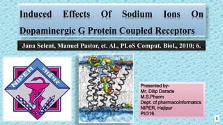
1. Induced Effects Of Sodium Ions On
Dopaminergic G Protein Coupled Receptors
1
Presented by-
Mr. Dilip Darade
M.S.Pharm
Dept. of pharmacoinformatics
NIPER, Hajipur
PI/316
Jana Selent, Manuel Pastor, et. Al., PLoS Comput. Biol., 2010; 6.
2. G protein coupled receptors
2
It is 7TM receptors, heptahelical receptors, serpentine receptor, and constitute a
large protein family of receptors, that detect molecules outside the cell and activate
internal signal transduction pathways and, ultimately, cellular responses.
It Coupling with G proteins, they are called seven-transmembrane receptors because
they pass through the cell membrane seven times.
Typically activated by orthosteric ligand binding and subject to allosteric
modulation.
At least 800 unique GPCRs, of which near about 460 are predicted to be olfactory
receptors.
It plays a significant role in controlling the sense of smell, taste, vision, hearing and
touch in humans.
4. Classification of GPCR
4
Dopaminergic
Receptors
Based on sequence similarity within the 7 transmembrane segments (TMs) & based on
sequence homology and functional similarity, GPCR can be divided into six families.
5. Dopaminergic Receptors
5
Used as drug targets for the treatment of CNS disorders (e.g. schizophrenia, Parkinson’s disease ).
D1 like family D2 like family
D1 receptor D5 receptor D2 receptor D3 receptor D4 receptor
Activates Adenylate
cyclase(Gs)
cAMP
Inhibition of
Adenylate cyclase(Gi)
cAMP
Dopamine receptors
Brain & Basal
Ganglia
Brain &
striatum
80% 75% 53%
7. Why
molecular
dynamic
simulation?
MD molecular dynamics
Allosteric modulation plays a central role in the GPCR signalling.
MD simulations is used for exploring the effect of sodium ion & mechanism
involved in their role as allosteric modulators.
X-ray crystallography & FRET
unable to provide structural
information of sufficient spatial
resolution & time scales for
describing specific atomic-level
aspects of allosteric interactions.
7
10. Methods
The TM region, it has a sequence homology of about 42% (calculation based on the
PAM 250 scoring matrix), which was suggested threshold of 30% acceptable for
transmembrane models of membrane proteins .
In their study, the high resolution X-ray structure of the β2 adrenergic receptor (
2RH1, 2.40 Å) was selected as a template for the D2 receptor modelling.
Superimposed- shows larger structural deviations in the intra- and extracellular loop
regions whereas high structural similarity was found in the TM region indicating that
the topology of the TM region is highly conserved .
Selected GPCRs closely-related phylogenetically to the D2 receptor (2RH1, 3D4S,
2VT4, 3EML).
10
11. Methods
NAME No. Of Runs Description
MD1 1 Single molecular dynamics trajectory of 1.1
microsecond using a GPU workstation
MD2 100 Multiple trajectories of approximately 50
ns/each on GPUGRID
MD3 25 Metadynamics runs of 14 ns/each on
GPUGRID
Molecular dynamics simulations was performed using ACEMD.
11
13. Allosteric modulation of Dopaminergic Receptors by sodium ions
Sodium ion’s pathway into the D2 receptor, receptor stability, and
conformational change of the toggle switch Trp 6.48. 13
Trp6.48 torsion
angle x2
180 ns 380 ns
MD2
Structural relaxation
Trp6.48 torsion
angle χ2
380 ns
14. Sodium Binding Sites in the TM Region
14
Sodium ion binding between the orthosteric and allosteric site.
15. Mechanism of the Sodium-Induced Effect
Mechanism of the sodium-induced effect on the rotamer switch (Trp 6.48). 15
16. Mechanism of the Sodium-Induced Effect
16Free energy profile of sodium binding
17. Conclusion
17
They showed the results of MD simulations at the microsecond scale to investigate the
sodium-induced effects on GPCRs, focusing on the dynamics and energetics of sodium
ions in the D2 receptor.
All-atom, unconstrained MD simulations show a two step binding of sodium ions to the
dopaminergic D2 receptor.
First, negatively charged residues (Asp400, Asp178, Glu181 and Glu95) in the
extracellular surface are involved in forming a large favourable volume for sodium at the
receptor entrance.
Second, the sodium ion visits three binding locations (a–c) between Asp3.32 (site a) and
Asp2.50 (site c). The computed energetics of the sodium ion binding indicate that site c is
energetically favoured over sites a and b.
The existence of the allosteric sodium binding site c is in agreement with experimental
data ,Most importantly, the computational results indicate that sodium ions that transit
from site a to c induce a conformational change acting like a fingertip toggling and
locking the rotamer switch (Trp6.48) in its inactive state.
PLoS(public library of science) ,In an effort to understand these effects, they performed microsecond scale all-atom molecular dynamics simulations on the dopaminergic D2 receptor, found that sodium ions enter the receptor from the extracellular side and bind at a deep allosteric site (Asp2.50).
Remarkably, the presence of a sodium ion at this allosteric site induces a conformational change of the rotamer toggle switch Trp6.48 which locks in a conformation identical to the one found in the partially inactive state of the crystallized human b2 adrenergic receptor.
This study provides detailed quantitative information about binding of sodium ions in the D2 receptor and reports a possibly important sodium-induced conformational change for modulation of D2 receptor function.
D receptors are grouped in two distinct families: D1-like receptors (which include D1 and D5 receptors) and D2-like receptors, like D2, D3, and D4 .
The main difference between those families of receptors concerns the action of G-proteins:
D1-like receptor has a Gs-protein whose activation results in an increase of cyclic AMP (cAMP) mediated by adenylate cyclase.
D2-like receptors are coupled to a Gi-protein which determines, instead, the inhibition of adenylate cyclase and, consequentially, a decrease of cAMP .
The D1 and D5 dopamine receptors are 80% homologous in their transmembrane domains, whereas the D3 and D4 dopamine receptors are 75 and 53% homologous,
respectively, with the D2 receptor.
Whereas the NH2- terminal domain has a similar number of amino acids in all of the dopamine receptors, the COOH-terminal for the D1-class receptors is seven times longer than that for the D2-class receptors.
FRET: fluorescence resonance energy transfer
An enzyme can catalyse the conversion of substrate to product.
However there are some drugs which are binds to a different site of enzymes called as allosteric site.
The bonding of inhibitor binding at allosteric site changes the shape of active site of an enzyme.
Such a way , substrate cannot binds with the enzyme.
When inhibitor leaves the allosteric site then enzyme agains catalyse the conversion of substrate to product.
2RH1-High resolution crystal structure of human B2-adrenergic G protein-coupled receptor.
2VT4-TURKEY BETA1 ADRENERGIC RECEPTOR WITH STABILISING MUTATIONS AND BOUND CYANOPINDOLOL.
3D4S-Cholesterol bound form of human beta2 adrenergic receptor.
3EML-The 2.6 A Crystal Structure of a Human A2A Adenosine Receptor bound to ZM241385.
PAM250- 250 mutations per 100 amino acid residues
In an effort to elucidate the mechanism of the sodium-induced effect, they have computationally investigated the mobility of sodium ions in the sodium-sensitive D2 receptor.
All-atom molecular dynamics (MD) simulations of the D2 receptor were performed to analyze more than 6 ms of simulation data.
This analysis comprises a single 1.1 ms long simulation (MD1), one hundred 50 ns simulations (MD2) and biased Metadynamics simulations (MD3) to compute accurate two-dimensional free energy profiles.
Metadynamics is a biased dynamics technique widely used to improve sampling for free energy calculations over a set of multidimensional reaction coordinates which would not be sampled exhaustively with normal unbiased simulations.
A 1.1 microsecond long-time MD simulation demonstrates that a sodium ion spontaneously penetrates the dopaminergic D2 receptor from the extracellular side.
Once inside the receptor, the sodium ion provokes a conformational change of the rotamer, switch Trp6.48.
Moreover, in the presence of a sodium ion site c (below the rotamer switch Trp6.48), a second sodium ion is able to enter at times the D2 receptor where it occupies the orthosteric site a (above the rotamer switch Trp6.48).
(A) Volumetric map of sodium ions (yellow isosurface) within the D2 receptor at chemical potential m=24kBT relative to the bulk concentration of 150 mM NaCl showing the sodium ions binding sites. The ions move from the extracellular loop (EL) along negatively charged residues (orange spheres) towards the receptor interior (sites a to c) as computed from the ion concentration over 4.7 ms (MD2) of data. The volumetric map of water (blue isosurface) computed at m=20.5kBT relative to bulk water illustrates that part of the receptor interior is filled with water molecules including sites a, b and c. (B) The RMSD of the D2 receptor model embedded in a hydrated lipid bilayer over MD1; grey line: TM region, blue line: whole receptor including TM and loop region. (C) Depiction of the sodium ion’s reaction coordinate z (blue line) and the Trp6.48 torsion angle x2, (grey line) over MD1. Sodium transition from Asp3.32 (site a) to Asp2.50 (site c) induces a conformational change of the Trp6.48 rotamer switch.
In site a (z,25.0 A ˚), the partial dehydration of the bound sodium ion and the receptor allows the formation of a salt bridge with the carboxylate group of Asp3.32.
At location b, rehydration of the ion and the protein side chain takes place. At this point, direct contacts between receptor and the ion are very limited due to the recovered ion’s hydration shell. The hydrated sodium ion occupies the space between Asp3.32, Gly7.42, Trp6.48, and Tyr7.43 for site b (z,27.5 A ˚).
The last binding location c is favoured by the electrostatic attraction between the positively charged bound sodium ion and the negatively charged Asp2.50. Both, the sodium ion and the carboxylate group of Asp2.50 partially dehydrate before forming a salt bridge, for site c .
In position c, the sodium ion is stabilized by a hydrogen-bonding network which involves interactions with Ser3.39, Asn7.45, Ser7.46 as well as the charge neutralizing interaction with Asp2.50.
Sodium-induced effect is mediated by cation-P interaction of the hydrated positively charged sodium ion and the negative charges of the Trp6.48 ring.
The sodium ion, water molecules as well as the Trp6.48 are colored according to their partial charges (blue: positive, red: negative).
Transition of a sodium ion from Asp3.32 (site a/b, x2 =0) to Asp2.50 (site c, x2 =100) induces a conformational change of Trp6.48 due to electrostatic interactions between the hydrated positively charged sodium ion and the negative charges of the indole ring of Trp6.48.
Two-dimensional free energy profile for (z,x2) reaction coordinates, where z is the z coordinate of the bound sodium ion and x2 is the dihedral angle (CA-CB-CG-CD1) of tryptophan Trp6.48. The binding site a, b, c of the receptor show the pathway for sodium in the receptor and the locking of the tryptophan into the 100 degrees position (inactive state). The vertical white line shows the approximate position of Trp6.48.
(B) Location of binding sites a, b, c in the D2 receptor.
(C) Associated absolute free energy error map of the free energy profile of (A) computed by 25 metadynamics runs of 14 ns each (MD3). All basins have sub-kcal/mol error while only higher hills in the free energy surface show more significant variations.