Case Review #24: 67 year old female with Degenerative Scoliosis
•
1 like•798 views

67 year old female presented with DeNovo Scoliosis, with significant rotation due to Adolescent Idiopathic Scoliosis. Dr. Pashman treated the patient with a posterior spinal fusion from T10-Pelvis. KIM/SRP Classification 2.

Recommended
Recommended
More Related Content
What's hot
What's hot (20)
Viewers also liked
Viewers also liked (19)
Similar to Case Review #24: 67 year old female with Degenerative Scoliosis
Similar to Case Review #24: 67 year old female with Degenerative Scoliosis (20)
More from Robert Pashman
More from Robert Pashman (11)
Recently uploaded
Recently uploaded (20)
Case Review #24: 67 year old female with Degenerative Scoliosis
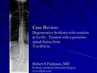
- 1. Case Review: Degenerative Scoliosis with rotation at L4-S1. Treated with a posterior spinal fusion from T10-Pelvis. Robert S Pashman, MD Scoliosis and Spinal Deformity Surgery www.eSpine.com
- 2. Patient History 67 year old female Status post intralaminar decompression on April 15, 2003, for primary complaints of radiculopathy in her right leg. Now she is having increased back pain, left pain radiating to her low back and laterally to her thigh. She has slight forward decompensation with gait, this due to lack of lumbar lordosis; 36 x 14 showed a collapsing degenerative scoliosis centered around L4 to S1 with significant rotation probably based on Adolescent Idiopathic Scoliosis with degenerative component. Low back pain especially with sitting On physical examination she is decompensated in the coronal plane and has thoracolumbar kyphosis up to approximately T12.
- 3. Indications for Surgery Adult Idiopathic/Denovo Scoliosis, thoracolumbar spine. Lumbar kyphosis, status post degeneration of the lumber spine. Status post interlaminar decompression, L3-4, L4-5 and L5-S1 on the right for radiculopathy. Now with increasing low back and radicular pain due to the above diagnoses. Multiple co-morbidities including osteopenia and pain medicine.
- 4. Surgical Strategy 1. Segmental spinal instrumentation at thoracic 10 to sacral pelvis using 1/4 inch stainless steel pedicle screw/rod construct. 2. Multiple level Smith-Peterson osteotomy, thoracolumbar spine T12 to L3 with bilateral radical facetectomy, under the microscope. 3. Interlaminar laminotomies with facetectomy, re-exploration and decompression, L3-4, L4-5, L5-S1 on the right. 4. Posterior spinal fusion using locally harvested autogenous bone and RH BMP, T10 to sacral pelvis. 5. Intraoperative somatosensory evoked potentials. 6. Intraoperative fluoroscopy.
- 5. Post-Op Films The patient is 6 months status post posterior instrumented fusion for Kim/SRP type 2 adult idiopathic curve. The patient is doing well. She has no radiculopathy, minimal low back pain, and is returning to tennis wearing a brace.