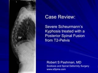
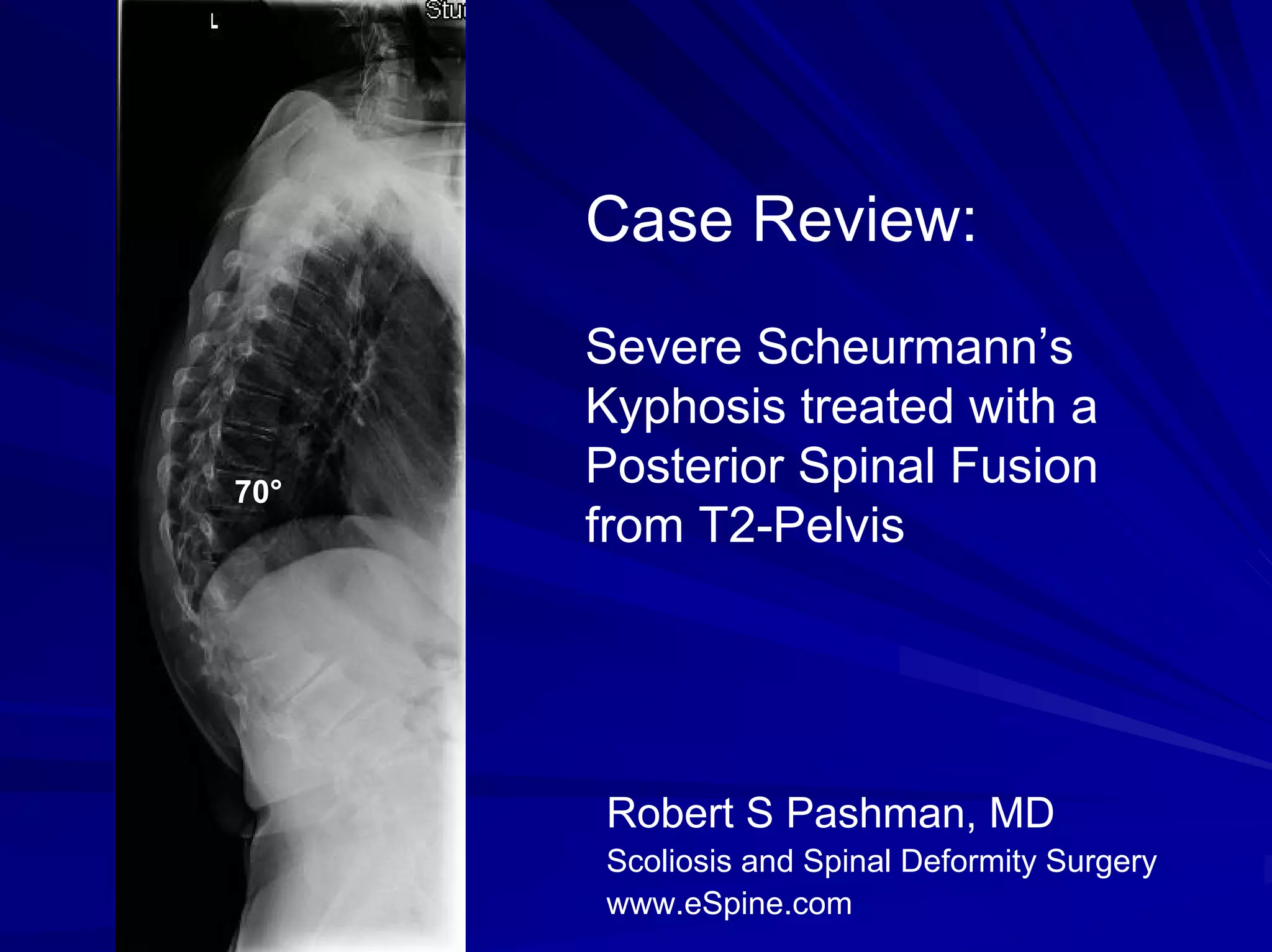
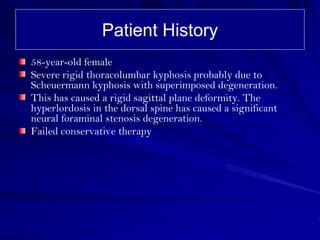
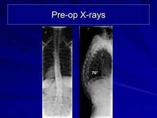
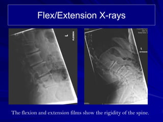
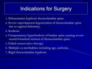
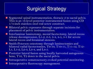
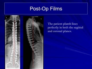
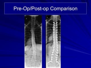
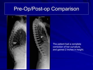
Case Review:
      Severe Scheurmann’s
      Kyphosis treated with a
70°
      Posterior Spinal Fusion
      from T2-Pelvis


      Robert S Pashman, MD
      Scoliosis and Spinal Deformity Surgery
      www.eSpine.com
Patient History
58-year-old female
Severe rigid thoracolumbar kyphosis probably due to
Scheuermann kyphosis with superimposed degeneration.
This has caused a rigid sagittal plane deformity. The
hyperlordosis in the dorsal spine has caused a significant
neural foraminal stenosis degeneration.
Failed conservative therapy
Pre-op X-rays


         70°
Flex/Extension X-rays


The flexion and extension films show the rigidity of the spine.
Indications for Surgery
1. Scheuermann kyphosis thoracolumbar spine.
2. Severe superimposed degeneration of thoracolumbar spine
   due to sagittal deformity.
3. Scoliosis.
4. Compensatory hyperlordosis of lumbar spine causing severe
   neural foraminal stenosis of thoracolumbar spine.
5. Failed conservative therapy.
6. Multiple co-morbidities including age, cachexia.
7. Rigid thoracolumbar kyphosis.
Surgical Strategy
Segmental spinal instrumentation, thoracic 2 to sacral pelvis.
This is an 18-level posterior instrumented fusion using CD
1/4-inch stainless steel rod screw construct.
Bilateral pelvic exposures through separate incisions for
placement of pelvic instrumentation.
Interlaminar laminotomy, mesial facetectomy, lateral recess
release decompression, L1-2, 2-3, 3-4, 4-5, 5-1 for severe
lateral recess and foraminal stenosis.
Smith-Peterson osteotomy through laminectomies and
bilateral radial facetectomies, T9-10, T10-11, T11-12, T12-
L1, L1-2, L2-3, L3-4, and L4-5.
Posterior lateral fusion using locally harvested autogenous
bone, T10, T2 down to the sacral pelvis.
Intraoperative somatosensory evoked potential monitoring.
Intraoperative fluoroscopy management.
Post-Op Films

       The patient plumb lines
       perfectly in both the sagittal
       and coronal planes.
Pre-Op/Post-op Comparison


          0°

70°            The patient had a complete
               correction of her curvature,
               and gained 2 inches in height.

Case Review 4: A 58 year old female presented with Scheurmann's Kyphosis

  • 1.
    Case Review: Severe Scheurmann’s Kyphosis treated with a 70° Posterior Spinal Fusion from T2-Pelvis Robert S Pashman, MD Scoliosis and Spinal Deformity Surgery www.eSpine.com
  • 2.
    Patient History 58-year-old female Severerigid thoracolumbar kyphosis probably due to Scheuermann kyphosis with superimposed degeneration. This has caused a rigid sagittal plane deformity. The hyperlordosis in the dorsal spine has caused a significant neural foraminal stenosis degeneration. Failed conservative therapy
  • 3.
  • 4.
    Flex/Extension X-rays The flexionand extension films show the rigidity of the spine.
  • 5.
    Indications for Surgery 1.Scheuermann kyphosis thoracolumbar spine. 2. Severe superimposed degeneration of thoracolumbar spine due to sagittal deformity. 3. Scoliosis. 4. Compensatory hyperlordosis of lumbar spine causing severe neural foraminal stenosis of thoracolumbar spine. 5. Failed conservative therapy. 6. Multiple co-morbidities including age, cachexia. 7. Rigid thoracolumbar kyphosis.
  • 6.
    Surgical Strategy Segmental spinalinstrumentation, thoracic 2 to sacral pelvis. This is an 18-level posterior instrumented fusion using CD 1/4-inch stainless steel rod screw construct. Bilateral pelvic exposures through separate incisions for placement of pelvic instrumentation. Interlaminar laminotomy, mesial facetectomy, lateral recess release decompression, L1-2, 2-3, 3-4, 4-5, 5-1 for severe lateral recess and foraminal stenosis. Smith-Peterson osteotomy through laminectomies and bilateral radial facetectomies, T9-10, T10-11, T11-12, T12- L1, L1-2, L2-3, L3-4, and L4-5. Posterior lateral fusion using locally harvested autogenous bone, T10, T2 down to the sacral pelvis. Intraoperative somatosensory evoked potential monitoring. Intraoperative fluoroscopy management.
  • 7.
    Post-Op Films The patient plumb lines perfectly in both the sagittal and coronal planes.
  • 8.
  • 9.
    Pre-Op/Post-op Comparison 0° 70° The patient had a complete correction of her curvature, and gained 2 inches in height.