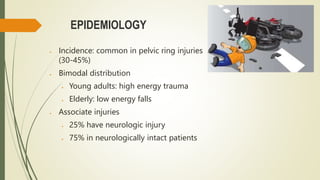
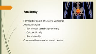
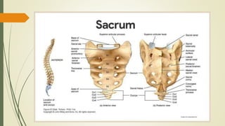
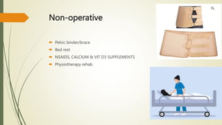
2. INTRODUCTION
Sacral fractures involve a break in one or more sacral bones (S1-S5)
Estimated incidence is about 1-3% of all spine fractures
3. EPIDEMIOLOGY
Incidence: common in pelvic ring injuries
(30-45%)
Bimodal distribution
Young adults: high energy trauma
Elderly: low energy falls
Associate injuries
25% have neurologic injury
75% in neurologically intact patients
4. Anatomy
Formed by fusion of 5 sacral vertebrae
Articulates with:
5th lumbar vertebra proximally
Coccyx distally
Ilium laterally
Contains 4 foramina for sacral nerves
5.
6. Nerves
L5 nerve root runs on top of sacral
ala
S1-S4 nerves through sacral
foramina
S1 and S2 nerves have higher injury
rates
Lower sacral nerves control:
Anal sphincter
Bulbocavernosus reflex
Perianal sensation
7. Presentation
- History: Trauma, falls in elderly
Symptoms: Peripelvic pain
Exam findings:
Palpate stability, swelling, open injuries
Neurological exam of lower extremities
Vascular exam of distal pulses
9. Denis classification
Zone 1
Fracture lateral to foramina
Most common (50%)
Nerve injury rare (5%).
-usually occurs to L5 nerve root
Zone 2
Fracture through foramina
May be stable vs. unstable
Zone 2 fracture with shear component highly unstable
Unstable fractures have higher risk of nonunion and poor functional
outcome
Zone 3
Fracture medial to foramina into the spinal canal
Highest rate of neurologic deficit (60%)
-bowel, bladder, and sexual dysfunction
14. Isler classification
Used for fractures that involve the lumbosacral
articulation:
• Isler 1: fracture occurs lateral to the L5/S1 facet
• Isler 2: fractures line involves the L5/S1 facet
• Isler 3: fracture line extends medially to the L5/S1
facet
15. Imaging
Radiographs show only 30% of fractures
CT is diagnostic study of choice
MRI for suspected neural compromise
19. Surgical Approaches for Sacral Fractures
Common approaches
Posterior approach
Anterior approach
Combined anterior and posterior approach
Factors determining choice of approach
Location and pattern of fracture
Associated injuries
Surgeon preference/experience
Operative
20. Fixation Options
Posterior fixation
Percutaneous iliosacral screws
Spinopelvic fixation
Sacroiliac plating
Anterior fixation
Sacral plating
Iliosacral screws
Lumbopelvic fixation
Decompression for neurologic injury
Goal is to achieve stabilization and allow bone healing
21. Post-operative Care & Rehabilitation
Early mobilization as tolerated
Physical therapy
Range of motion exercises
Strengthening
Balance and gait training
Management of bowel/bladder dysfunction if present
Follow up imaging to assess healing
Goal is to maximize function and quality of life
24. SUMMARY
o Sacral fractures are common pelvic ring injuries that are under-diagnosed and
often associated with neurologic compromise.
o Diagnosis can made with pelvis radiographs but frequently require pelvic CT
scan for full characterization.
o Treatment may be nonoperative or operative depending on fracture
displacement, associated pelvic ring instability and patient activity demands.