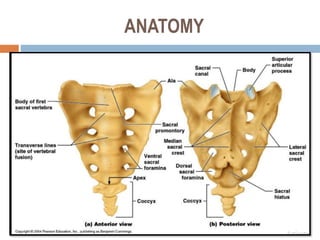
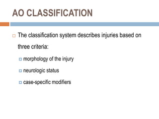
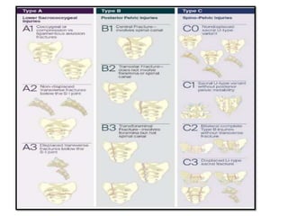
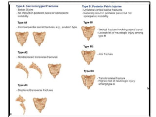
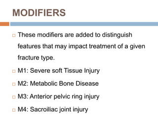
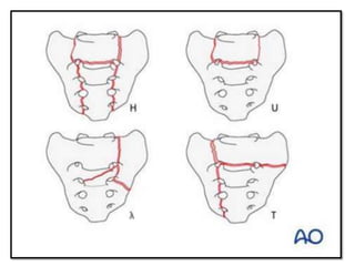
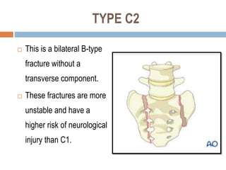
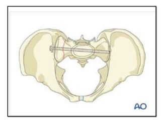
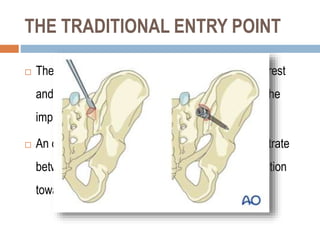
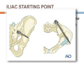
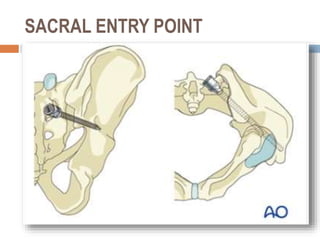
SACRAL FRACTURES
DR ASIF JATOI
SENIOR REGISTRAR
CMH RAWALPINDI
 The sacrum is an irregularly shaped bone, made up of a group of five fused
vertebrae
 The sacrum plays a central role in the stability of both the pelvis and the
spinal column.
 The important neurologic structures the lower sacral
roots and autonomic nerves that are important for continence of the bowel
and bladder and sexual function
ANATOMY
 frequently missed at presentation because these
associated with high-energy trauma and present with
multiple injuries & hemodynamically unstable.
 The neurologic evaluation must include DRE to assess
 rectal tone
 maximal contraction of the anal sphincter and
 rectal tears and anterior perineal lacerations.
 The L5 nerve is at risk at the anterior junction of the ala
and the sacral promontory, and the S1 nerve root can be
injured within the foramen.
 Extremity motor and sensory testing and rectal
examination
IMAGING STUDIOS
 Plain radiographs have not proven sensitive .
 CT scan of the pelvis with 2-mm slices and sagittal and
coronal reformatted images should be obtained
 When associated neurologic deficits with displaced
fractures, MRI also may be of value,
DENIS CLASSIFCATION
 Zone 1-lateral to the foramina
 50% of injuries with a 6% incidenceof L5 and S1
injuries
 Zone 2 -through the foramina
 34% of injuries, and 28% with deficits unilaterally at
the L5, S1, or S2 levels
 Zone 3- medial to the foramen
and involve the spinal canal
 Remaining 16% of injuries
ROY-CAMILLE CLASSIFICATION
ISLER CLASSIFICATION
AO CLASSIFICATION
 The classification system describes injuries based on
three criteria:
 morphology of the injury
 neurologic status
 case-specific modifiers
MORPHOLOGY OF THE INJURY
 TYPE A INJURIES
 are lower sacro-coccygeal fractures with no impact on the posterior pelvic or spino-pelvic
stability.
 TYPE B INJURIES
 are unilateral longitudinal (vertical) sacral fractures which result in posterior pelvic
instability but no impact on spino-pelvic stability.
 TYPE C INJURIES
 include unilateral B injuries with L5-S1 facet involvement, bilateral longitudinal
(vertical) sacral fractures and U fracture variations resulting in spino-pelvic
instability.
NEUROLOGY
 Neurological injuries are classified as follows:
 Nx: Cannot be examined
 N0: No neurological deficits
 N1: Transient neurological injury
 N2: Nerve root injury
 N3: Cauda Equina Syndrome/Incomplete SCI
 N4: Complete SCI*
MODIFIERS
 These modifiers are added to distinguish
features that may impact treatment of a given
fracture type.
 M1: Severe soft Tissue Injury
 M2: Metabolic Bone Disease
 M3: Anterior pelvic ring injury
 M4: Sacroiliac joint injury
TREATMENT
 Type A1 and A2 are managed conservatively
 Type A3, by sacral alar plating or laminectomy
 Type B1,B2, B3 are managed with illiosacral screws or
spinopelvic fixation.
C0 Nondisplaced U-fracture
 This is a nondisplaced
sacral U-type fracture,
result from low-energy
injuries.
 commonly seen as
insufficiency fractures in
patients with metabolic
bone disease.
 Treated conservatively
or by Iliosacral screws
(ISS)
TYPE C1
 This is any unilateral B-type
fracture involving a fracture of
the ipsilateral L5-S1 joint.
 This fracture type may impact
spino-pelvic instability and is
therefore classified as a C
fracture
TYPE C2
 This is a bilateral B-type
fracture without a
transverse component.
 These fractures are more
unstable and have a
higher risk of neurological
injury than C1.
TYPE C3
 This is a displaced U-type
fracture. It has a similar
instability profile as C2,
 but due to the transverse
fracture displacement it has
a higher likelihood of
neurological injury.

 Type C1, C2 and C3 are treated surgically by
spinopelvoc fixation and Fixation of associated pelvic
ring injuries.
ILIOSACRAL SCREW (ISS) FIXATION
 Iliosacral screw (ISS) fixation is a fluoroscopically
guided, percutaneous procedure.
 Its primary use is for fixation of satisfactorily reduced
sacral fractures or sacro-iliac joint disruptions.
 Anatomic reduction must be obtained before ISS
insertion.
REDUCTION
 entry point should be
anterior in S1 and
inferior to the iliac
cortical density (ICD),
which parallels the sacral
alar slope, usually
slightly caudal and
posterior
INSERTION OF ILIAC SCREW
 There are two standard
iliac screw starting points
within the ilium and one
within the sacrum.
THE TRADITIONAL ENTRY POINT
 The traditional entry point is in the posterior iliac crest
and countersunk to prevent pressure ulcers over the
implant.
 An oscillating drill (3.5 mm) or awl is used to penetrate
between the two cortices in a ventral, caudal direction
toward the anterior inferior iliac spine.
ILIAC STARTING POINT
 is referred to as anatomic starting point. It is more caudal
and medial than the traditional starting point and aligns
better with the lumbar pedicles.
 The more caudal position places this screw in a wider
cross section of bone above the sciatic notch
SACRAL ENTRY POINT
 The third starting point is at the inferolateral
aspect of the S1 foramen.

Sacral fractures.pptx

  • 1.
    SACRAL FRACTURES DR ASIFJATOI SENIOR REGISTRAR CMH RAWALPINDI
  • 2.
     The sacrumis an irregularly shaped bone, made up of a group of five fused vertebrae  The sacrum plays a central role in the stability of both the pelvis and the spinal column.  The important neurologic structures the lower sacral roots and autonomic nerves that are important for continence of the bowel and bladder and sexual function
  • 3.
  • 5.
     frequently missedat presentation because these associated with high-energy trauma and present with multiple injuries & hemodynamically unstable.  The neurologic evaluation must include DRE to assess  rectal tone  maximal contraction of the anal sphincter and  rectal tears and anterior perineal lacerations.
  • 7.
     The L5nerve is at risk at the anterior junction of the ala and the sacral promontory, and the S1 nerve root can be injured within the foramen.  Extremity motor and sensory testing and rectal examination
  • 8.
    IMAGING STUDIOS  Plainradiographs have not proven sensitive .  CT scan of the pelvis with 2-mm slices and sagittal and coronal reformatted images should be obtained  When associated neurologic deficits with displaced fractures, MRI also may be of value,
  • 9.
    DENIS CLASSIFCATION  Zone1-lateral to the foramina  50% of injuries with a 6% incidenceof L5 and S1 injuries  Zone 2 -through the foramina  34% of injuries, and 28% with deficits unilaterally at the L5, S1, or S2 levels  Zone 3- medial to the foramen and involve the spinal canal  Remaining 16% of injuries
  • 10.
  • 11.
  • 12.
    AO CLASSIFICATION  Theclassification system describes injuries based on three criteria:  morphology of the injury  neurologic status  case-specific modifiers
  • 13.
    MORPHOLOGY OF THEINJURY  TYPE A INJURIES  are lower sacro-coccygeal fractures with no impact on the posterior pelvic or spino-pelvic stability.  TYPE B INJURIES  are unilateral longitudinal (vertical) sacral fractures which result in posterior pelvic instability but no impact on spino-pelvic stability.  TYPE C INJURIES  include unilateral B injuries with L5-S1 facet involvement, bilateral longitudinal (vertical) sacral fractures and U fracture variations resulting in spino-pelvic instability.
  • 16.
    NEUROLOGY  Neurological injuriesare classified as follows:  Nx: Cannot be examined  N0: No neurological deficits  N1: Transient neurological injury  N2: Nerve root injury  N3: Cauda Equina Syndrome/Incomplete SCI  N4: Complete SCI*
  • 17.
    MODIFIERS  These modifiersare added to distinguish features that may impact treatment of a given fracture type.  M1: Severe soft Tissue Injury  M2: Metabolic Bone Disease  M3: Anterior pelvic ring injury  M4: Sacroiliac joint injury
  • 18.
    TREATMENT  Type A1and A2 are managed conservatively  Type A3, by sacral alar plating or laminectomy  Type B1,B2, B3 are managed with illiosacral screws or spinopelvic fixation.
  • 20.
    C0 Nondisplaced U-fracture This is a nondisplaced sacral U-type fracture, result from low-energy injuries.  commonly seen as insufficiency fractures in patients with metabolic bone disease.  Treated conservatively or by Iliosacral screws (ISS)
  • 22.
    TYPE C1  Thisis any unilateral B-type fracture involving a fracture of the ipsilateral L5-S1 joint.  This fracture type may impact spino-pelvic instability and is therefore classified as a C fracture
  • 23.
    TYPE C2  Thisis a bilateral B-type fracture without a transverse component.  These fractures are more unstable and have a higher risk of neurological injury than C1.
  • 24.
    TYPE C3  Thisis a displaced U-type fracture. It has a similar instability profile as C2,  but due to the transverse fracture displacement it has a higher likelihood of neurological injury. 
  • 25.
     Type C1,C2 and C3 are treated surgically by spinopelvoc fixation and Fixation of associated pelvic ring injuries.
  • 28.
    ILIOSACRAL SCREW (ISS)FIXATION  Iliosacral screw (ISS) fixation is a fluoroscopically guided, percutaneous procedure.  Its primary use is for fixation of satisfactorily reduced sacral fractures or sacro-iliac joint disruptions.  Anatomic reduction must be obtained before ISS insertion.
  • 29.
  • 30.
     entry pointshould be anterior in S1 and inferior to the iliac cortical density (ICD), which parallels the sacral alar slope, usually slightly caudal and posterior
  • 32.
    INSERTION OF ILIACSCREW  There are two standard iliac screw starting points within the ilium and one within the sacrum.
  • 33.
    THE TRADITIONAL ENTRYPOINT  The traditional entry point is in the posterior iliac crest and countersunk to prevent pressure ulcers over the implant.  An oscillating drill (3.5 mm) or awl is used to penetrate between the two cortices in a ventral, caudal direction toward the anterior inferior iliac spine.
  • 34.
    ILIAC STARTING POINT is referred to as anatomic starting point. It is more caudal and medial than the traditional starting point and aligns better with the lumbar pedicles.  The more caudal position places this screw in a wider cross section of bone above the sciatic notch
  • 35.
    SACRAL ENTRY POINT The third starting point is at the inferolateral aspect of the S1 foramen.