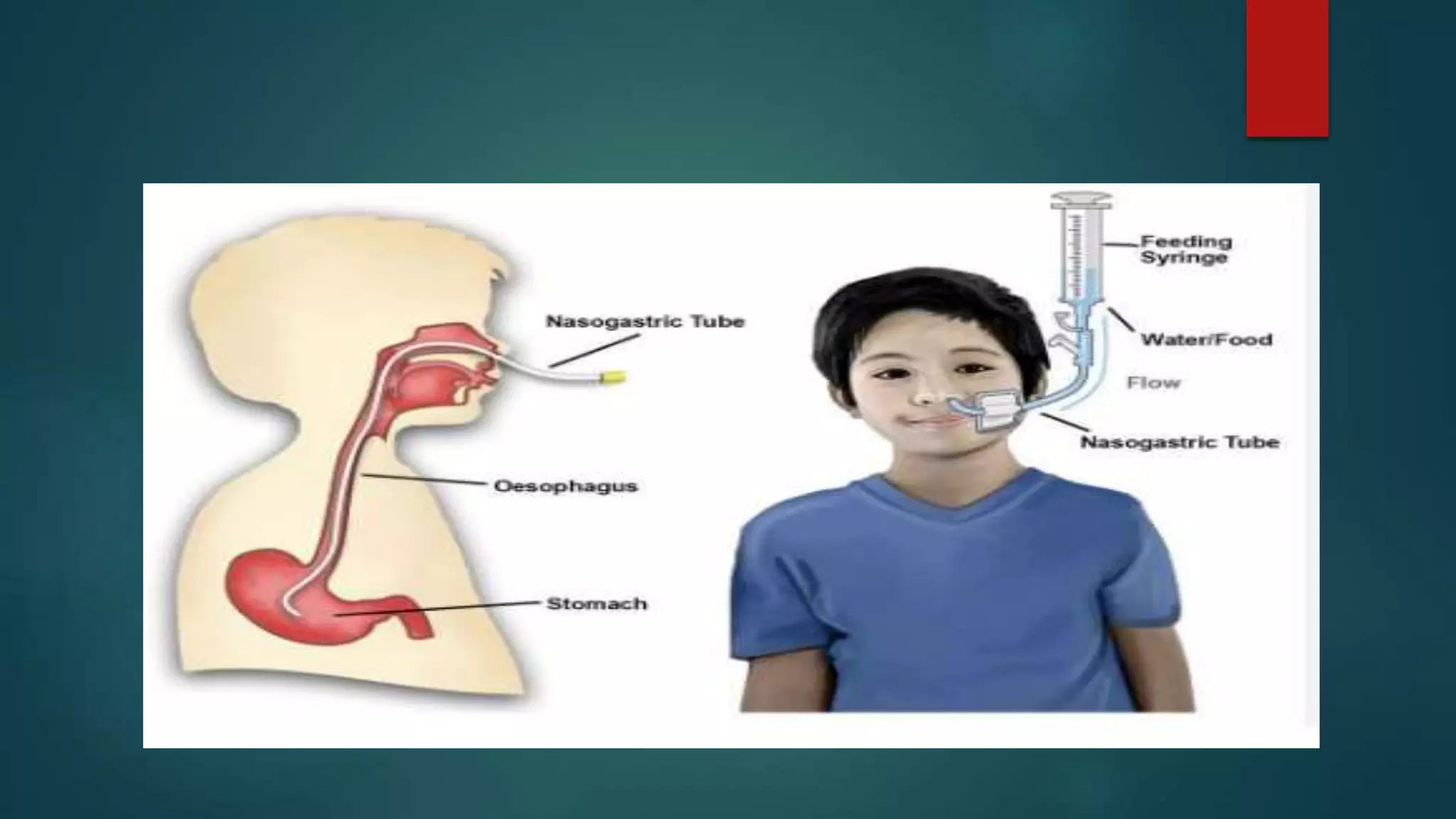
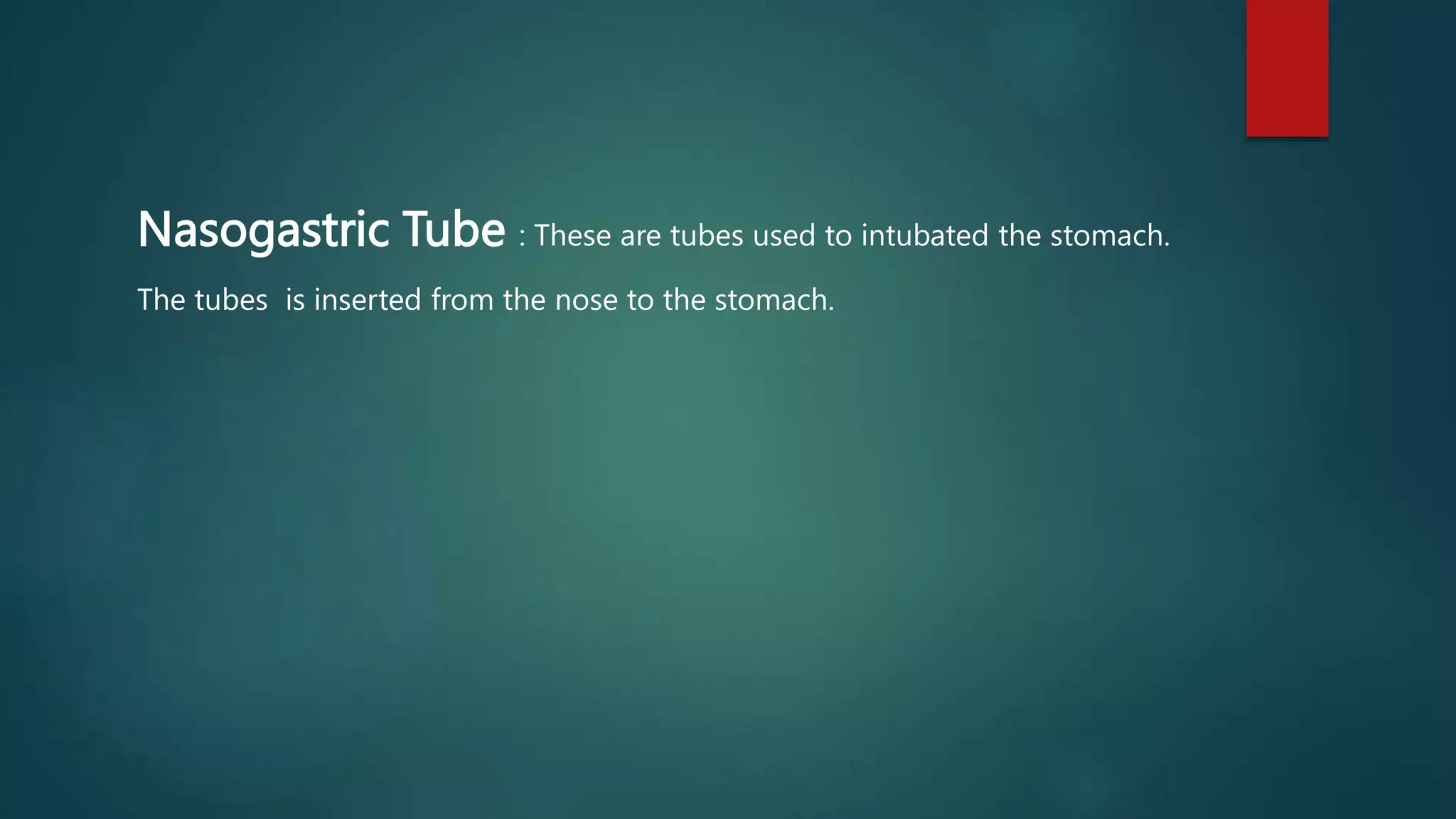
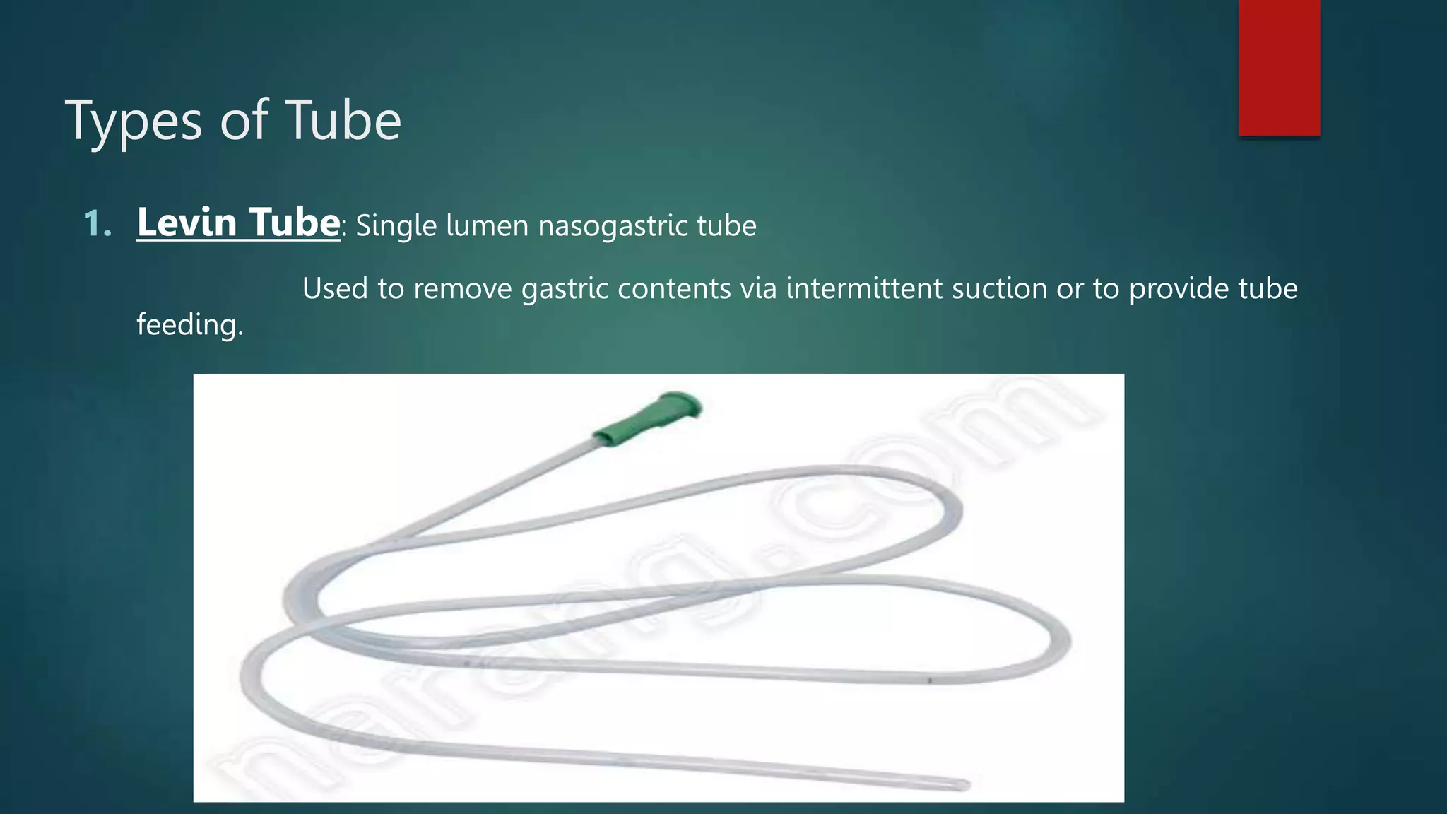
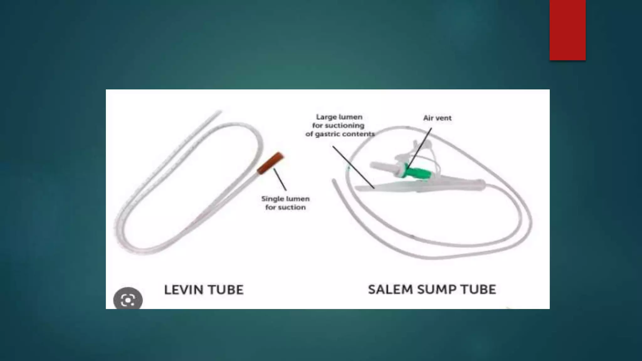
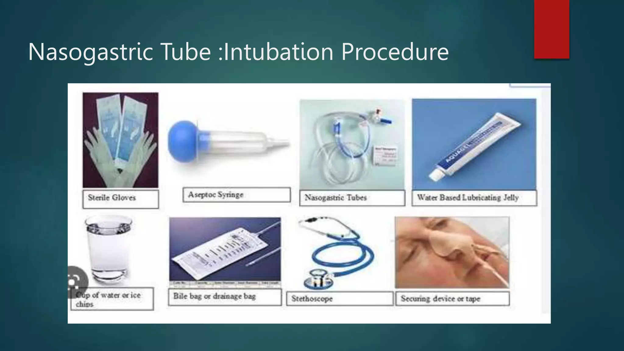
Ryles Tube Insertion
And Feeding
PRESENTED BY
ANKIT GEORGE
Nasogastric Tube : These are tubes used to intubated the stomach.
The tubes is inserted from the nose to the stomach.
Purpose:
 To decompress the stomach by removing fluids or gas to promote abdominal comfort .
 To allow surgical anastomoses to heal without distention
 To decrease the risk of aspiration
 To administer medication to client who are unable to swallow.
 To provide nutrition by acting as a temporary feeding tube.
 To irrigate the stomach and remove toxic substance such as in poisioning.
Types of Tube
1. Levin Tube: Single lumen nasogastric tube
Used to remove gastric contents via intermittent suction or to provide tube
feeding.
2 .Salem sump tube : A salem sump is a double lumen nasogastric tube with an air vent ( pigtail)
used for decompression with intermittent continuous suction .
Nasogastric Tube :Intubation Procedure
Nasogastric Tubes :Intubation Procedure
1. Follow agency procedures.
2. Explain the procedure and its potential discomfort to the
client.
3. Position the client in a high Fowler's position with pillows
behind the shoulders.
4. Determine which nostril is more patent.
5. Measure the length of the tube from the bridge of the
nose to
the earlobe to the xiphoid process and indicate this length
with a piece of tape on the tube (remember the
abbreviation
NEX, which stands for nose, earlobe, and xiphoid process).
6. If the client is conscious and alert, have him or her swallow
or drink water (follow agency procedure).
7. Lubricate the tip of the tube with water-soluble lubricant.
8. Gently insert the tube into the nasopharynx and advance
the tube.
9. When the tube nears the back of the throat (first black mea-
surement on the tube), instruct the client to swallow or
drink sips of water (unless contraindicated). If resistance
is met, slowly rotate and aim the tube downward and
toward the closer ear; in the intubated or semiconscious cli-
ent, flex the head toward the chest while passing the tube
10. Immediately withdraw the tube if any change is noted in the
client's respiratory status.
11. Following insertion, obtain an abdominal x-ray study to
confirm placement of the tube.
12. Connect the tube to suction, to either the intermittent or the
continuous suction setting, as prescribed if the purpose o
the tube is for decompression.
13. Secure the tube to the client's nose with adhesive tape and
to the client's gown (follow agency procedure and check for
in client allergy to tape).
14. Observe the client for nausea, vomiting, abdominal full-
ness, or distention and monitor gastric output.
15. Check residual volumes every 4 hours, before each feeding,
and before giving medications. Aspirate all stomach con-
tents (residual) and measure the amount. Reinstill residual
contents to prevent excessive fluid and electrolyte losses,
unless the residual contents appear abnormal or the vol-
ume is large (greater than 250 mL). Always follow agency
procedure. Withhold a feeding if the residual amount is
more than 100 mL or according to agency or nutritional
consult recommendations.
16. Before the instillation of any substance through the tube
(i.e., irrigation solution, feeding, medications), aspirate
stomach contents and test the pH (a pH of 3.5 or lower indi-
cates that the tip of the tube is in a gastric location).
17. If irrigation is indicated, use normal saline solution .
18. Observe the client for fluid and electrolyte balance.
19. Instruct the client about movement to prevent nasal irrita-
tion and dislodgment of the tube.
20. On a daily basis, remove the adhesive tape that is securing
the tube to the nose and clean and dry the skin, assessing
for excoriation; then reapply the tape.
Administration Of Feeding
1. Check the HCP’s prescription and hospital policy regarding residual
amount usually, if the residual is less than 100 ml feeding is
administered, large volume aspirates indicated dealayed gastric
emptying and place the client at risk of aspiration.
2. Assess bowel sounds, hold th feeding and notify HCP if bowel sound
are absent .
3. Position the client in a high Fowler’s position ;if comatose.
4. Assess tube placement by aspirating gastric contents and measuring
the pH(should be 3.5 or lower)
4. Shake the formula well before pouring it into the container
(feeding bag). Some feedings require the use of a bag in which
formula is added, or require the use of bottles that feeding tubing
can be attached to directly. The tubing some- times has a Y-site
connection so a regular flush can be programmed using the pump
rather than using a piston syringe.
5. Always assess bowel sounds; do not administer
any feedings if bowel sounds are absent.
6. Administer the feeding at the prescribed rate or
via gravity flow (intermittent bolus feedings)
with a 50- to 60-mL syringe with the plunger
removed.
7. Gently flush with 30 to 50 mL of water or NS
(depending on agency policy) using the irriga-
tion syringe after the feeding.
Precautions:
1. Change the feeding container and tubing every 24
hours or per agency policy.
2. Do not hang more solution than is required for a
4-hour period; this prevents bacterial growth.
3. Check the expiration date on the formula before
administering.
5. Aspirates all stomach contents ( residual),measure the amount and
return the contents to the stomach to prevent electrolyte imbalance
(unless the color or characteristics of the residual is abnormal or the
amount is greater than 250ml)
6.Warm the feeding to room temperature to prevent diarrhea and cramps.
7. For bolus feeding maintain the client in a High Fowler’s position for 30
minutes after the feeding
8. For continuous feeding, keep the client in a semi-Fowler,s position at all
time.
Prevention of Complication
1. Diarrhea
a. Assess the client for lactose intolerance.
b. Use fiber-containing feedings.
c. Administer feeding slowly and at room
temperature.
2. Aspiration
a. Verify tube placement.
b. Do not administer the feeding if residual is
more than 100 mL (check HCP's prescription
and agency policy).
c. Keep the head of the bed elevated.
d. If aspiration occurs, suction as needed, assess respiratory rate,
auscultate lung sounds, monitor temperature for aspiration
pneumonia, and prepare to obtain a chest radiograph.
3. Clogged tube
a. Use liquid forms of medication, if possible.
b. Flush the tube with 30 to 50 mL of water or NS before and after
medication administration and before and after bolus feeding
c. Flush with water every 4 hours for continuous
feeding.
4. Vomiting
a. Administer feedings slowly and, for bolus feedings, make feeding
last for at least 30 minutes.
b. Measure abdominal girth.
c. Do not allow the feeding bag to empty.
d. Do not allow air to enter the tubing.
e. Administer the feeding at room temperature.
f. Elevate the head of the bed.
g. Administer antiemetics as prescribed.
Ryles Tube Insertion And Feeding.pptx

Ryles Tube Insertion And Feeding.pptx

  • 1.
    Ryles Tube Insertion AndFeeding PRESENTED BY ANKIT GEORGE
  • 3.
    Nasogastric Tube :These are tubes used to intubated the stomach. The tubes is inserted from the nose to the stomach.
  • 4.
    Purpose:  To decompressthe stomach by removing fluids or gas to promote abdominal comfort .  To allow surgical anastomoses to heal without distention  To decrease the risk of aspiration  To administer medication to client who are unable to swallow.  To provide nutrition by acting as a temporary feeding tube.  To irrigate the stomach and remove toxic substance such as in poisioning.
  • 5.
    Types of Tube 1.Levin Tube: Single lumen nasogastric tube Used to remove gastric contents via intermittent suction or to provide tube feeding.
  • 6.
    2 .Salem sumptube : A salem sump is a double lumen nasogastric tube with an air vent ( pigtail) used for decompression with intermittent continuous suction .
  • 8.
  • 9.
    Nasogastric Tubes :IntubationProcedure 1. Follow agency procedures. 2. Explain the procedure and its potential discomfort to the client. 3. Position the client in a high Fowler's position with pillows behind the shoulders. 4. Determine which nostril is more patent.
  • 10.
    5. Measure thelength of the tube from the bridge of the nose to the earlobe to the xiphoid process and indicate this length with a piece of tape on the tube (remember the abbreviation NEX, which stands for nose, earlobe, and xiphoid process).
  • 11.
    6. If theclient is conscious and alert, have him or her swallow or drink water (follow agency procedure). 7. Lubricate the tip of the tube with water-soluble lubricant. 8. Gently insert the tube into the nasopharynx and advance the tube.
  • 12.
    9. When thetube nears the back of the throat (first black mea- surement on the tube), instruct the client to swallow or drink sips of water (unless contraindicated). If resistance is met, slowly rotate and aim the tube downward and toward the closer ear; in the intubated or semiconscious cli- ent, flex the head toward the chest while passing the tube
  • 13.
    10. Immediately withdrawthe tube if any change is noted in the client's respiratory status. 11. Following insertion, obtain an abdominal x-ray study to confirm placement of the tube. 12. Connect the tube to suction, to either the intermittent or the continuous suction setting, as prescribed if the purpose o the tube is for decompression.
  • 14.
    13. Secure thetube to the client's nose with adhesive tape and to the client's gown (follow agency procedure and check for in client allergy to tape). 14. Observe the client for nausea, vomiting, abdominal full- ness, or distention and monitor gastric output.
  • 15.
    15. Check residualvolumes every 4 hours, before each feeding, and before giving medications. Aspirate all stomach con- tents (residual) and measure the amount. Reinstill residual contents to prevent excessive fluid and electrolyte losses, unless the residual contents appear abnormal or the vol- ume is large (greater than 250 mL). Always follow agency procedure. Withhold a feeding if the residual amount is more than 100 mL or according to agency or nutritional consult recommendations.
  • 16.
    16. Before theinstillation of any substance through the tube (i.e., irrigation solution, feeding, medications), aspirate stomach contents and test the pH (a pH of 3.5 or lower indi- cates that the tip of the tube is in a gastric location). 17. If irrigation is indicated, use normal saline solution .
  • 17.
    18. Observe theclient for fluid and electrolyte balance. 19. Instruct the client about movement to prevent nasal irrita- tion and dislodgment of the tube. 20. On a daily basis, remove the adhesive tape that is securing the tube to the nose and clean and dry the skin, assessing for excoriation; then reapply the tape.
  • 18.
    Administration Of Feeding 1.Check the HCP’s prescription and hospital policy regarding residual amount usually, if the residual is less than 100 ml feeding is administered, large volume aspirates indicated dealayed gastric emptying and place the client at risk of aspiration. 2. Assess bowel sounds, hold th feeding and notify HCP if bowel sound are absent . 3. Position the client in a high Fowler’s position ;if comatose. 4. Assess tube placement by aspirating gastric contents and measuring the pH(should be 3.5 or lower)
  • 19.
    4. Shake theformula well before pouring it into the container (feeding bag). Some feedings require the use of a bag in which formula is added, or require the use of bottles that feeding tubing can be attached to directly. The tubing some- times has a Y-site connection so a regular flush can be programmed using the pump rather than using a piston syringe.
  • 20.
    5. Always assessbowel sounds; do not administer any feedings if bowel sounds are absent. 6. Administer the feeding at the prescribed rate or via gravity flow (intermittent bolus feedings) with a 50- to 60-mL syringe with the plunger removed.
  • 21.
    7. Gently flushwith 30 to 50 mL of water or NS (depending on agency policy) using the irriga- tion syringe after the feeding.
  • 22.
    Precautions: 1. Change thefeeding container and tubing every 24 hours or per agency policy. 2. Do not hang more solution than is required for a 4-hour period; this prevents bacterial growth. 3. Check the expiration date on the formula before administering.
  • 23.
    5. Aspirates allstomach contents ( residual),measure the amount and return the contents to the stomach to prevent electrolyte imbalance (unless the color or characteristics of the residual is abnormal or the amount is greater than 250ml) 6.Warm the feeding to room temperature to prevent diarrhea and cramps. 7. For bolus feeding maintain the client in a High Fowler’s position for 30 minutes after the feeding 8. For continuous feeding, keep the client in a semi-Fowler,s position at all time.
  • 24.
    Prevention of Complication 1.Diarrhea a. Assess the client for lactose intolerance. b. Use fiber-containing feedings. c. Administer feeding slowly and at room temperature.
  • 25.
    2. Aspiration a. Verifytube placement. b. Do not administer the feeding if residual is more than 100 mL (check HCP's prescription and agency policy). c. Keep the head of the bed elevated.
  • 26.
    d. If aspirationoccurs, suction as needed, assess respiratory rate, auscultate lung sounds, monitor temperature for aspiration pneumonia, and prepare to obtain a chest radiograph.
  • 27.
    3. Clogged tube a.Use liquid forms of medication, if possible. b. Flush the tube with 30 to 50 mL of water or NS before and after medication administration and before and after bolus feeding c. Flush with water every 4 hours for continuous feeding.
  • 28.
    4. Vomiting a. Administerfeedings slowly and, for bolus feedings, make feeding last for at least 30 minutes. b. Measure abdominal girth. c. Do not allow the feeding bag to empty. d. Do not allow air to enter the tubing.
  • 29.
    e. Administer thefeeding at room temperature. f. Elevate the head of the bed. g. Administer antiemetics as prescribed.