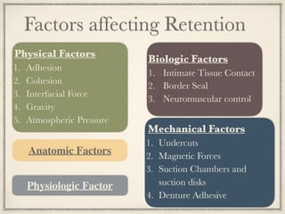
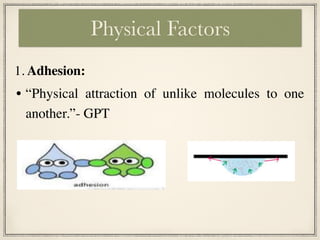
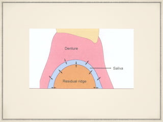
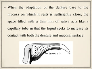
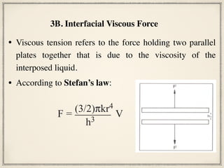
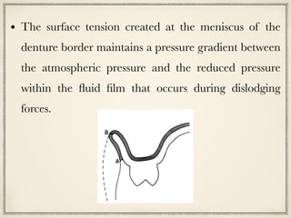
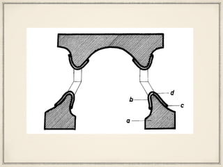
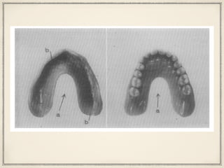
Retention in Complete
Denture
Dr Aastha Subba
Junior Resident
Dept. of Prosthodontics and Crown-Bridge
BPKIHS, Dharan.
Introduction
Factors Affecting Retention
Alternative Treatment plan for Retention
Conclusion
Contents
Psychologic Physiologic
Comfort
....::::::::+
x7
.:.:.::.:.!g
::::::::.
..::
Comfort
i,‘~ J;
ee
% .
‘/*
e
.....xc:.:.:.
‘$$$$::y 5
.A&.....
‘>;:+$.
‘.:.’
vi9
Support
Success t
Longevity
Fig. 1. Retention, stability, and support are important
• Complete denture retention is the resistance to
displacement of the denture base away from the ridge.
• Boucher describes retention as the most spectacular
yet probably the least important of all complete
denture objectives.
This property may indeed be
least important; however, it
provides psychologic
comfort to the patient.
Factors affecting Retention
Physical Factors
1. Adhesion
2. Cohesion
3. Interfacial Force
4. Gravity
5. Atmospheric Pressure
Biologic Factors
1. Intimate Tissue Contact
2. Border Seal
3. Neuromuscular control
Mechanical Factors
1. Undercuts
2. Magnetic Forces
3. Suction Chambers and
suction disks
4. Denture Adhesive
Anatomic Factors
Physiologic Factor
Physical Factors
• “Physical attraction of unlike molecules to one
another.”- GPT
1. Adhesion:
• In oral cavity, adhesion is seen between the saliva and
mucous membrane and between saliva and denture
base.
• This adhesion is achieved through ionic forces between
charged salivary glycoproteins and surface epithelium
or acrylic resin.
• Adhesion works to enhance further the retentive forces
of interfacial surface tension.
2. Cohesion:
“Physical attraction of like molecules for each other”
-GPT.
Cohesive forces within the saliva
Adhesion and Cohesion are the fundamental
molecular forces involved in denture retention,
and these give rise to the macroscopic
properties such as viscosity and surface
tension and through these properties,
intermolecular forces produce retentive effects.
Interfacial force is the resistance to separation of two
parallel surfaces that is imparted by a film of liquid
between them.
Interfacial forces is best broken into separate titles:
A. Interfacial surface tension and
B. Viscous tension.
3. Interfacial Force
3 A. Interfacial surface tension:
Page stated that “interfacial surface tension operates by
virtue of a thin fluid film between two intimately contacted
object.”
He described interfacial surface tension as a forces
involved in maintaining the attraction of two opposed
ground solid plates with an intervening fluid film that
resists displacing forces applied at right angles to the fluid
film surface.
If the surface is convex the pressure
is higher within the liquid than
without — therefore, positive
pressure.
If the total curvature is negative or
concave, the pressure is negative.
One of the consequences of the surface tension of liquids is
the tendency to minimize the area of the free surface,
generating the familiar curved surfaces of raindrops and
menisci.
Separation
proach
pat
(Fig. 4). Notice that if the bead of liqui
the edge were bulging out, the force wo
ow that although it is an
of successful complete
d
gher
e
Separation
Approach
pat + Δp pat – Δp
Separation
• Thus, on the assumption that the denture base is wetted
by saliva, an attempt to withdraw the denture generates
along its periphery a narrow, highly negatively-curved
saliva surface.
• There is therefore a lowered pressure in the liquid-filled
space and a retentive force is experienced.
• Interfacial surface tension depends on the ability of the
fluid to wet the rigid surrounding material.
• If the surrounding material has low surface tension, as
the oral mucosa does, fluid will maximize its contact,
thereby wetting it readily and spreading out in thin film.
• Denture base materials vary in their surface tension, with
processed material displaying greater wettability than
autocured products.
Comparison of the wettability of saliva, a saliva substitute,
and distilled water was done with three different denture
base material: Heat cure polymethylmethacrylate (DPI heat
cure), high-impact polymethylmethacrylate (Trevalon HI),
and nylon (Valplast).
Ramanna PK. Wettability of three denture base materials to human saliva,
saliva substitute, and distilled water: A comparative in vitro study. J Indian
Prosthodont Soc. 2018;18(3):248-256. doi:10.4103/jips.jips_301_17
The study concluded that:
• The wettability of the commercially available saliva
substitute tested was comparable to that of human saliva.
• High-impact heat-polymerized polymethylmethacrylate
denture base material (Trevalon HI) was the most easily
wetted.
• Nylon denture base material (Valplast) could possibly
provide the best retention of the three denture base
materials tested.
FIG 1.-Capillary rise between plates at an
angle.
equation for (ysv - ysv) gives the follow-
ing equation
v* = 'Y +2s y cos
6-
7 n
zb (5)
The work necessary to stretch the menis-
cus film by an amount ib is equat
increase in surface energy of the
F b = y* n2sbb,
where n2 S 8 b is the increase in surf
If the value of y* from equation
stituted, the following is obtained
2 y cos a t S2
b
At angles of V of a few degrees
52= 2Rb,
which gives
F = y s2s + 4 ntR y cos 0.
a b C
Capillary action or capillarity is the ability of a
liquid to flow in narrow spaces without the assistance
of, or even in opposition to, external forces like
gravity.
Capillarity and Interfacial surface tension
When the adaptation of the denture base to the
mucosa on which it rests is sufficiently close, the
space filled with a thin film of saliva acts like a
capillary tube in that the liquid seeks to increase its
contact with both the denture and mucosal surface.
straighter mandibular incisors. A partially
.................. :::Nx)AMEL.)
xx
..............................
..................... ::cccc)xx
..................... ::)XX))))))XXX
..................... ::XXXXXXXXX
............................ XXXX)XX
- X@ @s .o*sA A A A A . . .................
@-------------------------------------
* vvXXXXXXXX- -A
-***--
*- v*v vvv-- --v-
-A-A
A -A
-A
-A-A
. -A -v -A
e
v**
-................................---
*-..--- FILL----I --- ---.....-
-- --.. .--
--- *..-. . .
..-. ------
* ............
......--
::::::::::::::::::::::::::::::: =--------)
A--..*----....-*..------------.-----
------------------------------------
::::::::::::::: ..::-=- ---
FIG 10.-Capillary penetration around filling.
ner the layer of liquid, the tighter the
peripheral seal (this creates the isolated
capillary) and the greater the wettability,
the greater the adhesion. This component of
denture retention, therefore, is a result of a
strengthening of saliva caused by an in-
creased intermolecular attraction at the
liquid-vapor interface.
Conclusions
Capillary penetration of liquids into nar-
row crevices depends on the wetting of the
solid by the liquid, surface tension of the
liquid, and inversely, on the size of the
opening. The properties of capillary liquid
change when the liquid is trapped in isolated
contact with a solid. The surface tension in-
creases to a value y*. As a result, the
strength of the liquid becomes much greater
and the vapor pressure decreases. The
strength of liquid bridges between solids
may be derived on the basis of this surface
tension increase, rather than on the basis
of a negative pressure within the liquid film.
Human saliva was found to have the tensile
%
I so c a p
FIG 11.-Isocap space between denture and mucosa.
-
• Viscous tension refers to the force holding two parallel
plates together that is due to the viscosity of the
interposed liquid.
• According to Stefan’s law:
3B. Interfacial Viscous Force
l plates together that is due to the viscosity of
interposed liquid. Viscous tension is described
Stefan’s law.* For two parallel, circular plates of
ius (r) that are separated by a newtonian
compressible) liquid of viscosity (k) and thick-
s (h), this principle states that the force (F) nec-
ary to pull the plates apart at a velocity (V) in a
ection perpendicular to the radius will be
F = (3/2)πkr4
V
h3
The relationship expressed by Stefan’s law
kes it clear that the viscous force increases pro-
be
bra
pa
sal
to
an
eff
to
the
pa
ch
tai
ing
car
The relationship expressed by Stefan’s law makes it
clear that:
• The viscous force increases proportionally to
increase in the viscosity of the interposed fluid.
• The viscous force drops off readily as the distance
between the plates (i.e., the thickness of the
interposed medium) increases.
• The force increases proportionally to the square of
the area of the opposing surfaces.
When applied to denture retention, the equation
demonstrates the essential importance of
• an optimal adaptation between denture and basal seat
(a minimal h),
• the advantage of maximizing the surface area covered
by the denture (a maximum r)
• the theoretical improvement in retention made
possible by increasing the viscosity of the medium
between the denture and its seat.
• This physical force primarily concerns the mandibular
prosthesis.
4. Gravity
• Sometimes, when it is difficult
to bring the other factors of
retention into play when
constructing a lower denture,
gravity aids in providing the
necessary force to maintain
the prosthesis in place at rest.
• Atmospheric pressure can act to resist dislodging forces
applied to dentures, if the dentures have an effective seal
around their borders.
• It could only operate by way of a pressure difference, that
is, beneath the denture there must be a lower pressure.
• However, the full effect could only be felt if there were a
vacuum there.
5. Atmospheric pressure
that is, beneath the denture there must be
lower pressure, and the full effect coul
only be felt if there were a vacuum there
Fig. 1 Displacement of a sealed bellows-like device results in a vacuum
in the emergent space, the displacing force being balanced by that from
atmospheric pressure. There is no static retaining force otherwise.
Comparable conditions are unlikely to occur in the mouth
Dislodging
force
Pressure
zero
Atmospheric
pressure
Seal
attention to detail on the part of the
practitioner.
•Under normal conditions, therefore, there is no pressure
difference, no static retaining force, and atmospheric
pressure as such has no bearing on retention.
• “Denture retention could not be explained merely
in terms of simple physical equations but that the
human element entered into the system.”
• The quest for a solution to the problem of
complete denture retention could only be solved
through understanding of the biological factors.
Biologic Factors
• refers to the close adaptation of the denture base to
the underlying soft tissues.
• The impression technique will determine the degree
of intimate tissue contact obtained with the tissues at
rest and during function.
1. Intimate tissue contact
• To be effective air must be excluded from the
intaglio, and the fluid film must be as thin as
possible.
• Intimate tissue contact is the biologic factor that
promotes these conditions by eliminating air
entrapment.
• The surface tension created at the meniscus of the
denture border maintains a pressure gradient between
the atmospheric pressure and the reduced pressure
within the fluid film that occurs during dislodging
forces.
COMPLETE DENTURE RETENTION
surface. In reality some gas always exists due to the
partial pressure of gases dissolved in the saliva. The
presence of dissolved gases or air inclusions serves to
decreasethe effectiveness of atmospheric pressure pro-
portionately.
Clinical observations of the authors are also in
agreement with the research results cited. The intro-
duction of a small palatal perforation or the presenceof
an inadequate posterior palatal seal markedly reduces
the physical retention of most maxillary complete
dentures. Such effects would not be observed if the
forces of adhesion and cohesion, which depend primar-
ily on the surface area of intimate contact, were the
critical retentive factors. Certainly removal of a poste-
rior palatal seal or placement of a 1 mm perforation
does not significantly alter the surface area.
THE ROLE OF PHYSICAL FACTORS
The results of these studies and observations clearly
explain the physical retention of complete dentures.
Fig. 5. Thin fluid film exists (shaded area) between
denture base and tissues of residual ridges. Meniscus
that develops at border of denture is similar to that
• involves intimate contact of the denture borders
with the surrounding soft tissue.
• The seal encompasses the circumference of the
denture and includes features such as posterior
palatal seal to enhance its effectiveness.
2. Border seal
TION
gas always exists due to the
s dissolved in the saliva. The
ses or air inclusions serves to
s of atmospheric pressure pro-
of the authors are also in
arch results cited. The intro-
perforation or the presenceof
palatal seal markedly reduces
of most maxillary complete
would not be observed if the
hesion, which depend primar-
of intimate contact, were the
Certainly removal of a poste-
ement of a 1 mm perforation
er the surface area.
CAL FACTORS
udies and observations clearly
ention of complete dentures.
eated at the meniscus of the
a pressure gradient between
e and the reduced pressure
hat occurs during dislodging
Fig. 5. Thin fluid film exists (shaded area) between
denture base and tissues of residual ridges. Meniscus
that develops at border of denture is similar to that
shown in Fig. 4 between two glass slabs. Note that
position of meniscus will depend on where soft tissue
loses contact with denture border. Draping effect of
cheeks may provide a meniscus along polished surface
of denture border CA). When cheek is retracted, menis-
The border seal prevents the ingress of air once the
denture is seated.
It also maintains the thin fluid film at the denture
border, allowing a meniscus to develop in response to
displacing forces.
• Neuromuscular control refers to the functional
forces exerted by the musculature of the patient
that can affect retention.
• This is primarily a learned biologic
phenomenon.
3. Neuromuscular Control
• Brill et al asserted that touch receptors in the mucosa of
the oral cavity and tongue are particularly concerned
with denture retention.
• He observed that a patient wearing complete dentures
lost control of them when surface anesthesia was
applied to the mucosa.
For the oral and facial musculature to be most effective in
providing retention for complete dentures
(1)the denture bases must be properly extended.
(2)the occlusal plane must be at the correct level; and
(3)the arch form of the teeth must be in the neutral
zone between the tongue and the cheeks.
The shape of the buccal and lingual flanges must make it
possible for the musculature to fit automatically against
the denture and thereby to reinforce the border seal
If the buccal flanges of the maxillary denture slope up
and out from the occlusal surfaces of the teeth and the
buccal flanges of the mandibular denture slope down
and out from the occlusal plane, the contraction of the
buccinators will tend to retain both dentures on their
basal seats.
e teeth and the buccal flanges of the mandibu- should slope toward the center of the mou
A
B
C
Figure 22-2 Frontal section showing dentures properly filling the available space. A, The buc-
cinator. B, The lingual flange and border are placed under the tongue. C, The mylohyoid ridge.
Notice that both upper and lower dentures are so shaped that the action of the tongue and cheeks
The lingual surfaces of the lingual flanges should
slope toward the center of the mouth so the
tongue can fit against them and perfect the border
seal on the lingual side of the denture.
The base of the tongue is guided on top of the
lingual flange by the lingual side of the distal end
of the flange, which turns laterally toward the
ramus.
• Encroachment of the oral musculature by an over-
extended periphery of a denture, or by incorrectly
shaped polished surfaces, will undoubtedly lead to
dislodgement of the denture, if not ulceration of the
peripheral tissues.
• However it must be noted that dislodgement of
the denture is not seen in certain cases, even in
ill-fitting or broken denture worn for long
duration as they are retained mainly by muscle
function.
• The biologic factor of neuromuscular control
gradually becomes a major determinant in
complete denture retention as experienced
patients learn to alter their muscular function to
harmonize with the prosthesis.
ANATOMIC INFLUENCES ON MAXILLARY DENTURE
RETENTION
1. Posterior Palatal seal: The posterior palatal seal
maintains tissue contact during base movement or soft
palate function and compensates for processing changes.
• This area allows moderate tissue displacement to maintain
the thin fluid film.
Anatomic Factors
• In these situations a metal base
or subsequent bench-cure
reline procedure would be
incorporated into the initial
treatment plan.
• Patients exhibiting highly tapered steep palatal vaults
present processing error which may be so severe that no
amount of posterior palatal seal can compensate for the
resulting deficiency in intimate tissue contact.
This varies in size and shape but must be filled to avoid
ingress of air beneath the denture base.
2. Buccal space or retrozygomatic space:
ANATOMIC INFLUENCES ON MANDIBULAR
DENTURE RETENTION
Mylohoid Muscle
Retromylohyoid
fossa
Genioglossus Muscle
Mylohoid Muscle:
the fibers join those from the mylohyoid muscle of
the opposite side, and posteriorly they continue to
the hyoid base (Figure 14-10). The muscle lies deep
to the sublingual gland and other structures in the
anterior region and so does not affect the border of
the denture in this region except indirectly.
However, the posterior part of the mylohyoid
muscle in the molar region affects the lingual impres-
A
A
B
B
C
C
D
D
Figure 14-10 Relationships of the mylohyoid muscle in various regions. The letters with
prime signs denote cross sections of the designated areas. A, Canine region. B, Premolar
region. C, First molar. D, Third molar. At point D, notice that the mylohyoid ridge approaches
• The posterior part of the mylohyoid muscle in the
molar region affects the lingual impression border in
swallowing and in moving the tongue.
• Extension of the lingual flange under the mylohyoid
ridge cannot be tolerated in function because it will
interfere with the action of the mylohyoid muscle
when it contracts, and this will displace the denture,
causing soreness.
Fig. 9. A, Clinical appearance of retromylohyoid curtain.
muscular structures that lie deep to mucosa of retromylohyo
Fig. 10. Lingual flange of mandibular denture must
incline medially to allow for contraction of mylohyoid
muscle, which lies beneath mucosa covering lingual
slope of residual ridge. Dotted lines represent an acti-
border. The
extension of
pear-shaped
shaped pad,
residual sca
associated re
pear-shaped
and firmly
Immediately
more resilien
contains glan
can tolerate
suggest bea
pear-shaped
seal along th
LINGUAL
INFLUENCE
The bord
lingual flang
If the border stops above the mylohyoid ridge, vertical
forces will cause soreness, and the border seal will be
easily broken.
If the flange is properly shaped and extended, it will
provide border seal and guide the tongue to rest on top of
the flange.
For the denture to be successful, the flange must be made
parallel to the mylohyoid muscle when it is contracted.
Retromylohyoid Fossa
The retromylohyoid fossa is bounded
by the retromylohyoid curtain.
The posterolateral portion of the
retromylohyoid curtain overlies the
superior constrictor muscle.
The posteromedial portion covers
the palatoglossal muscle plus the
lateral surface of the tongue.
The inferior wall overlies the
submandibular gland.
242 Part Three Rehabilitation of the Edentulous Patient: Fabrication of Complete Dentures
PR
B
M
RM
MP
RMC
SC
Figure 14-11 Diagram shows the relationship of the medial pterygoid musc
superior constrictor muscle. B, Buccinator muscle; M, masseter muscle; MP, med
muscle; PR, pterygomandibular raphe; RM, ramus of the mandible; RMC, postero
portion of the retromylohyoid curtain formed by the mucous membrane coverin
superior constrictor muscle (SC).
The extension of the denture into this area can
resist horizontal forces, increases border seal and
contribute in the neuromuscular control
mechanism.
(P S Huang et al 2007)
MANDIBULAR ANTERIOR LINGUAL
INFLUENCES
• The most difficult region to obtain a border seal is the
anterior lingual border.
• It is utmost important to establish and maintain
border seal throughout the functional range of
movement of the anterior floor of the mouth.
Genioglossus
Fig. li. Genioglossus muscle in cross section, illus-
trating two main divisions. Superior fibers, A, are
contracted, depressing central body of tongue .and
causing tip to be retracted and floor of mouth to reach
its most superior position. B, Inferior fibers of genio-
glossus.
Superior most floor
of mouth
Fig. 13. A technique for establish
seal is diagrammed. First, dentu
molded to contact superiormost
mouth. Extension posteriorly t
folds should maintain a border
mouth is at rest.
Several methods may be used to establish and maintain
border seal.
• Some techniques recommend the horizontal extension
of the anterior lingual flange sublingually.
• The flange can then be extended posteriorly to contact
the sublingual folds and thereby establish a seal when
the tongue is at rest and the floor of the mouth drops
JACOBSON AND KROL
Superior most floor
of mouth
Rest
Fig. 13. A technique for establishing anterior lingual
seal is diagrammed. First, denture must be border-
molded to contact superiormost level of floor of
mouth. Extension posteriorly to contact sublingual
folds should maintain a border seal when floor of
• Another technique involves border molding to
determine the inferior extension of the flange.
• Then a slight additional amount of softened border-
molding material is added to the inner surface of the
previously molded anterior lingual area and custom
tray is reseated.
section, illus-
ibers, A, are
tongue .and
outh to reach
ers of genio-
UAL
btain a border
he mylohyoid
y to raise the
seal is diagrammed. First, denture must be border-
molded to contact superiormost level of floor of
mouth. Extension posteriorly to contact sublingual
folds should maintain a border seal when floor of
mouth is at rest.
Fig. 14. A second technique. to establish anterior lin-
gual seal. A, Denture is extended to most superior
level of floor of mouth. B, Slight pressure on mucosa
overlying lingual slope of anterior mandible ensures a
border seal when tongue is at rest.
Mandibular dentures cover less surface area than
maxillary prostheses and therefore are subject to a
lower magnitude of retentive forces.
Similarly, patients with small jaws or very flat alveolar
ridges (small basal seats) cannot expect retention to be
as great as patients with large jaws or prominent
alveoli.
Saliva is critical for retention and
comfort in wearing removable
prostheses.
Physiologic Factor (Saliva)
In the denture wearing population, salivary wetting
mechanics are necessary to create adhesion, cohesion
and surface tension that ultimately lead to increased
retention of prostheses.
Total salivary output may have less relevance to the
denture- wearing population than the flow from a
specific gland.
There is only a slight correlation between the
secretion from parotid glands and the retention of
maxillary dentures and a significant relationship has
been discovered between palatal salivary gland
function and denture retention.
Various studies have shown that mucous saliva is necessary
for undisturbed function of the denture bearing mucosa
and for comfort with dentures and it is the palatal saliva
that improves the retention of upper full dentures.
The covering of the mucosa with adequate mucous saliva
has an anti-xerostomic effect and reduces the risk of the
incidence of oral burning sensations or other complaints of
oral dysaesthesia.
• Exaggerated bony undercuts may compromise denture
retention by necessitating extensive internal
adjustment of the denture.
• Less severe undercuts can be extremely helpful to the
retention of the prosthesis.
Mechanical Factor
1.Undercuts, Rotational Insertion Paths, and
Parallel Walls
• Some “undercuts” are only undercut in relationship to
a linear path of insertion.
• However, if the undercut area is seated first and the
remainder of the denture base can be brought into
proximity with the basal seat on rotation of the
prosthesis around the undercut part that is already
seated and this “rotational path” will provide
resistance to vertical displacement.
• Prominent alveolar ridges with parallel buccal and
lingual walls may also provide significant
retention.
• Prominent ridges also resist denture movement by
limiting the range of displacing force directions
possible.
• Magnets have been used as an aid for denture
retention for many years with some success.
• The reason of the popularity of magnets is their small
size and their attraction or repulsion forces, which
allow them to be placed within prosthesis without
being obtrusive in the mouth.
Magnets
In a magnetic material, a large portion of these atoms
which have a tiny magnetic field, align in small regions
called “domains”.
In an un-magnetized state, the orientation of these
domains is random and no overall magnetization is
experienced.
On the application of a magnetic field, these domains
align and reach a saturation point, where the material is
said to have been magnetized.
How do magnets work?
Magnets are implanted in the jaw and in otherwise
conventional complete denture.
The attraction between these magnets helps retain the
denture in position.
Magnets in Complete dentures
Retention via magnets occur by
two mechanism:
a)  Magnet Repulsion
b)  Magnet Attraction.
• uses the forces of repulsion of like poles of magnets to
maintain and improve the seating of complete dentures.
• The magnets had been magnetized from end to end, in
the customary fashion, so that the north pole was at one
end and the south pole at the opposite end.
little loss of energy.
Changes in Magnet Design.-To accompany the change in direction
netization a change in design was made. By flattening the magnets, m
lines of force between the two magnets were shortened and greater e
made available. As shown in Fig. 11, A, with a cross section of cylindric
the central line of force is the shortest; those at the periphery are longer
sequently, weaker. With a cross section of flattened magnets (Fig. 11,
of the lines of force are short and, consequently, stronger. The shape of
now is that of a flattened cylinder, l/4 inch in length, less than rA inch
and a little more than G inch in width. To be precise, it measures 0.25
0.10 inch (6.35 by 3.55 by 2.54 mm.) (Fig. 12, B).
Changes in the Mesh Overlay.-The second time the new flattened
magnetized through the diameter, were used, an interesting phenom
curred (Table I, case 3). The magnet was so strong that it was a
the needle used to suture the mucoperiosteum. It followed the directio
1”“’
t+q
I I
1 1
t t
a)  Magnet Repulsion
Here, the entire top surface is one pole and bottom surface
the opposite pole.
All the lines of force between the magnets are attracting
and are shorter when compared to previous variant.
ween the two magnets were shortened and greater energy was
s shown in Fig. 11, A, with a cross section of cylindric magnets,
force is the shortest; those at the periphery are longer and, con-
With a cross section of flattened magnets (Fig. 11, B), more
e are short and, consequently, stronger. The shape of the magnet
lattened cylinder, l/4 inch in length, less than rA inch in height,
han G inch in width. To be precise, it measures 0.25 by 0.14 by
3.55 by 2.54 mm.) (Fig. 12, B).
he Mesh Overlay.-The second time the new flattened magnets,
h the diameter, were used, an interesting phenomenon oc-
case 3). The magnet was so strong that it was attracted to
o suture the mucoperiosteum. It followed the direction of the
“’
q I
1 1
t t
b)  Magnet Attraction.
The major problem associated with these magnets
as retentive devices are corrosion by oral fluids,
which limits their use.
Drawback of Magnets
• suggested in 1883 by the Greene brothers and Ames.
Suction chambers and suction disk
• Suction disk provides retention
and stability by inducing negative
pressure on the mucosal surface.
• Use of suction disk in maxillary denture is one of the
techniques to improve retention.
• Resorption of the alveolar bone leads to a situation
where the denture although well retained by the
rubber disc, rocks around the mid palatal area,
exerting excessive pressure.
• This negative pressure leads to the loss of palatal bone
“Atmospheric pressure manifesting itself in emergency
vacuum is most desirable. However, chambers and
patented suction cups are not recommended, as nature
will tolerate vacuum under a denture only a short time.”
In the functional vacuum method, retention is based
neither on cohesion nor on local vacuums, but on a
general vacuum created between the entire tissue
surface of the denture and the mucosa.
Functional Vacuum
The principle of the functional vacuum method is similar
to that of a reciprocating pump which consists of :
A. Cylinder,
B. Piston that can move in the cylinder,
C. packing ring, that stops leakage between the cylinder
and the piston, and
D. an admission valve.
676 KUBALI J. Pros.
July-August,
Fig. 4.-A section of the reciprocating puma: la) the cylinder; (b) the piston; (c) the packin
ring belt; (d) the valve.
• In oral cavity, the denture-bearing tissue plays the
same function as the pump piston.
• A denture constructed from a mucostatic impression
becomes the cylinder.
• The packing ring, put on the inner borders of the
denture as a seal.
and ( 5) psychologic factors.
Stability.-Soft tissues and border extensions which take part in ensuring
stability in other systems do not play any significant role in the functional vacuum
Fig. 6.-The pump system is transferred to the denture: (a) the maxillae and mandible
(piston); (b) the denture (cylinder); (c) the packing ring seal; (d) the gap between the denture
and the mucosa.
680 KUBALI 5. Pros. Den.
Julz+August, 1960
Fig. 10. Fig. 11.
Fig. lO.-The tissue surface of a denture constructed by the functional vacuum method: (a)
the palatal base material is not present; (b) the packing ring belt is placed around the borders.
Fig. Il.-The palatal portion of the denture is not needed in the functional vacuum method.
According to Zarb et al, the term “denture adhesive”
refers to a commercially available, nontoxic, soluble
material that is applied to the tissue surface of the
denture to enhance retention, stability, and function.
Denture Adhesive
Denture Adhesive enhance retention through
(1) increasing the adhesive and cohesive properties and
viscosity of the medium lying between the denture and its
basal seat and
(2) eliminating voids between the denture base and its
basal seat.
How does Denture Adhesive work?
According to Grasso, denture adhesives can be
categorized into
A. Soluble group: The soluble category includes
creams, powders, and pastes.
B. Insoluble groups: The insoluble group consists of
wafers and pads.
Classification of Denture Adhesive
On the basis of their composition, denture adhesives
can be divided into
A. Natural: based on vegetable gums (e.g., karaya,
tragacanth, xanthan, and acacia).
B. Synthetic: consist of mixtures of the salts of short-
acting (carboxymethylcellulose) and long-acting
(polyvinyl methyl ether maleate, or “gantrez”)
polymers.
Types of denture adhesives tested according to
the selected articles.
Figure 2
Current Trends and Future Perspectives in the Development of Denture Adhesives:
An Overview Based on Technological Monitoring Process and Systematic Review
According to Kumar et al., the main ingredients of
denture adhesives could be classified into three groups:
• adhesive agents delivering the adhesion between
denture and mucosa: methyl-cellulose, sodium
carboxy-methyl cellulose, and synthetic polymers like
acrylamides, acetic polyvinyl and polyethylene oxide 

Composition
Guidelines for the Use of Denture Adhesives and their
benefits for Oral and General Health.
• anti-microbial agents reducing microbial growth in
the adhesive: sodium tetraborate, ethanol,
hexachlorophene and sodium borate,
• other agents to help with application, storage and the
delivery of freshness to the mouth: plasticizing agents;
flavouring agents like oil of peppermint, oil of
wintergreen, and spearmint.
Guidelines for the Use of Denture Adhesives and their
benefits for Oral and General Health.
1. Retention
Grasso et al. measured denture movements.
measurements of denture movements were made at
baseline (no adhesive) and at 0, 2, 4 hours post-
adhesive application.
Advantages of Denture Adhesives
The results of the measurements showed that:
• mandibular (lower) denture movements, both with and
without an adhesive, were significantly greater than
maxillary (upper) denture movements
• the adhesive significantly reduced movement of the
maxillary and mandibular dentures during both chewing
and biting,
• the improvement happened immediately after the
adhesive was applied and was maintained for the four
hours of follow-up.
Munoz et al. confirmed in their study that subjects had
significantly fewer dislodgements while eating an apple
after adhesive was applied to dentures.
Significant increases in subjective ratings of confidence
and comfort, as well as decreases in denture wobble, were
associated with the use of adhesive.
2. Stability
A study by Fujimori et al. found that the use of the denture
adhesive increased maximum biting force and provided
rhythmic masseter muscle activity during mastication for
both good and poor denture-bearing tissues.
3. Masticatory (chewing) performance
• A crossover randomised clinical trial by Marin et al.
showed that there was increase in vertical movements
during chewing and less intrusion of maxillary complete
dentures.
A recognized secondary benefit of denture adhesives in
patients with complete dentures is their ability to act as a
barrier to help prevent the migration and accumulation of
food particles under the dentures.
4. Food trapping
Since, denture adhesives add to retention and so
improve chewing ability, reduce any instability,
provide comfort and eliminate the accumulation of
food debris beneath the dentures, they increase the
patient’s sense of security and satisfaction.
5. Psychological confidence
Nicolas et al. assessed the oral health-related quality of life
of complete-denture wearers who used a denture adhesive
over a 6-month period.
The results showed that using a denture adhesive may
improve subjects’ ability to manage conventional dentures
and enhance their oral health quality of life.
6. Quality of life
Polyzois et al. in a study showed that there is a definite
improvement in 2 weeks after using an adhesive and
adhesives do help in the improvement of OHQoL in
patients with new complete dentures and may be used
to shorten the adaptation period for new dentures.


Bogucki conducted a study to estimate the retention
capability of denture adhesives in complete maxillary
denture patients with previously diagnosed xerostomia.
The study concluded that:
• All patients had poor retention of their maxillary
dentures without DAs.
• The majority of DA used showed the highest retention
ability after 1 h.
7. Xerostomia
Challenges with the use of
Denture Adhesives
1. Cytotoxicity:
Several studies have examined the cytotoxicity of denture
adhesives on monolayer cell cultures and varied degrees of
cytotoxicity have been reported.
However, further in vivo tests or long-term clinical trials are
needed to determine the biological safety of denture
adhesives.
The most serious health issue so far reported as a result
of long-term and excessive use of denture adhesives is
potential neurotoxicity related to the presence of
zinc in certain denture adhesives.
2. Toxicity of Zinc-containing adhesives
A study by Hedera et al. looked at the different sources
of zinc intake among patients suffering from
progressive myelopolyneuropathy and who had
unexplained hypocupremia with hyperzincemia.
• All had a history of ill-fitting dentures which
needed large amounts of denture cream, resulting
in significant zinc exposure.
• Their copper and zinc normalised after they
stopped using zinc-containing denture adhesive,
further confirming that this is the source of high
zinc.
Manufacturer
• “Zinc overload” and its consequences will not occur
by occasional or moderate use of denture adhesives.
The consequences reported are associated with the
use of large amounts of denture adhesive over years.
• The best advice is to recommend zinc-free denture
adhesives to denture patients.
• Dentists need to stress the need for periodically relining
denture bases to minimize the need for denture
adhesives.
• Endosseous implants can also improve denture stability.


Denture adhesives often include antimicrobial agents such
as hexachlorophene, sodium tetra borate, methyl salicylate
and sodium borate.
Therefore, the long-term use of adhesives may affect the
oral microflora by selectively supporting the growth of some
micro-organisms and inhibiting others.
3. Microbial growth
Özkan et al. compared the presence of Candida Albicans and
α-haemolytic streptococci in the saliva and on the palate and
dentures of a group not using a denture adhesive, and another
group using a denture adhesive and found no statistically
significant difference.
They therefore concluded that prolonged use of the denture
adhesive (tested up to 2 months) did not lead to an increase in
micro-organisms of the oral flora.
Leite et al. found similar colony counts with or without the
use of adhesive for both the mucosa and internal surfaces of
maxillary dentures after 15 days.
However, there are no longitudinal trials of longer than 6
months on the continual use of denture adhesives by the
same patients, the effects of long-term use of adhesives on
oral tissues are currently unknown.


Although oral cancer is not directly related to the use of
denture adhesives, there is a link between ill-fitting dentures
and the risk of developing oral cancer.
Manoharan et al. through a meta-analysis found that the use
of ill-fitting dentures substantially increased this risk.
5. Oral cancer
Since, use of denture adhesives with ill-fitting
dentures may lead to a false sense of adequate
fitting, this may lead to patients not seeing a dental
professional regularly, allowing oral cancer to
develop unchecked.
• Denture adhesive should be used only in sufficient
quantities (three or four pea-sized drops) on each denture.
• Adhesive is applied to clean dentures, which are then
positioned in the mouth and held in place for a few seconds,
according to the manufacturer’s instructions.
• If increasing amounts of adhesives are required to achieve
the same level of denture retention, the patient should see a
dentist or dental professional to evaluate.
American Dental Association
Application guidelines for Denture adhesives
• Patients who wear dentures should be checked annually by
the dentist, prosthodontist or dental professional for
maintenance of optimum denture fit and function, for
evaluation for oral lesions and bone loss, and for assessment
of oral health status.
•  Denture adhesives should be completely removed from the
prosthesis and the oral cavity on a daily basis.
• During denture cleaning, adhesive should be removed with
gentle scrubbing to prevent contamination.
American Dental Association
An implant-retained overdenture is a removable dental
prosthesis supported by the residual oral tissues and
retained by dental implants.
They demonstrate improved retention and stability
when compared to conventional dentures.
This type of prosthesis should be considered in all
patients unable to tolerate conventional dentures
Implant Retained Overdenture
A prospective randomized clinical trial was performed to
evaluate 10 years of treatment of patients receiving a
mandibular implant-retained overdenture or a conventional
complete denture (CD) where 121 edentulous patients were
treated with an IRO (n = 61) or a conventional CD (n = 60).
Clinical aspects and patient satisfaction were evaluated. The
study concluded that patients in the IRO group were
significantly more satisfied than patients in the CD group
Meijer HJ, Raghoebar GM, Van 't Hof MA. Comparison of implant-retained mandibular
overdentures and conventional complete dentures: a 10-year prospective study of clinical
aspects and patient satisfaction. Int J Oral Maxillofac Implants. 2003 Nov-Dec;18(6):879-85.
Conclusion
• Establishing optimal complete denture retention
requires an understanding of the factors discussed.
• Incorporation of these determinants into the
prosthesis through proper design and technique
contributes to the success of complete dentures.
–Bohannan
“Technique itself is merely the practical
application of principles, and if the
principles are unsound, the most
elaborate and painstaking technique
certainly is doomed to failure.”
• Prosthodontic Treatment for Edentulous Patients- Zarb & Bolender,Twelfth
edition
• Darvell BW, Clark RK. The physical mechanisms of complete denture
retention. Br Dent J. 2000 Sep 9;189(5):248-52.
• Murray MD, Darvell BW. The evolution of the complete denture base. Theories
of complete denture retention--a review. Part 1. Aust Dent J. 1993 Jun;38(3):
216-9.
• Murray MD, Darvell BW. The evolution of the complete denture base. Theories
of complete denture retention--a review. Part 2. Aust Dent J. 1993 Aug;38(4):
299-305.
• Murray MD, Darvell BW. The evolution of the complete denture base. Theories
of complete denture retention--a review. Part 3. Aust Dent J. 1993 Oct;38(5):
389-93.
References
• Murray MD, Darvell BW. The evolution of the complete denture base. Theories
of complete denture retention--a review. Part 4. Aust Dent J. 1993 Dec;38(6):
450-5.
• Kawazoe Y, Hamada T. The role of saliva in retention of maxillary complete
dentures. J Prosthet Dent. 1978 Aug;40(2):131-6.
• Niedermeier WH, Krämer R. Salivary secretion and denture retention. J Prosthet
Dent. 1992 Feb;67(2):211-6.
• Turner M, Jahangiri L, Ship JA. Hyposalivation, xerostomia and the complete
denture: a systematic review. J Am Dent Assoc. 2008 Feb;139(2):146-50.
• Niedermeier W, Huber M, Fischer D, Beier K, Müller N, Schuler R, Brinninger
A, Fartasch M, Diepgen T, Matthaeus C, Meyer C, Hector MP. Significance of
saliva for the denture-wearing population. Gerodontology. 2000 Dec;17(2):
104-18.
• David Bartlett, Nigel Carter, David Felton, Guy Goffin, Yasuhiko Kawai,
Frauke Muller, Grigorios Polyzois, Angus Walls White paper on
guidelines for the use of denture adhesives and their benefits for oral and
general health. Global Task Force for Development of Guidelines on Use
of Denture Adhesives.
• Rassawet Reena Roy, Mittal Sanjeev, Kalra Himanshi. Magnets – Role in
prosthodontic rehabilitation: A reviewYear : Indian Journal of Dental
sciences.2020 ;12 (3);168-171
• Vere J, Bhakta S, Patel R. Implant-retained overdentures: a review. Dent
Update. 2012 Jun;39(5):370-2, 374-5
• Meijer HJ, Raghoebar GM, Van 't Hof MA. Comparison of implant-
retained mandibular overdentures and conventional complete dentures: a
10-year prospective study of clinical aspects and patient satisfaction. Int
J Oral Maxillofac Implants. 2003 Nov-Dec;18(6):879-85.
Retention in cd

Retention in cd

  • 1.
    Retention in Complete Denture DrAastha Subba Junior Resident Dept. of Prosthodontics and Crown-Bridge BPKIHS, Dharan.
  • 2.
    Introduction Factors Affecting Retention AlternativeTreatment plan for Retention Conclusion Contents
  • 3.
    Psychologic Physiologic Comfort ....::::::::+ x7 .:.:.::.:.!g ::::::::. ..:: Comfort i,‘~ J; ee %. ‘/* e .....xc:.:.:. ‘$$$$::y 5 .A&..... ‘>;:+$. ‘.:.’ vi9 Support Success t Longevity Fig. 1. Retention, stability, and support are important
  • 4.
    • Complete dentureretention is the resistance to displacement of the denture base away from the ridge. • Boucher describes retention as the most spectacular yet probably the least important of all complete denture objectives. This property may indeed be least important; however, it provides psychologic comfort to the patient.
  • 5.
    Factors affecting Retention PhysicalFactors 1. Adhesion 2. Cohesion 3. Interfacial Force 4. Gravity 5. Atmospheric Pressure Biologic Factors 1. Intimate Tissue Contact 2. Border Seal 3. Neuromuscular control Mechanical Factors 1. Undercuts 2. Magnetic Forces 3. Suction Chambers and suction disks 4. Denture Adhesive Anatomic Factors Physiologic Factor
  • 6.
    Physical Factors • “Physicalattraction of unlike molecules to one another.”- GPT 1. Adhesion:
  • 7.
    • In oralcavity, adhesion is seen between the saliva and mucous membrane and between saliva and denture base. • This adhesion is achieved through ionic forces between charged salivary glycoproteins and surface epithelium or acrylic resin. • Adhesion works to enhance further the retentive forces of interfacial surface tension.
  • 9.
    2. Cohesion: “Physical attractionof like molecules for each other” -GPT.
  • 10.
  • 11.
    Adhesion and Cohesionare the fundamental molecular forces involved in denture retention, and these give rise to the macroscopic properties such as viscosity and surface tension and through these properties, intermolecular forces produce retentive effects.
  • 12.
    Interfacial force isthe resistance to separation of two parallel surfaces that is imparted by a film of liquid between them. Interfacial forces is best broken into separate titles: A. Interfacial surface tension and B. Viscous tension. 3. Interfacial Force
  • 13.
    3 A. Interfacialsurface tension: Page stated that “interfacial surface tension operates by virtue of a thin fluid film between two intimately contacted object.” He described interfacial surface tension as a forces involved in maintaining the attraction of two opposed ground solid plates with an intervening fluid film that resists displacing forces applied at right angles to the fluid film surface.
  • 14.
    If the surfaceis convex the pressure is higher within the liquid than without — therefore, positive pressure. If the total curvature is negative or concave, the pressure is negative. One of the consequences of the surface tension of liquids is the tendency to minimize the area of the free surface, generating the familiar curved surfaces of raindrops and menisci.
  • 15.
    Separation proach pat (Fig. 4). Noticethat if the bead of liqui the edge were bulging out, the force wo ow that although it is an of successful complete d gher e Separation Approach pat + Δp pat – Δp Separation
  • 16.
    • Thus, onthe assumption that the denture base is wetted by saliva, an attempt to withdraw the denture generates along its periphery a narrow, highly negatively-curved saliva surface. • There is therefore a lowered pressure in the liquid-filled space and a retentive force is experienced.
  • 17.
    • Interfacial surfacetension depends on the ability of the fluid to wet the rigid surrounding material. • If the surrounding material has low surface tension, as the oral mucosa does, fluid will maximize its contact, thereby wetting it readily and spreading out in thin film. • Denture base materials vary in their surface tension, with processed material displaying greater wettability than autocured products.
  • 18.
    Comparison of thewettability of saliva, a saliva substitute, and distilled water was done with three different denture base material: Heat cure polymethylmethacrylate (DPI heat cure), high-impact polymethylmethacrylate (Trevalon HI), and nylon (Valplast). Ramanna PK. Wettability of three denture base materials to human saliva, saliva substitute, and distilled water: A comparative in vitro study. J Indian Prosthodont Soc. 2018;18(3):248-256. doi:10.4103/jips.jips_301_17
  • 19.
    The study concludedthat: • The wettability of the commercially available saliva substitute tested was comparable to that of human saliva. • High-impact heat-polymerized polymethylmethacrylate denture base material (Trevalon HI) was the most easily wetted. • Nylon denture base material (Valplast) could possibly provide the best retention of the three denture base materials tested.
  • 20.
    FIG 1.-Capillary risebetween plates at an angle. equation for (ysv - ysv) gives the follow- ing equation v* = 'Y +2s y cos 6- 7 n zb (5) The work necessary to stretch the menis- cus film by an amount ib is equat increase in surface energy of the F b = y* n2sbb, where n2 S 8 b is the increase in surf If the value of y* from equation stituted, the following is obtained 2 y cos a t S2 b At angles of V of a few degrees 52= 2Rb, which gives F = y s2s + 4 ntR y cos 0. a b C Capillary action or capillarity is the ability of a liquid to flow in narrow spaces without the assistance of, or even in opposition to, external forces like gravity. Capillarity and Interfacial surface tension
  • 21.
    When the adaptationof the denture base to the mucosa on which it rests is sufficiently close, the space filled with a thin film of saliva acts like a capillary tube in that the liquid seeks to increase its contact with both the denture and mucosal surface. straighter mandibular incisors. A partially .................. :::Nx)AMEL.) xx .............................. ..................... ::cccc)xx ..................... ::)XX))))))XXX ..................... ::XXXXXXXXX ............................ XXXX)XX - X@ @s .o*sA A A A A . . ................. @------------------------------------- * vvXXXXXXXX- -A -***-- *- v*v vvv-- --v- -A-A A -A -A -A-A . -A -v -A e v** -................................--- *-..--- FILL----I --- ---.....- -- --.. .-- --- *..-. . . ..-. ------ * ............ ......-- ::::::::::::::::::::::::::::::: =--------) A--..*----....-*..------------.----- ------------------------------------ ::::::::::::::: ..::-=- --- FIG 10.-Capillary penetration around filling. ner the layer of liquid, the tighter the peripheral seal (this creates the isolated capillary) and the greater the wettability, the greater the adhesion. This component of denture retention, therefore, is a result of a strengthening of saliva caused by an in- creased intermolecular attraction at the liquid-vapor interface. Conclusions Capillary penetration of liquids into nar- row crevices depends on the wetting of the solid by the liquid, surface tension of the liquid, and inversely, on the size of the opening. The properties of capillary liquid change when the liquid is trapped in isolated contact with a solid. The surface tension in- creases to a value y*. As a result, the strength of the liquid becomes much greater and the vapor pressure decreases. The strength of liquid bridges between solids may be derived on the basis of this surface tension increase, rather than on the basis of a negative pressure within the liquid film. Human saliva was found to have the tensile % I so c a p FIG 11.-Isocap space between denture and mucosa. -
  • 22.
    • Viscous tensionrefers to the force holding two parallel plates together that is due to the viscosity of the interposed liquid. • According to Stefan’s law: 3B. Interfacial Viscous Force l plates together that is due to the viscosity of interposed liquid. Viscous tension is described Stefan’s law.* For two parallel, circular plates of ius (r) that are separated by a newtonian compressible) liquid of viscosity (k) and thick- s (h), this principle states that the force (F) nec- ary to pull the plates apart at a velocity (V) in a ection perpendicular to the radius will be F = (3/2)πkr4 V h3 The relationship expressed by Stefan’s law kes it clear that the viscous force increases pro- be bra pa sal to an eff to the pa ch tai ing car
  • 23.
    The relationship expressedby Stefan’s law makes it clear that: • The viscous force increases proportionally to increase in the viscosity of the interposed fluid. • The viscous force drops off readily as the distance between the plates (i.e., the thickness of the interposed medium) increases. • The force increases proportionally to the square of the area of the opposing surfaces.
  • 24.
    When applied todenture retention, the equation demonstrates the essential importance of • an optimal adaptation between denture and basal seat (a minimal h), • the advantage of maximizing the surface area covered by the denture (a maximum r) • the theoretical improvement in retention made possible by increasing the viscosity of the medium between the denture and its seat.
  • 25.
    • This physicalforce primarily concerns the mandibular prosthesis. 4. Gravity • Sometimes, when it is difficult to bring the other factors of retention into play when constructing a lower denture, gravity aids in providing the necessary force to maintain the prosthesis in place at rest.
  • 26.
    • Atmospheric pressurecan act to resist dislodging forces applied to dentures, if the dentures have an effective seal around their borders. • It could only operate by way of a pressure difference, that is, beneath the denture there must be a lower pressure. • However, the full effect could only be felt if there were a vacuum there. 5. Atmospheric pressure
  • 27.
    that is, beneaththe denture there must be lower pressure, and the full effect coul only be felt if there were a vacuum there Fig. 1 Displacement of a sealed bellows-like device results in a vacuum in the emergent space, the displacing force being balanced by that from atmospheric pressure. There is no static retaining force otherwise. Comparable conditions are unlikely to occur in the mouth Dislodging force Pressure zero Atmospheric pressure Seal attention to detail on the part of the practitioner.
  • 28.
    •Under normal conditions,therefore, there is no pressure difference, no static retaining force, and atmospheric pressure as such has no bearing on retention.
  • 29.
    • “Denture retentioncould not be explained merely in terms of simple physical equations but that the human element entered into the system.” • The quest for a solution to the problem of complete denture retention could only be solved through understanding of the biological factors. Biologic Factors
  • 30.
    • refers tothe close adaptation of the denture base to the underlying soft tissues. • The impression technique will determine the degree of intimate tissue contact obtained with the tissues at rest and during function. 1. Intimate tissue contact
  • 31.
    • To beeffective air must be excluded from the intaglio, and the fluid film must be as thin as possible. • Intimate tissue contact is the biologic factor that promotes these conditions by eliminating air entrapment.
  • 32.
    • The surfacetension created at the meniscus of the denture border maintains a pressure gradient between the atmospheric pressure and the reduced pressure within the fluid film that occurs during dislodging forces. COMPLETE DENTURE RETENTION surface. In reality some gas always exists due to the partial pressure of gases dissolved in the saliva. The presence of dissolved gases or air inclusions serves to decreasethe effectiveness of atmospheric pressure pro- portionately. Clinical observations of the authors are also in agreement with the research results cited. The intro- duction of a small palatal perforation or the presenceof an inadequate posterior palatal seal markedly reduces the physical retention of most maxillary complete dentures. Such effects would not be observed if the forces of adhesion and cohesion, which depend primar- ily on the surface area of intimate contact, were the critical retentive factors. Certainly removal of a poste- rior palatal seal or placement of a 1 mm perforation does not significantly alter the surface area. THE ROLE OF PHYSICAL FACTORS The results of these studies and observations clearly explain the physical retention of complete dentures. Fig. 5. Thin fluid film exists (shaded area) between denture base and tissues of residual ridges. Meniscus that develops at border of denture is similar to that
  • 33.
    • involves intimatecontact of the denture borders with the surrounding soft tissue. • The seal encompasses the circumference of the denture and includes features such as posterior palatal seal to enhance its effectiveness. 2. Border seal
  • 34.
    TION gas always existsdue to the s dissolved in the saliva. The ses or air inclusions serves to s of atmospheric pressure pro- of the authors are also in arch results cited. The intro- perforation or the presenceof palatal seal markedly reduces of most maxillary complete would not be observed if the hesion, which depend primar- of intimate contact, were the Certainly removal of a poste- ement of a 1 mm perforation er the surface area. CAL FACTORS udies and observations clearly ention of complete dentures. eated at the meniscus of the a pressure gradient between e and the reduced pressure hat occurs during dislodging Fig. 5. Thin fluid film exists (shaded area) between denture base and tissues of residual ridges. Meniscus that develops at border of denture is similar to that shown in Fig. 4 between two glass slabs. Note that position of meniscus will depend on where soft tissue loses contact with denture border. Draping effect of cheeks may provide a meniscus along polished surface of denture border CA). When cheek is retracted, menis-
  • 35.
    The border sealprevents the ingress of air once the denture is seated. It also maintains the thin fluid film at the denture border, allowing a meniscus to develop in response to displacing forces.
  • 36.
    • Neuromuscular controlrefers to the functional forces exerted by the musculature of the patient that can affect retention. • This is primarily a learned biologic phenomenon. 3. Neuromuscular Control
  • 37.
    • Brill etal asserted that touch receptors in the mucosa of the oral cavity and tongue are particularly concerned with denture retention. • He observed that a patient wearing complete dentures lost control of them when surface anesthesia was applied to the mucosa.
  • 38.
    For the oraland facial musculature to be most effective in providing retention for complete dentures (1)the denture bases must be properly extended. (2)the occlusal plane must be at the correct level; and (3)the arch form of the teeth must be in the neutral zone between the tongue and the cheeks.
  • 39.
    The shape ofthe buccal and lingual flanges must make it possible for the musculature to fit automatically against the denture and thereby to reinforce the border seal If the buccal flanges of the maxillary denture slope up and out from the occlusal surfaces of the teeth and the buccal flanges of the mandibular denture slope down and out from the occlusal plane, the contraction of the buccinators will tend to retain both dentures on their basal seats.
  • 40.
    e teeth andthe buccal flanges of the mandibu- should slope toward the center of the mou A B C Figure 22-2 Frontal section showing dentures properly filling the available space. A, The buc- cinator. B, The lingual flange and border are placed under the tongue. C, The mylohyoid ridge. Notice that both upper and lower dentures are so shaped that the action of the tongue and cheeks
  • 41.
    The lingual surfacesof the lingual flanges should slope toward the center of the mouth so the tongue can fit against them and perfect the border seal on the lingual side of the denture. The base of the tongue is guided on top of the lingual flange by the lingual side of the distal end of the flange, which turns laterally toward the ramus.
  • 42.
    • Encroachment ofthe oral musculature by an over- extended periphery of a denture, or by incorrectly shaped polished surfaces, will undoubtedly lead to dislodgement of the denture, if not ulceration of the peripheral tissues.
  • 43.
    • However itmust be noted that dislodgement of the denture is not seen in certain cases, even in ill-fitting or broken denture worn for long duration as they are retained mainly by muscle function.
  • 44.
    • The biologicfactor of neuromuscular control gradually becomes a major determinant in complete denture retention as experienced patients learn to alter their muscular function to harmonize with the prosthesis.
  • 45.
    ANATOMIC INFLUENCES ONMAXILLARY DENTURE RETENTION 1. Posterior Palatal seal: The posterior palatal seal maintains tissue contact during base movement or soft palate function and compensates for processing changes. • This area allows moderate tissue displacement to maintain the thin fluid film. Anatomic Factors
  • 47.
    • In thesesituations a metal base or subsequent bench-cure reline procedure would be incorporated into the initial treatment plan. • Patients exhibiting highly tapered steep palatal vaults present processing error which may be so severe that no amount of posterior palatal seal can compensate for the resulting deficiency in intimate tissue contact.
  • 48.
    This varies insize and shape but must be filled to avoid ingress of air beneath the denture base. 2. Buccal space or retrozygomatic space:
  • 49.
    ANATOMIC INFLUENCES ONMANDIBULAR DENTURE RETENTION Mylohoid Muscle Retromylohyoid fossa Genioglossus Muscle
  • 50.
    Mylohoid Muscle: the fibersjoin those from the mylohyoid muscle of the opposite side, and posteriorly they continue to the hyoid base (Figure 14-10). The muscle lies deep to the sublingual gland and other structures in the anterior region and so does not affect the border of the denture in this region except indirectly. However, the posterior part of the mylohyoid muscle in the molar region affects the lingual impres- A A B B C C D D Figure 14-10 Relationships of the mylohyoid muscle in various regions. The letters with prime signs denote cross sections of the designated areas. A, Canine region. B, Premolar region. C, First molar. D, Third molar. At point D, notice that the mylohyoid ridge approaches
  • 51.
    • The posteriorpart of the mylohyoid muscle in the molar region affects the lingual impression border in swallowing and in moving the tongue. • Extension of the lingual flange under the mylohyoid ridge cannot be tolerated in function because it will interfere with the action of the mylohyoid muscle when it contracts, and this will displace the denture, causing soreness.
  • 52.
    Fig. 9. A,Clinical appearance of retromylohyoid curtain. muscular structures that lie deep to mucosa of retromylohyo Fig. 10. Lingual flange of mandibular denture must incline medially to allow for contraction of mylohyoid muscle, which lies beneath mucosa covering lingual slope of residual ridge. Dotted lines represent an acti- border. The extension of pear-shaped shaped pad, residual sca associated re pear-shaped and firmly Immediately more resilien contains glan can tolerate suggest bea pear-shaped seal along th LINGUAL INFLUENCE The bord lingual flang
  • 53.
    If the borderstops above the mylohyoid ridge, vertical forces will cause soreness, and the border seal will be easily broken. If the flange is properly shaped and extended, it will provide border seal and guide the tongue to rest on top of the flange. For the denture to be successful, the flange must be made parallel to the mylohyoid muscle when it is contracted.
  • 54.
    Retromylohyoid Fossa The retromylohyoidfossa is bounded by the retromylohyoid curtain. The posterolateral portion of the retromylohyoid curtain overlies the superior constrictor muscle. The posteromedial portion covers the palatoglossal muscle plus the lateral surface of the tongue. The inferior wall overlies the submandibular gland. 242 Part Three Rehabilitation of the Edentulous Patient: Fabrication of Complete Dentures PR B M RM MP RMC SC Figure 14-11 Diagram shows the relationship of the medial pterygoid musc superior constrictor muscle. B, Buccinator muscle; M, masseter muscle; MP, med muscle; PR, pterygomandibular raphe; RM, ramus of the mandible; RMC, postero portion of the retromylohyoid curtain formed by the mucous membrane coverin superior constrictor muscle (SC).
  • 55.
    The extension ofthe denture into this area can resist horizontal forces, increases border seal and contribute in the neuromuscular control mechanism. (P S Huang et al 2007)
  • 56.
    MANDIBULAR ANTERIOR LINGUAL INFLUENCES •The most difficult region to obtain a border seal is the anterior lingual border. • It is utmost important to establish and maintain border seal throughout the functional range of movement of the anterior floor of the mouth.
  • 57.
    Genioglossus Fig. li. Genioglossusmuscle in cross section, illus- trating two main divisions. Superior fibers, A, are contracted, depressing central body of tongue .and causing tip to be retracted and floor of mouth to reach its most superior position. B, Inferior fibers of genio- glossus. Superior most floor of mouth Fig. 13. A technique for establish seal is diagrammed. First, dentu molded to contact superiormost mouth. Extension posteriorly t folds should maintain a border mouth is at rest.
  • 58.
    Several methods maybe used to establish and maintain border seal. • Some techniques recommend the horizontal extension of the anterior lingual flange sublingually. • The flange can then be extended posteriorly to contact the sublingual folds and thereby establish a seal when the tongue is at rest and the floor of the mouth drops
  • 59.
    JACOBSON AND KROL Superiormost floor of mouth Rest Fig. 13. A technique for establishing anterior lingual seal is diagrammed. First, denture must be border- molded to contact superiormost level of floor of mouth. Extension posteriorly to contact sublingual folds should maintain a border seal when floor of
  • 60.
    • Another techniqueinvolves border molding to determine the inferior extension of the flange. • Then a slight additional amount of softened border- molding material is added to the inner surface of the previously molded anterior lingual area and custom tray is reseated.
  • 61.
    section, illus- ibers, A,are tongue .and outh to reach ers of genio- UAL btain a border he mylohyoid y to raise the seal is diagrammed. First, denture must be border- molded to contact superiormost level of floor of mouth. Extension posteriorly to contact sublingual folds should maintain a border seal when floor of mouth is at rest. Fig. 14. A second technique. to establish anterior lin- gual seal. A, Denture is extended to most superior level of floor of mouth. B, Slight pressure on mucosa overlying lingual slope of anterior mandible ensures a border seal when tongue is at rest.
  • 62.
    Mandibular dentures coverless surface area than maxillary prostheses and therefore are subject to a lower magnitude of retentive forces. Similarly, patients with small jaws or very flat alveolar ridges (small basal seats) cannot expect retention to be as great as patients with large jaws or prominent alveoli.
  • 63.
    Saliva is criticalfor retention and comfort in wearing removable prostheses. Physiologic Factor (Saliva) In the denture wearing population, salivary wetting mechanics are necessary to create adhesion, cohesion and surface tension that ultimately lead to increased retention of prostheses.
  • 64.
    Total salivary outputmay have less relevance to the denture- wearing population than the flow from a specific gland. There is only a slight correlation between the secretion from parotid glands and the retention of maxillary dentures and a significant relationship has been discovered between palatal salivary gland function and denture retention.
  • 65.
    Various studies haveshown that mucous saliva is necessary for undisturbed function of the denture bearing mucosa and for comfort with dentures and it is the palatal saliva that improves the retention of upper full dentures. The covering of the mucosa with adequate mucous saliva has an anti-xerostomic effect and reduces the risk of the incidence of oral burning sensations or other complaints of oral dysaesthesia.
  • 66.
    • Exaggerated bonyundercuts may compromise denture retention by necessitating extensive internal adjustment of the denture. • Less severe undercuts can be extremely helpful to the retention of the prosthesis. Mechanical Factor 1.Undercuts, Rotational Insertion Paths, and Parallel Walls
  • 67.
    • Some “undercuts”are only undercut in relationship to a linear path of insertion. • However, if the undercut area is seated first and the remainder of the denture base can be brought into proximity with the basal seat on rotation of the prosthesis around the undercut part that is already seated and this “rotational path” will provide resistance to vertical displacement.
  • 68.
    • Prominent alveolarridges with parallel buccal and lingual walls may also provide significant retention. • Prominent ridges also resist denture movement by limiting the range of displacing force directions possible.
  • 69.
    • Magnets havebeen used as an aid for denture retention for many years with some success. • The reason of the popularity of magnets is their small size and their attraction or repulsion forces, which allow them to be placed within prosthesis without being obtrusive in the mouth. Magnets
  • 70.
    In a magneticmaterial, a large portion of these atoms which have a tiny magnetic field, align in small regions called “domains”. In an un-magnetized state, the orientation of these domains is random and no overall magnetization is experienced. On the application of a magnetic field, these domains align and reach a saturation point, where the material is said to have been magnetized. How do magnets work?
  • 72.
    Magnets are implantedin the jaw and in otherwise conventional complete denture. The attraction between these magnets helps retain the denture in position. Magnets in Complete dentures Retention via magnets occur by two mechanism: a)  Magnet Repulsion b)  Magnet Attraction.
  • 73.
    • uses theforces of repulsion of like poles of magnets to maintain and improve the seating of complete dentures. • The magnets had been magnetized from end to end, in the customary fashion, so that the north pole was at one end and the south pole at the opposite end. little loss of energy. Changes in Magnet Design.-To accompany the change in direction netization a change in design was made. By flattening the magnets, m lines of force between the two magnets were shortened and greater e made available. As shown in Fig. 11, A, with a cross section of cylindric the central line of force is the shortest; those at the periphery are longer sequently, weaker. With a cross section of flattened magnets (Fig. 11, of the lines of force are short and, consequently, stronger. The shape of now is that of a flattened cylinder, l/4 inch in length, less than rA inch and a little more than G inch in width. To be precise, it measures 0.25 0.10 inch (6.35 by 3.55 by 2.54 mm.) (Fig. 12, B). Changes in the Mesh Overlay.-The second time the new flattened magnetized through the diameter, were used, an interesting phenom curred (Table I, case 3). The magnet was so strong that it was a the needle used to suture the mucoperiosteum. It followed the directio 1”“’ t+q I I 1 1 t t a)  Magnet Repulsion
  • 74.
    Here, the entiretop surface is one pole and bottom surface the opposite pole. All the lines of force between the magnets are attracting and are shorter when compared to previous variant. ween the two magnets were shortened and greater energy was s shown in Fig. 11, A, with a cross section of cylindric magnets, force is the shortest; those at the periphery are longer and, con- With a cross section of flattened magnets (Fig. 11, B), more e are short and, consequently, stronger. The shape of the magnet lattened cylinder, l/4 inch in length, less than rA inch in height, han G inch in width. To be precise, it measures 0.25 by 0.14 by 3.55 by 2.54 mm.) (Fig. 12, B). he Mesh Overlay.-The second time the new flattened magnets, h the diameter, were used, an interesting phenomenon oc- case 3). The magnet was so strong that it was attracted to o suture the mucoperiosteum. It followed the direction of the “’ q I 1 1 t t b)  Magnet Attraction.
  • 75.
    The major problemassociated with these magnets as retentive devices are corrosion by oral fluids, which limits their use. Drawback of Magnets
  • 76.
    • suggested in1883 by the Greene brothers and Ames. Suction chambers and suction disk • Suction disk provides retention and stability by inducing negative pressure on the mucosal surface. • Use of suction disk in maxillary denture is one of the techniques to improve retention.
  • 77.
    • Resorption ofthe alveolar bone leads to a situation where the denture although well retained by the rubber disc, rocks around the mid palatal area, exerting excessive pressure. • This negative pressure leads to the loss of palatal bone
  • 78.
    “Atmospheric pressure manifestingitself in emergency vacuum is most desirable. However, chambers and patented suction cups are not recommended, as nature will tolerate vacuum under a denture only a short time.”
  • 79.
    In the functionalvacuum method, retention is based neither on cohesion nor on local vacuums, but on a general vacuum created between the entire tissue surface of the denture and the mucosa. Functional Vacuum
  • 80.
    The principle ofthe functional vacuum method is similar to that of a reciprocating pump which consists of : A. Cylinder, B. Piston that can move in the cylinder, C. packing ring, that stops leakage between the cylinder and the piston, and D. an admission valve.
  • 81.
    676 KUBALI J.Pros. July-August, Fig. 4.-A section of the reciprocating puma: la) the cylinder; (b) the piston; (c) the packin ring belt; (d) the valve.
  • 82.
    • In oralcavity, the denture-bearing tissue plays the same function as the pump piston. • A denture constructed from a mucostatic impression becomes the cylinder. • The packing ring, put on the inner borders of the denture as a seal.
  • 83.
    and ( 5)psychologic factors. Stability.-Soft tissues and border extensions which take part in ensuring stability in other systems do not play any significant role in the functional vacuum Fig. 6.-The pump system is transferred to the denture: (a) the maxillae and mandible (piston); (b) the denture (cylinder); (c) the packing ring seal; (d) the gap between the denture and the mucosa.
  • 84.
    680 KUBALI 5.Pros. Den. Julz+August, 1960 Fig. 10. Fig. 11. Fig. lO.-The tissue surface of a denture constructed by the functional vacuum method: (a) the palatal base material is not present; (b) the packing ring belt is placed around the borders. Fig. Il.-The palatal portion of the denture is not needed in the functional vacuum method.
  • 85.
    According to Zarbet al, the term “denture adhesive” refers to a commercially available, nontoxic, soluble material that is applied to the tissue surface of the denture to enhance retention, stability, and function. Denture Adhesive
  • 86.
    Denture Adhesive enhanceretention through (1) increasing the adhesive and cohesive properties and viscosity of the medium lying between the denture and its basal seat and (2) eliminating voids between the denture base and its basal seat. How does Denture Adhesive work?
  • 87.
    According to Grasso,denture adhesives can be categorized into A. Soluble group: The soluble category includes creams, powders, and pastes. B. Insoluble groups: The insoluble group consists of wafers and pads. Classification of Denture Adhesive
  • 88.
    On the basisof their composition, denture adhesives can be divided into A. Natural: based on vegetable gums (e.g., karaya, tragacanth, xanthan, and acacia). B. Synthetic: consist of mixtures of the salts of short- acting (carboxymethylcellulose) and long-acting (polyvinyl methyl ether maleate, or “gantrez”) polymers.
  • 89.
    Types of dentureadhesives tested according to the selected articles. Figure 2 Current Trends and Future Perspectives in the Development of Denture Adhesives: An Overview Based on Technological Monitoring Process and Systematic Review
  • 90.
    According to Kumaret al., the main ingredients of denture adhesives could be classified into three groups: • adhesive agents delivering the adhesion between denture and mucosa: methyl-cellulose, sodium carboxy-methyl cellulose, and synthetic polymers like acrylamides, acetic polyvinyl and polyethylene oxide 
 Composition Guidelines for the Use of Denture Adhesives and their benefits for Oral and General Health.
  • 91.
    • anti-microbial agentsreducing microbial growth in the adhesive: sodium tetraborate, ethanol, hexachlorophene and sodium borate, • other agents to help with application, storage and the delivery of freshness to the mouth: plasticizing agents; flavouring agents like oil of peppermint, oil of wintergreen, and spearmint. Guidelines for the Use of Denture Adhesives and their benefits for Oral and General Health.
  • 92.
    1. Retention Grasso etal. measured denture movements. measurements of denture movements were made at baseline (no adhesive) and at 0, 2, 4 hours post- adhesive application. Advantages of Denture Adhesives
  • 93.
    The results ofthe measurements showed that: • mandibular (lower) denture movements, both with and without an adhesive, were significantly greater than maxillary (upper) denture movements • the adhesive significantly reduced movement of the maxillary and mandibular dentures during both chewing and biting,
  • 94.
    • the improvementhappened immediately after the adhesive was applied and was maintained for the four hours of follow-up.
  • 95.
    Munoz et al.confirmed in their study that subjects had significantly fewer dislodgements while eating an apple after adhesive was applied to dentures. Significant increases in subjective ratings of confidence and comfort, as well as decreases in denture wobble, were associated with the use of adhesive. 2. Stability
  • 96.
    A study byFujimori et al. found that the use of the denture adhesive increased maximum biting force and provided rhythmic masseter muscle activity during mastication for both good and poor denture-bearing tissues. 3. Masticatory (chewing) performance
  • 97.
    • A crossoverrandomised clinical trial by Marin et al. showed that there was increase in vertical movements during chewing and less intrusion of maxillary complete dentures.
  • 98.
    A recognized secondarybenefit of denture adhesives in patients with complete dentures is their ability to act as a barrier to help prevent the migration and accumulation of food particles under the dentures. 4. Food trapping
  • 99.
    Since, denture adhesivesadd to retention and so improve chewing ability, reduce any instability, provide comfort and eliminate the accumulation of food debris beneath the dentures, they increase the patient’s sense of security and satisfaction. 5. Psychological confidence
  • 100.
    Nicolas et al.assessed the oral health-related quality of life of complete-denture wearers who used a denture adhesive over a 6-month period. The results showed that using a denture adhesive may improve subjects’ ability to manage conventional dentures and enhance their oral health quality of life. 6. Quality of life
  • 101.
    Polyzois et al.in a study showed that there is a definite improvement in 2 weeks after using an adhesive and adhesives do help in the improvement of OHQoL in patients with new complete dentures and may be used to shorten the adaptation period for new dentures.
  • 102.
    
 Bogucki conducted astudy to estimate the retention capability of denture adhesives in complete maxillary denture patients with previously diagnosed xerostomia. The study concluded that: • All patients had poor retention of their maxillary dentures without DAs. • The majority of DA used showed the highest retention ability after 1 h. 7. Xerostomia
  • 103.
    Challenges with theuse of Denture Adhesives
  • 104.
    1. Cytotoxicity: Several studieshave examined the cytotoxicity of denture adhesives on monolayer cell cultures and varied degrees of cytotoxicity have been reported. However, further in vivo tests or long-term clinical trials are needed to determine the biological safety of denture adhesives.
  • 105.
    The most serioushealth issue so far reported as a result of long-term and excessive use of denture adhesives is potential neurotoxicity related to the presence of zinc in certain denture adhesives. 2. Toxicity of Zinc-containing adhesives
  • 106.
    A study byHedera et al. looked at the different sources of zinc intake among patients suffering from progressive myelopolyneuropathy and who had unexplained hypocupremia with hyperzincemia.
  • 107.
    • All hada history of ill-fitting dentures which needed large amounts of denture cream, resulting in significant zinc exposure. • Their copper and zinc normalised after they stopped using zinc-containing denture adhesive, further confirming that this is the source of high zinc.
  • 109.
  • 110.
    • “Zinc overload”and its consequences will not occur by occasional or moderate use of denture adhesives. The consequences reported are associated with the use of large amounts of denture adhesive over years.
  • 111.
    • The bestadvice is to recommend zinc-free denture adhesives to denture patients. • Dentists need to stress the need for periodically relining denture bases to minimize the need for denture adhesives. • Endosseous implants can also improve denture stability.
  • 112.
    
 Denture adhesives ofteninclude antimicrobial agents such as hexachlorophene, sodium tetra borate, methyl salicylate and sodium borate. Therefore, the long-term use of adhesives may affect the oral microflora by selectively supporting the growth of some micro-organisms and inhibiting others. 3. Microbial growth
  • 113.
    Özkan et al.compared the presence of Candida Albicans and α-haemolytic streptococci in the saliva and on the palate and dentures of a group not using a denture adhesive, and another group using a denture adhesive and found no statistically significant difference. They therefore concluded that prolonged use of the denture adhesive (tested up to 2 months) did not lead to an increase in micro-organisms of the oral flora.
  • 114.
    Leite et al.found similar colony counts with or without the use of adhesive for both the mucosa and internal surfaces of maxillary dentures after 15 days. However, there are no longitudinal trials of longer than 6 months on the continual use of denture adhesives by the same patients, the effects of long-term use of adhesives on oral tissues are currently unknown.
  • 115.
    
 Although oral canceris not directly related to the use of denture adhesives, there is a link between ill-fitting dentures and the risk of developing oral cancer. Manoharan et al. through a meta-analysis found that the use of ill-fitting dentures substantially increased this risk. 5. Oral cancer
  • 116.
    Since, use ofdenture adhesives with ill-fitting dentures may lead to a false sense of adequate fitting, this may lead to patients not seeing a dental professional regularly, allowing oral cancer to develop unchecked.
  • 117.
    • Denture adhesiveshould be used only in sufficient quantities (three or four pea-sized drops) on each denture. • Adhesive is applied to clean dentures, which are then positioned in the mouth and held in place for a few seconds, according to the manufacturer’s instructions. • If increasing amounts of adhesives are required to achieve the same level of denture retention, the patient should see a dentist or dental professional to evaluate. American Dental Association Application guidelines for Denture adhesives
  • 118.
    • Patients whowear dentures should be checked annually by the dentist, prosthodontist or dental professional for maintenance of optimum denture fit and function, for evaluation for oral lesions and bone loss, and for assessment of oral health status. •  Denture adhesives should be completely removed from the prosthesis and the oral cavity on a daily basis. • During denture cleaning, adhesive should be removed with gentle scrubbing to prevent contamination. American Dental Association
  • 120.
    An implant-retained overdentureis a removable dental prosthesis supported by the residual oral tissues and retained by dental implants. They demonstrate improved retention and stability when compared to conventional dentures. This type of prosthesis should be considered in all patients unable to tolerate conventional dentures Implant Retained Overdenture
  • 121.
    A prospective randomizedclinical trial was performed to evaluate 10 years of treatment of patients receiving a mandibular implant-retained overdenture or a conventional complete denture (CD) where 121 edentulous patients were treated with an IRO (n = 61) or a conventional CD (n = 60). Clinical aspects and patient satisfaction were evaluated. The study concluded that patients in the IRO group were significantly more satisfied than patients in the CD group Meijer HJ, Raghoebar GM, Van 't Hof MA. Comparison of implant-retained mandibular overdentures and conventional complete dentures: a 10-year prospective study of clinical aspects and patient satisfaction. Int J Oral Maxillofac Implants. 2003 Nov-Dec;18(6):879-85.
  • 122.
    Conclusion • Establishing optimalcomplete denture retention requires an understanding of the factors discussed. • Incorporation of these determinants into the prosthesis through proper design and technique contributes to the success of complete dentures.
  • 123.
    –Bohannan “Technique itself ismerely the practical application of principles, and if the principles are unsound, the most elaborate and painstaking technique certainly is doomed to failure.”
  • 124.
    • Prosthodontic Treatmentfor Edentulous Patients- Zarb & Bolender,Twelfth edition • Darvell BW, Clark RK. The physical mechanisms of complete denture retention. Br Dent J. 2000 Sep 9;189(5):248-52. • Murray MD, Darvell BW. The evolution of the complete denture base. Theories of complete denture retention--a review. Part 1. Aust Dent J. 1993 Jun;38(3): 216-9. • Murray MD, Darvell BW. The evolution of the complete denture base. Theories of complete denture retention--a review. Part 2. Aust Dent J. 1993 Aug;38(4): 299-305. • Murray MD, Darvell BW. The evolution of the complete denture base. Theories of complete denture retention--a review. Part 3. Aust Dent J. 1993 Oct;38(5): 389-93. References
  • 125.
    • Murray MD,Darvell BW. The evolution of the complete denture base. Theories of complete denture retention--a review. Part 4. Aust Dent J. 1993 Dec;38(6): 450-5. • Kawazoe Y, Hamada T. The role of saliva in retention of maxillary complete dentures. J Prosthet Dent. 1978 Aug;40(2):131-6. • Niedermeier WH, Krämer R. Salivary secretion and denture retention. J Prosthet Dent. 1992 Feb;67(2):211-6. • Turner M, Jahangiri L, Ship JA. Hyposalivation, xerostomia and the complete denture: a systematic review. J Am Dent Assoc. 2008 Feb;139(2):146-50. • Niedermeier W, Huber M, Fischer D, Beier K, Müller N, Schuler R, Brinninger A, Fartasch M, Diepgen T, Matthaeus C, Meyer C, Hector MP. Significance of saliva for the denture-wearing population. Gerodontology. 2000 Dec;17(2): 104-18.
  • 126.
    • David Bartlett,Nigel Carter, David Felton, Guy Goffin, Yasuhiko Kawai, Frauke Muller, Grigorios Polyzois, Angus Walls White paper on guidelines for the use of denture adhesives and their benefits for oral and general health. Global Task Force for Development of Guidelines on Use of Denture Adhesives. • Rassawet Reena Roy, Mittal Sanjeev, Kalra Himanshi. Magnets – Role in prosthodontic rehabilitation: A reviewYear : Indian Journal of Dental sciences.2020 ;12 (3);168-171 • Vere J, Bhakta S, Patel R. Implant-retained overdentures: a review. Dent Update. 2012 Jun;39(5):370-2, 374-5 • Meijer HJ, Raghoebar GM, Van 't Hof MA. Comparison of implant- retained mandibular overdentures and conventional complete dentures: a 10-year prospective study of clinical aspects and patient satisfaction. Int J Oral Maxillofac Implants. 2003 Nov-Dec;18(6):879-85.