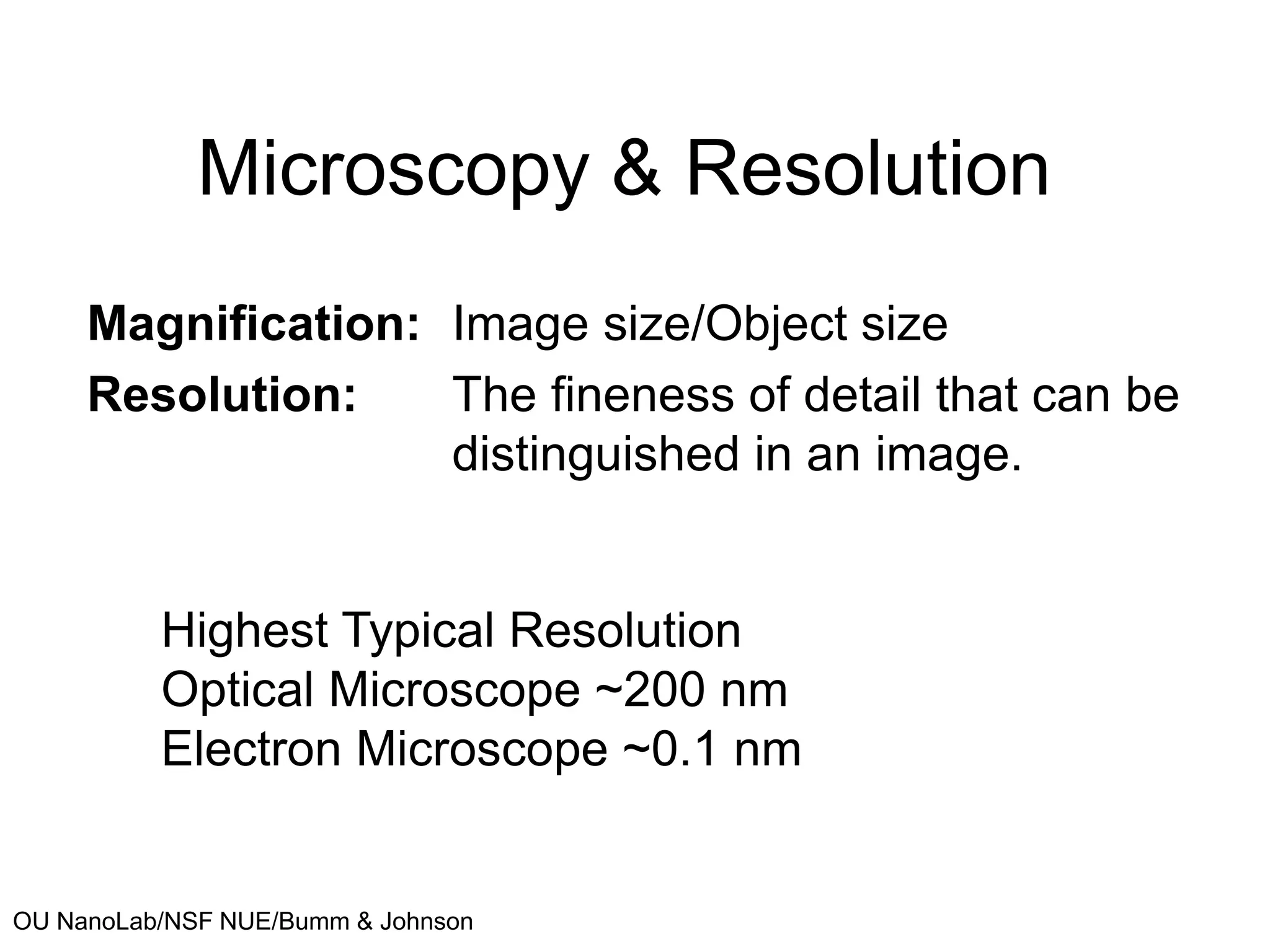
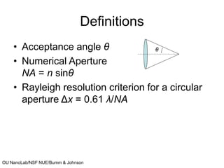
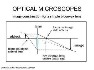
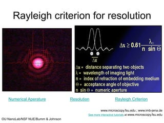
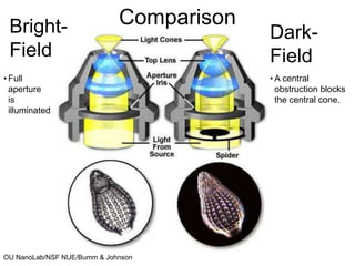
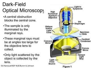
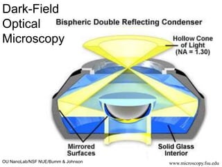
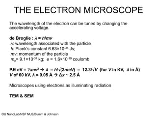
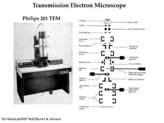
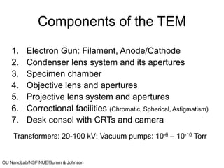
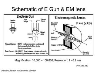
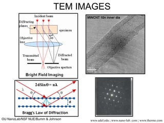
OU NanoLab/NSF NUE/Bumm & Johnson
Microscopy & Resolution
Magnification: Image size/Object size
Resolution: The fineness of detail that can be
distinguished in an image.
Highest Typical Resolution
Optical Microscope ~200 nm
Electron Microscope ~0.1 nm
OU NanoLab/NSF NUE/Bumm & Johnson
Definitions
• Acceptance angle θ
• Numerical Aperture
NA = n sinθ
• Rayleigh resolution criterion for a circular
aperture Δx = 0.61 λ/NA
θ
OU NanoLab/NSF NUE/Bumm & Johnson
OPTICAL MICROSCOPES
Image construction for a simple biconvex lens
OU NanoLab/NSF NUE/Bumm & Johnson
Rayleigh criterion for resolution
www.microscopy.fsu.edu ; www.imb-jena.de
See more interactive tutorials at www.microscopy.fsu.edu
Numerical Aperature Resolution Rayleigh Criterion
OU NanoLab/NSF NUE/Bumm & Johnson
Comparison
Bright-
Field
Dark-
Field
• Full
aperture
is
illuminated
• A central
obstruction blocks
the central cone.
OU NanoLab/NSF NUE/Bumm & Johnson www.microscopy.fsu.edu
Dark-Field
Optical Microscopy
•A central obstruction
blocks the central cone.
•The sample is only
illuminated by the
marginal rays.
•These marginal rays must
be at angles too large for
the objective lens to
collect.
•Only light scattered by the
object is collected by the
lens.
OU NanoLab/NSF NUE/Bumm & Johnson www.microscopy.fsu.edu
Dark-Field
Optical
Microscopy
OU NanoLab/NSF NUE/Bumm & Johnson
THE ELECTRON MICROSCOPE
The wavelength of the electron can be tuned by changing the
accelerating voltage.
de Broglie : λ = h/mv
λ: wavelength associated with the particle
h: Plank’s constant 6.63×10⁻³⁴ Js;
mv: momentum of the particle
me= 9.1×10⁻³¹ kg; e = 1.6×10⁻¹⁹ coulomb
P.E eV = ½mv2  λ = h/(2meV) = 12.3/V (for V in KV, λ in Å)
V of 60 kV, λ = 0.05 Å  Δx ~ 2.5 Å
Microscopes using electrons as illuminating radiation
TEM & SEM
OU NanoLab/NSF NUE/Bumm & Johnson
Components of the TEM
1. Electron Gun: Filament, Anode/Cathode
2. Condenser lens system and its apertures
3. Specimen chamber
4. Objective lens and apertures
5. Projective lens system and apertures
6. Correctional facilities (Chromatic, Spherical, Astigmatism)
7. Desk consol with CRTs and camera
Transformers: 20-100 kV; Vacuum pumps: 10-6 – 10-10 Torr
OU NanoLab/NSF NUE/Bumm & Johnson
Schematic of E Gun & EM lens
Magnification: 10,000 – 100,000; Resolution: 1 - 0.2 nm
www.udel.edu
OU NanoLab/NSF NUE/Bumm & Johnson
TEM IMAGES
www.udel.edu ; www.nano-lab. com ; www.thermo.com

Resolution Theory.ppt

  • 1.
    OU NanoLab/NSF NUE/Bumm& Johnson Microscopy & Resolution Magnification: Image size/Object size Resolution: The fineness of detail that can be distinguished in an image. Highest Typical Resolution Optical Microscope ~200 nm Electron Microscope ~0.1 nm
  • 2.
    OU NanoLab/NSF NUE/Bumm& Johnson Definitions • Acceptance angle θ • Numerical Aperture NA = n sinθ • Rayleigh resolution criterion for a circular aperture Δx = 0.61 λ/NA θ
  • 3.
    OU NanoLab/NSF NUE/Bumm& Johnson OPTICAL MICROSCOPES Image construction for a simple biconvex lens
  • 4.
    OU NanoLab/NSF NUE/Bumm& Johnson Rayleigh criterion for resolution www.microscopy.fsu.edu ; www.imb-jena.de See more interactive tutorials at www.microscopy.fsu.edu Numerical Aperature Resolution Rayleigh Criterion
  • 5.
    OU NanoLab/NSF NUE/Bumm& Johnson Comparison Bright- Field Dark- Field • Full aperture is illuminated • A central obstruction blocks the central cone.
  • 6.
    OU NanoLab/NSF NUE/Bumm& Johnson www.microscopy.fsu.edu Dark-Field Optical Microscopy •A central obstruction blocks the central cone. •The sample is only illuminated by the marginal rays. •These marginal rays must be at angles too large for the objective lens to collect. •Only light scattered by the object is collected by the lens.
  • 7.
    OU NanoLab/NSF NUE/Bumm& Johnson www.microscopy.fsu.edu Dark-Field Optical Microscopy
  • 8.
    OU NanoLab/NSF NUE/Bumm& Johnson THE ELECTRON MICROSCOPE The wavelength of the electron can be tuned by changing the accelerating voltage. de Broglie : λ = h/mv λ: wavelength associated with the particle h: Plank’s constant 6.63×10⁻³⁴ Js; mv: momentum of the particle me= 9.1×10⁻³¹ kg; e = 1.6×10⁻¹⁹ coulomb P.E eV = ½mv2  λ = h/(2meV) = 12.3/V (for V in KV, λ in Å) V of 60 kV, λ = 0.05 Å  Δx ~ 2.5 Å Microscopes using electrons as illuminating radiation TEM & SEM
  • 9.
  • 10.
    OU NanoLab/NSF NUE/Bumm& Johnson Components of the TEM 1. Electron Gun: Filament, Anode/Cathode 2. Condenser lens system and its apertures 3. Specimen chamber 4. Objective lens and apertures 5. Projective lens system and apertures 6. Correctional facilities (Chromatic, Spherical, Astigmatism) 7. Desk consol with CRTs and camera Transformers: 20-100 kV; Vacuum pumps: 10-6 – 10-10 Torr
  • 11.
    OU NanoLab/NSF NUE/Bumm& Johnson Schematic of E Gun & EM lens Magnification: 10,000 – 100,000; Resolution: 1 - 0.2 nm www.udel.edu
  • 12.
    OU NanoLab/NSF NUE/Bumm& Johnson TEM IMAGES www.udel.edu ; www.nano-lab. com ; www.thermo.com