1) The document describes a study identifying new genes involved in gliding motility in Flavobacterium johnsoniae.
2) Transposon mutagenesis of an F. johnsoniae sprB mutant identified 8 mutants with increased phage resistance and reduced motility. 4 mutants had transposon insertions in remA, which encodes a cell surface protein with a lectin domain.
3) RemA was shown to localize to the cell surface and move rapidly along the cell surface, suggesting it acts as a mobile adhesin involved in gliding motility.
![30°C, as previously described (23). To observe colony spreading, F. john-
soniae was grown at 25°C on PY2 medium (1) or EC medium (4) supple-
mented with 10 g of agar per liter. Motility medium (MM) (17) and EC
medium were used to observe movement of individual cells in wet
mounts. The bacteriophages active against F. johnsoniae that were used
here were Cj1, Cj13, Cj23, Cj28, Cj29, Cj42, Cj48, and Cj54
(4, 28, 42). Sensitivity to bacteriophages was determined essentially as
previously described by spotting 5 l of phage lysates (109
PFU/ml) onto
lawns of cells in CYE overlay agar (11). The plates were incubated for 24 h
at 25°C to observe lysis. The strains and plasmids used in the present study
are listed in Table 1. The plasmids used for complementation were all
derived from pCP1 and have copy numbers of approximately 10 in F.
johnsoniae (1, 13, 23). Antibiotics were used at the following concentra-
tions when needed: ampicillin, 100 g/ml; cefoxitin, 100 g/ml; chloram-
phenicol, 30 g/ml; erythromycin, 100 g/ml; kanamycin, 35 g/ml; and
tetracycline, 20 g/ml.
Isolation of phage-resistant mutants of F. johnsoniae CJ1584
[⌬(sprC sprD sprB)] by HimarEm1 mutagenesis and identification of
sites of insertion. pHimarEm1 was introduced into F. johnsoniae CJ1584
by conjugation from Escherichia coli S17-1 pir essentially as previously
described (2). HimarEm1 mutants were selected by plating cells on CYE
agar containing erythromycin. Cells from 800 random erythromycin-re-
sistant colonies were transferred to CYE agar (master plate) and to CYE
agar overlaid with 4 ml of CYE top agar containing ϳ109
PFU of Cj42,
followed by incubation for 24 h at 25°C. Colonies that grew in the pres-
ence of Cj42 were picked from the corresponding colonies on the master
plate and streaked for isolation on CYE with erythromycin. Colonies were
tested again for phage sensitivity, and those with increased resistance were
selected for further analyses.
Chromosomal DNA was isolated from each of the phage-resistant
mutants, and the HimarEm1 transposons and adjacent DNA from each
were cloned in E. coli EC100D pirϩ
. Sequences of F. johnsoniae DNA
disrupted by HimarEm1 were determined as previously described (2).
Strain construction. Unmarked deletions were made as previously
described (32). To delete remA, a 1.8-kbp fragment spanning fjoh_0809
and the final 72 bp of remA was amplified by PCR using the primers 1061
(introducing a SalI site) and 760 (introducing a PstI site). The fragment
was digested with SalI and PstI and ligated into pRR51 that had been
digested with the same enzymes to generate pRR76. A 2.1-kbp fragment
spanning fjoh_0807 and the first 150 bp of remA was amplified by PCR
with primers 1059 (introducing a BamHI site) and 1060 (introducing a
SalI site). The fragment was digested with BamHI and SalI and fused to the
region downstream of remA by ligation with pRR76 that had been di-
gested with the same enzymes to generate the deletion construct pRR78.
Plasmid pRR78 was introduced into the streptomycin-resistant wild-type
F. johnsoniae strain CJ1827 and into the sprB deletion mutant CJ1922 by
triparental conjugation, and remA deletion mutants were isolated as pre-
viously described (32). Deletion of remA was confirmed by PCR amplifi-
cation using the primers 778 and 1062, which flank remA, and sequencing
the resulting 1.8-kbp product. All other deletion strains listed in Table 1
except for CJ2089 [rpsL2 ⌬(gldN gldO) remA::myc-tag-1] and CJ2090
[rpsL2 ⌬(gldN gldO)] were constructed in the same way, using the primers
listed in Table S1 in the supplemental material and the plasmids listed in
Table 1. CJ2089 and CJ2090 were constructed as previously described (33)
by introducing pNap3 into CJ2083 or CJ1827, respectively, selecting for
antibiotic-resistant colonies that had the plasmid inserted in the genome
and then selecting for resistance to Cj1 to obtain the gldNO deletions.
The sprF mutant CJ2097 was constructed by integration of pRR47, which
carries an internal fragment of sprF, into the chromosome of CJ2083
essentially as previously described (31).
To observe RemA on the cell surface, four strains expressing RemA
containing the myc tag sequence (EQKLISEEDL) at different locations
were generated using a previously described allelic-exchange method
(32). As an example, to generate the strain carrying remA::myc-tag-1
(CJ2083), the primers 1059 (introducing a BamHI site) and 1112 (intro-
ducing the myc tag) were used to amplify a 2.1-kbp fragment spanning
fjoh_0807 and the first 150 bp of remA. Similarly, the primers 761 (intro-
ducing a SalI site) and 1110 (introducing the myc tag) were used to amplify
the 2.4-kbp fragment beginning at bp 151 of remA. The two PCR products
were then used as templates in a crossover PCR with the primers 761 and
1059. The resulting 4.5-kbp PCR product was digested with BamHI and
SalI and ligated into the pRR51 suicide vector to generate pRR92. Plasmid
pRR92 was introduced into the streptomycin-resistant wild-type strain
CJ1827 and the sprB deletion mutant CJ1922 by triparental conjugation,
and allelic exchange was performed as previously described (32). Replace-
ment of wild-type remA with the remA::myc-tag-1 allele was confirmed by
PCR amplification using primers 1142 (myc-tag sequence) and 1063
(complementary to the remA sequence), and sequencing the resulting
0.6-kbp product. Four different remA::myc-tag strains were generated,
with remA::myc-tag-1, remA::myc-tag-3, remA::myc-tag-4, and remA::
myc-tag-5 inserted 150, 2,058, 2,286, and 2,514 bp downstream of the “A”
in the start codon, respectively.
Cloning of remA and complementation of remA mutants. Attempts
to clone remA amplified by PCR were not successful, so an alternative
approach was used. The plasmids pMM332 and pMM336 that were used
to sequence remA HimarEm1 insertions served as raw material to recon-
struct the intact remA gene. The 3= end of remA was obtained as a SphI-
XbaI fragment from pMM336. This fragment was ligated into pUC18 that
had been digested with the same enzymes to generate pRR36. The 5= end
of remA and upstream sequences were obtained as a HindIII-SphI frag-
ment from pMM332. This fragment was inserted into pRR36 that had
been digested with HindIII and SphI to generate pRR37, which carries the
intact remA gene. remA was transferred to pBC SKϩ as a 4.9-kbp HindIII-
XbaI fragment generating pRR38. This provided convenient restriction
sites (KpnI and XbaI) to allow transfer of remA into the shuttle vector
pCP23, generating pRR39. pRR39 was introduced into remA mutants by
conjugation as previously described (11, 21), except that pRK2013 (5) was
used for triparental conjugations.
Expression of recombinant RemA in E. coli and generation of anti-
bodies. A 1,966-bp fragment encoding the 561-amino-acid C-terminal
region of RemA was amplified using Phusion DNA polymerase (New
England Biolabs, Ipswich, MA) and the primers 745 and 762. The PCR
product was digested with BamHI and SalI and cloned into the pMAL-c2
expression vector (New England Biolabs) that had been digested with the
same enzymes, generating pSP18. pSP18 was introduced into E. coli Ro-
setta 2 (DE3) cells (Novagen, Madison, WI), which expressed seven rare
tRNAs required for the efficient expression of RemA. To isolate recombi-
nant RemA, cells were grown to mid-log phase at 37°C in rich medium
containing glucose (10 g of tryptone, 5 g of yeast extract, 5 g of NaCl, and
2 g of glucose/liter), induced by the addition of 0.3 mM IPTG (isopropyl-
-D-thiogalactopyranoside), and incubated for 8 h at 25°C. Cells were
disrupted using a French press, and recombinant RemA was purified us-
ing an amylose resin column (New England Biolabs). Polyclonal antibod-
ies against recombinant RemA were produced and affinity purified using
the recombinant protein by ProteinTech Group, Inc. (Chicago, IL).
Immunodetection and localization of RemA. F. johnsoniae cells were
grown to mid-log phase in CYE at 25°C. Whole cells were centrifuged at
4,000 ϫ g, resuspended in SDS-PAGE loading buffer, and boiled for 5
min. Proteins were separated by SDS-PAGE, and Western blot analyses
were performed essentially as previously described (33) using affinity-
purified antisera against RemA (1:1,000 dilution) or antisera against the
c-myc epitope (1:10,000 dilution; AbCam, Cambridge, MA). To deter-
mine the localization of RemA, cells were disrupted with a French press
and fractionated into soluble and insoluble fractions as described previ-
ously by centrifugation at 352,900 ϫ g for 30 min (34), and Western
blotting was performed as described above.
Microscopic observations of cell movement. Wild-type and mutant
cells of F. johnsoniae were examined for movement over glass by phase-
contrast microscopy. Tunnel slides were prepared essentially as described
previously (38) using Nichiban NW-5 double-sided tape (Nichiban Co.,
F. johnsoniae Gliding Motility Protein RemA
July 2012 Volume 194 Number 14 jb.asm.org 3679
onAugust28,2015byguesthttp://jb.asm.org/Downloadedfrom](https://image.slidesharecdn.com/261c21a2-b256-4fab-bdaf-3a11ad5b1a09-160305181539/85/RemA-copy-2-320.jpg)
![others (Cj28, Cj42, Cj48, and Cj54) (see Fig. S1 in the sup-
plemental material). A total of 800 randomly chosen erythromy-
cin-resistant colonies containing HimarEm1 insertions were
screened for resistance to Cj42, and eight mutants with partial or
complete resistance were obtained. In addition to resistance to
Cj42, the mutants exhibited increased resistance to other phages
to which CJ1584 is susceptible (see Fig. S1 in the supplemental
material). Microscopic examination revealed that each mutant
also had a more severe motility defect than did the parent strain
(see Movie S1 in the supplemental material, and data not shown).
The sites of the transposon insertions were determined by
cloning HimarEm1 and adjacent DNA and sequencing across the
junction. Four of the mutants had insertions in fjoh_0808, which
we named remA (for redundant motility gene A) (Fig. 1). remA
encodes a predicted 152-kDa protein (after removal of its signal
peptide) that exhibits limited similarity to the much larger (669-
kDa) cell surface motility protein SprB (26). Similarity to SprB
was confined to 5 regions of RemA (amino acids 158 to 225, 453 to
494, 580 to 642, 976 to 1022, and 1282 to 1370) and ranged from
26 to 43% identity. BLASTP analysis also revealed similarity to
two conserved domains of known function. The C-terminal 80
amino acids exhibited similarity to a Por secretion system C-ter-
minal sorting domain (TIGR04183; E-value 6.93e-05), suggesting
that, like SprB, RemA may be secreted across the outer membrane
by the F. johnsoniae PorSS. In addition, the region between amino
acids 725 and 800 exhibited similarity to SUEL-related galactose/
rhamnose-binding lectins (pfam02140; E-value 2.44e-17), sug-
gesting that RemA may interact with sugars. The remaining four
mutants had insertions in fjoh_1657 (remB), fjoh_0216 (remC),
fjoh_0361 (wza), and fjoh_0360 (wzc). RemB is predicted to be an
outer membrane protein of unknown function, RemC is a pre-
dicted glycosyltransferase, and Wza and Wzc are predicted com-
ponents of a polysaccharide synthesis and secretion system. Se-
creted or cell surface polysaccharides have previously been
implicated in gliding of F. johnsoniae and of other bacteria (7, 8,
16, 18, 41, 43).
SprB, SprC, and SprD are each required for efficient motility
on agar and for the formation of spreading colonies (26, 31). SprB
is a mobile cell surface adhesin, and SprC and SprD are thought to
support SprB function. Disruption of sprB results in increased
resistance to Cj1, Cj13, Cj23, and Cj29, whereas the disrup-
tion of sprC and sprD does not result in resistance to phages that
have been tested (31). CJ1985 (⌬sprB ⌬remA) and CJ1984
(⌬remA) were constructed to simplify determination of the roles
of remA and sprB in phage resistance and motility. Cells of CJ1985
(⌬sprB ⌬remA) exhibited the same phage resistance phenotypes as
those of CJ1601 [⌬(sprC sprD sprB) remA::HimarEm1] (Fig. 2; see
Fig. S1 in the supplemental material). Cells of CJ1984 (⌬remA)
exhibited increased resistance to Cj42, Cj48, and Cj54, and
the introduction of remA on pRR39 restored sensitivity to these
phages (Fig. 2).
Deletion of remA in an sprB mutant results in decreased mo-
tility. Cells of CJ1984 (⌬remA), CJ1922 (⌬sprB), and CJ1985
(⌬sprB ⌬remA) were examined for motility (see Movies S2 and S3
in the supplemental material). Cells of CJ1984 (⌬remA) were in-
distinguishable from wild-type cells, whereas cells of CJ1922
(⌬sprB) exhibited a partial motility defect similar to that of CJ1584
[⌬(sprC sprD sprB)], as previously reported (26, 31, 32). CJ1922
formed nonspreading colonies on agar, but individual cells re-
tained some ability to move on glass surfaces. Cells of CJ1985
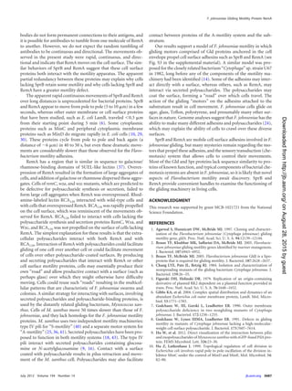
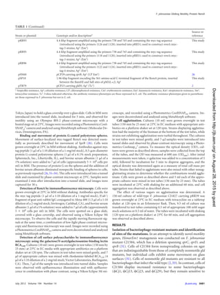
FIG 1 Map of the remA region. Numbers below the map refer to kilobase pairs of sequence. The sites of HimarEm1 insertions in remA are indicated by triangles,
with orientations indicated by the direction in which the triangle is pointing. Triangles pointing to the right in this diagram (CJ1597 for example) have IR2 on
the right side and the kanamycin resistance gene of the transposon reading toward the right. The regions of DNA carried by plasmids used in the present study
are indicated beneath the map.
Shrivastava et al.
3682 jb.asm.org Journal of Bacteriology
onAugust28,2015byguesthttp://jb.asm.org/Downloadedfrom](https://image.slidesharecdn.com/261c21a2-b256-4fab-bdaf-3a11ad5b1a09-160305181539/85/RemA-copy-5-320.jpg)
![(⌬sprB ⌬remA) exhibited more dramatic motility defects, with
most cells exhibiting little if any movement. The motility behavior
of CJ1985 was similar to that of CJ1601 [⌬(sprC sprD sprB) remA::
HimarEm1] (see Movie S1 in the supplemental material). Com-
plementation of CJ1985 (⌬sprB ⌬remA) with pRR39, which car-
ries remA, restored motility comparable to or exceeding that
exhibited by the parent strain CJ1922 (⌬sprB). The decreased mo-
tility of CJ1985 (⌬sprB ⌬remA) compared to CJ1922 (⌬sprB) sug-
gests that SprB and RemA may be partially redundant compo-
nents of the motility apparatus. sprB mutants form nonspreading
colonies as a result of motility defects, but colonies of the remA
deletion mutant CJ1984 (⌬remA) were indistinguishable from
those of the wild type (see Fig. S2 in the supplemental material),
indicating that, unlike SprB, RemA is not required for movement
of cells on agar.
RemA moves rapidly along the cell surface. RemA was pre-
dicted to be a cell-surface-exposed outer membrane protein. An-
tiserum against recombinant RemA was used to detect RemA in F.
johnsoniae cell extracts. RemA was found in the insoluble fraction
of cell extracts and migrated with an apparent molecular mass of
ϳ150 kDa (Fig. 3A), which is close to the size predicted for the
mature protein. Antiserum against RemA detected denatured
protein in Western blots but failed to detect native RemA on the
cell surface or in cell extracts. Four myc-tagged versions of remA
were constructed and inserted into the genome in place of wild-
type remA. Two of the strains, CJ2083 (remA::myc-tag-1; myc-tag
inserted 150 bp downstream of the “A” in the start codon) and
CJ2112 (remA::myc-tag-4; myc-tag inserted 2,286 bp downstream
of the start codon), produced stable myc-tagged RemA protein, as
detected by Western blotting (Fig. 3). These strains were sensitive
to the same phages as were wild-type cells, suggesting that the
myc-tagged versions of RemA localized properly and supported
phage infection. Protein-G-coated latex spheres carrying antibod-
ies against the myc-tag peptide bound specifically to cells of
CJ2083 (remA::myc-tag-1) and CJ2112 (remA::myc-tag-4), indi-
cating that RemA was exposed on the cell surface (Table 2). The
bound spheres moved rapidly along the cell surface (see Movie S4
in the supplemental material), suggesting that RemA is propelled
along the cell by the gliding “motor,” as previously suggested for
SprB (26). As previously reported (26, 31–34), and as shown in
Table 2 and in Movie S4 in the supplemental material, Protein
G-coated spheres did not bind to cells unless specific antiserum
was added. They also failed to bind to wild type cells that did not
express Myc-tagged RemA. Cells of CJ2077 (remA::myc-tag-3;
myc-tag inserted 2,058 bp downstream of the start codon) and of
CJ2073 (remA::myc-tag-5; myc-tag inserted 2,514 bp downstream
of the start codon) produced little if any RemA and failed to bind
protein-G-coated latex spheres carrying antibodies against the
myc-tag peptide. To eliminate potential artifacts resulting from
the relatively large (0.5-m) latex spheres, the localization and
movement of RemA-myc-tag-1 were also examined by immuno-
fluorescence microscopy using antibodies against the myc-tag.
Cells of CJ2083 expressed RemA-myc-tag-1 and were fluores-
cently labeled, whereas cells of CJ1827 (wild type) were not. The
fluorescent antibodies moved smoothly along the cell surface at
speeds of 1 to 2 m/sec (Fig. 4; see Movie S5 in the supplemental
material). The fluorescent signals traveled the length of the cell,
looped around the pole, and returned to a location near the start-
ing point, all within about 10 s. The movements of spheres and
fluorescent antibodies are consistent with a model for gliding in
which the gliding motor in the cell envelope propels cell-surface
adhesins such as RemA. SprB, which also moves on the cell sur-
face, was not required for RemA movement, since cells of CJ2072
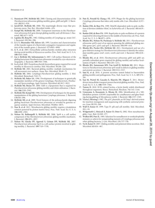
FIG 2 Effect of mutations on bacteriophage resistance. Bacteriophages (5 l
of lysates containing ϳ109
PFU/ml) were spotted onto lawns of cells in CYE
overlay agar. The plates were incubated at 25°C for 24 h to observe lysis.
Bacteriophages were spotted in the following order from left to right, as indi-
cated also by the numbers in panel A: top row, Cj1, Cj13, and Cj23;
middle row, Cj28, Cj29, and Cj42; bottom row, Cj48 and Cj54. (A)
Wild-type F. johnsoniae CJ1827; (B) CJ1984 (⌬remA); (C) CJ1984 comple-
mented with pRR39, which carries remA; (D) CJ1922 (⌬sprB); (E) CJ1985
(⌬sprB ⌬remA); (F) CJ1985 complemented with pRR39.
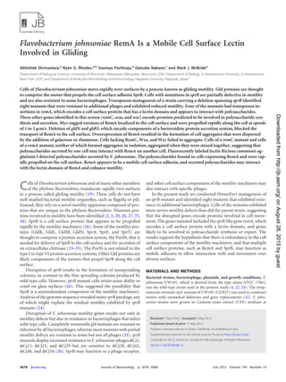
FIG 3 Immunodetection of RemA and RemA-myc-tag. (A) Cell extracts (15
g of protein) were examined by Western blotting with antiserum against
RemA. Lane 1, wild-type F. johnsoniae CJ1827; lane 2, CJ1984 (⌬remA); lane 3,
CJ1984 complemented with pRR39 which carries remA; lane 4, CJ2090
[⌬(gldN-gldO)]; lane 5, CJ2083 (remA::myc-tag-1); lane 6, CJ2089 [⌬(gldN-
gldO) remA::myc-tag-1]; lane 7, soluble (cytoplasmic and periplasmic) frac-
tion of wild-type cell extract; lane 8, particulate (membrane) fraction of wild-
type cell extract. (B) Cell extracts (20 g of protein) were examined by Western
blotting with antiserum against myc-tag peptide. Lane 1, wild-type F. john-
soniae CJ1827; lane 2, CJ2083 (remA::myc-tag-1); lane 3, CJ2077 (remA::myc-
tag-3); lane 4, CJ2112 (remA::myc-tag-4); lane 5, CJ2073 (remA::myc-tag-5);
lane 6, CJ2089 [⌬(gldN-gldO) remA::myc-tag-1].
F. johnsoniae Gliding Motility Protein RemA
July 2012 Volume 194 Number 14 jb.asm.org 3683
onAugust28,2015byguesthttp://jb.asm.org/Downloadedfrom](https://image.slidesharecdn.com/261c21a2-b256-4fab-bdaf-3a11ad5b1a09-160305181539/85/RemA-copy-6-320.jpg)
![(⌬sprB remA::myc-tag-1) also propelled fluorescently labeled an-
ti-myc antibodies (see Movie S5 in the supplemental material).
Mutations in porSS genes disrupt secretion of RemA. A novel
protein secretion system, the Por secretion system (PorSS), is re-
quired for delivery of SprB to the cell surface, and also for secre-
tion of an extracellular chitinase (33–35). A strain carrying myc-
tagged remA and carrying a deletion of the region spanning the
porSS genes gldN and gldO was constructed to determine whether
the PorSS is involved in secretion of RemA. Cells lacking gldN and
gldO produced myc-tagged RemA (Fig. 3) but failed to secrete it to
the cell surface, as determined by the failure of latex spheres car-
rying antibodies against the myc-tag peptide to bind to the cells
(Table 2). Complementation with pTB79, which carries gldN, re-
stored surface exposure of myc-tagged RemA. The results suggest
that the PorSS is required for secretion of RemA, in addition to its
previously demonstrated roles in secretion of SprB and chitinase
(33–35). SprB requires an additional protein, SprF, for secretion
to the cell surface (31). SprF is thought to be an adapter to the
PorSS that is specific for SprB, since SprF is not required for se-
cretion of chitinase. Many of the F. johnsoniae sprB paralogs are
adjacent to sprF paralogs, and the predicted SprF-like proteins
may function in secretion of their cognate SprB-like proteins.
remA does not have an sprF-like gene nearby. Cells of CJ2097
(remA::myc-tag-1 sprF) bound and propelled spheres carrying an-
tibodies against the myc-tag peptide, indicating that SprF is not
required for secretion of RemA (Table 2).
Analysis of the remA paralogs fjoh_0803, fjoh_0804,
fjoh_0805, and fjoh_0806. Four remA paralogs lie near remA on
the F. johnsoniae genome (Fig. 1). The products of fjoh_0803,
fjoh_0804, fjoh_0805, and fjoh_0806 exhibited 57, 47, 93, and 64%
identities, respectively, to the C-terminal 500 amino acids of
RemA and exhibited more limited similarity over the rest of
RemA. The region of RemA between 725 and 830 amino acids,
which corresponds to the lectin domain, was not conserved in the
four remA paralogs. To determine whether the remA paralogs
function in gliding and phage sensitivity, the region spanning
fjoh_0803-fjoh_0806 was deleted in wild-type cells, in CJ1984
(⌬remA) cells, and in CJ1985 (⌬sprB ⌬remA) cells. Deletion of
fjoh_0803-fjoh_0806 from any of these strains had no effect on
phage sensitivity, motility, or colony spreading (see Fig. S2 in the
supplemental material and data not shown).
Involvement of RemA and cell-surface polysaccharides in
cell-cell interactions. Wild-type F. johnsoniae cells grown in EC
medium formed small cell aggregates (Fig. 5A). Cells of CJ1984
(⌬remA) were deficient in aggregate formation and complemen-
tation with remA on pRR39 restored the ability to form aggregates.
The aggregates formed by the complemented strain were much
larger than those formed by wild-type cells, suggesting that mod-
erate overexpression of RemA from pRR39, which has a copy
number of ϳ10, enhanced aggregation. Introduction of pRR39
into wild-type cells also enhanced aggregation. The control vector
pCP23 did not cause cell aggregation, indicating that remA was
responsible for this phenomenon. The large cell aggregates
formed for strains carrying pRR39 resulted in rapid settling of the
TABLE 2 Binding of protein G-coated polystyrene spheres carrying antibodies against myc-tag peptide
Strain Description Antibody added
Avg (SD) % of cells with
spheres attacheda
CJ1827 Wild type No antibody 0.6 (0.5)
CJ1827 Wild type Anti-myc 0.6 (1.1)
CJ2083 remA::myc-tag-1 No antibody 0.0 (0.0)
CJ2083 remA::myc-tag-1 Anti-myc 52.3 (3.2)
CJ2077 remA::myc-tag-3 Anti-myc 0.0 (0.0)
CJ2112 remA::myc-tag-4 Anti-myc 33.3 (3.1)
CJ2073 remA::myc-tag-5 Anti-myc 0.3 (0.5)
CJ2089 ⌬(gldN-gldO) remA::myc-tag-1 Anti-myc 0.0 (0.0)
CJ2089 with pTB79 carrying gldN ⌬(gldN-gldO) remA::myc-tag-1/pTB79 (gldN) Anti-myc 52.0 (3.0)
CJ2097 remA::myc-tag-1 sprF Anti-myc 52.6 (4.0)
a
Purified anti-myc-tag antiserum and 0.5-m-diameter protein G-coated polystyrene spheres were added to cells as described in Materials and Methods. Samples were introduced
into a tunnel slide, incubated for 1 min at 25°C, and examined using a phase-contrast microscope. Images were recorded for 30 s, and 100 randomly selected cells were examined
for the presence of spheres that remained attached to the cells during this time. The numbers in parentheses are standard deviations calculated from three measurements.
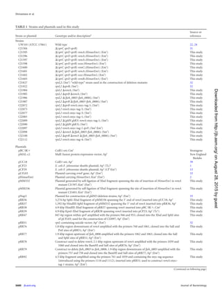
FIG 4 Movement of RemA on the cell surface. (A) Cells of CJ2083, which
expressed RemA-myc-tag-1, were examined by a combination of immunoflu-
orescence and phase-contrast microscopy. Low-light phase contrast was used
to detect the cells, and antisera against the myc-tag peptide and goat anti-
rabbit IgG conjugated to Alexa 488 were used to detect RemA-myc-tag-1. The
sequence shown corresponds to the first 3.9 s of the third sequence (CJ2083
[RemA-myc-tag-1], with anti-myc-tag antibody) in Movie S5 in the supple-
mental material. The full movie includes controls demonstrating the specific-
ity of the antiserum for the myc-tag peptide. Numbers in each panel indicate
time in seconds. Arrows outline the apparent movements of two selected fluo-
rescent signals. Bar, 5 m. (B) Diagram illustrating the apparent movements of
the two selected fluorescent signals highlighted in panel A over 3.9 s.
Shrivastava et al.
3684 jb.asm.org Journal of Bacteriology
onAugust28,2015byguesthttp://jb.asm.org/Downloadedfrom](https://image.slidesharecdn.com/261c21a2-b256-4fab-bdaf-3a11ad5b1a09-160305181539/85/RemA-copy-7-320.jpg)
![cells from suspension, allowing macroscopic observation and
quantitation of aggregation (Fig. 5B and C). The predicted lectin
domain of RemA suggested the possibility that RemA bound to
polysaccharides on the surface of neighboring cells. Addition of 5
mM D-galactose, L-rhamnose, or D-lactose resulted in the rapid
dissolution of the aggregates, whereas the addition of other sugars
(D-glucose, D-mannose, D-fructose, D-ribose, D-sorbitol, D-su-
crose, D-maltose, and D-trehalose) at 5 to 100 mM did not (Fig. 6
and data not shown). The ability to completely disperse aggregates
by adding 5 mM galactose allowed us to rapidly estimate total
biomass by measuring the OD600 (Fig. 5C). Not surprisingly, the
formation of large aggregates as a result of the overexpression of
RemA correlated with decreased total biomass (Fig. 5C), probably
because of reduced availability of nutrients or O2 in the dense
aggregates. As mentioned earlier, RemA moves rapidly on the cell
surface (Fig. 4). The addition of galactose or rhamnose had no
effect on the movement of RemA, as observed using antibody-
coated spheres or by immunofluorescence microscopy (data not
shown).
remC, wza, and wzc were identified in the same genetic screen
that resulted in the identification of remA, and the proteins en-
coded by these genes are predicted to be involved in polysaccha-
ride synthesis and secretion. Secreted polysaccharides might inter-
act with RemA and be involved in cell-cell or cell-substratum
interactions. pRR39 was introduced into cells of CJ1584 [⌬(sprC
sprD sprB)], CJ1598 [⌬(sprC sprD sprB) wza::HimarEm1], CJ1602
[⌬(sprC sprD sprB) wzc::HimarEm1], and CJ1600 [⌬(sprC sprD
sprB) remC::HimarEm1] in order to determine whether cells that
overexpressed RemA but that were deficient in polysaccharide
synthesis or secretion would form aggregates. Cell aggregates
formed with CJ1584 carrying pRR39 but not with any of the
strains with mutations in the predicted polysaccharide synthesis
and secretion genes (Fig. 7). This suggests that both RemA and
secreted polysaccharides are required for aggregate formation.
RemA may function as a cell surface adhesin that interacts with
polysaccharides synthesized and secreted by RemC, Wza, and
Wzc. SprB, SprC, and SprD may also affect aggregate formation,
FIG 5 Effect of RemA on cell aggregation. Cells of CJ1827 (wild type) and
CJ1984 (⌬remA) containing either control plasmid pCP23 (images on the left
in panels A and B) or remA-expressing plasmid pRR39 (images on the right in
panels A and B) were incubated in EC medium. (A) Observation of cells or cell
clumps by phase-contrast microscopy. Bar, 50 m. (B) Macroscopic observa-
tion of cell clumps in culture tubes. (C) Turbidity (OD600) of samples taken
from the top of the culture tubes. Measurements were obtained from samples
taken directly from the tubes and from samples taken after addition of 5 mM
D-galactose and incubation for 5 min to cause cell dispersal.
FIG 6 Effect of sugars on RemA-mediated cell aggregation. Cells of CJ1827
(wild type) carrying pRR39 to overexpress RemA were transferred into test
tubes containing various sugars at final concentrations of 5 mM. Cultures were
incubated with gentle shaking for 1 h at 23°C and examined for aggregation.
(A) Observation of cells or cell clumps by phase-contrast microscopy. Bar, 50
m. (B) Macroscopic observation of cell clumps in culture tubes. (C) Turbid-
ity (OD600) of samples taken from the top of the culture tubes.
F. johnsoniae Gliding Motility Protein RemA
July 2012 Volume 194 Number 14 jb.asm.org 3685
onAugust28,2015byguesthttp://jb.asm.org/Downloadedfrom](https://image.slidesharecdn.com/261c21a2-b256-4fab-bdaf-3a11ad5b1a09-160305181539/85/RemA-copy-8-320.jpg)
![since the aggregates formed by cells of CJ1584 [⌬(sprC sprD sprB)]
carrying pRR39 were smaller than those formed by wild-type cells
carrying the same plasmid (Fig. 5A and 7A). However, although
the overexpression of RemA (from pRR39) resulted in increased
cell aggregation, the overexpression of SprB (from pSN60) did not
(data not shown). Polysaccharide production and RemA expres-
sion do not need to occur in the same cell for aggregation to occur.
Cells of CJ1984 (⌬remA), and of CJ1600 [⌬(sprC sprD sprB) rem-
C::HimarEm1] carrying pRR39, neither of which formed aggre-
gates in isolation, resulted in aggregate formation when they were
mixed together (Fig. 7). Similar results were obtained when cells of
CJ1984 were mixed with cells of the wza mutant CJ1598 carrying
pRR39.
A fluorescent lectin was used to visualize secreted or cell sur-
face polysaccharides or oligosaccharides on the cells of F. john-
soniae. Rhodamine-labeled Ricinus communis agglutinin I
(RCA120), which binds galactose, labeled cell clumps and individ-
ual cells. The fluorescent lectin was propelled rapidly on the cell
surface (see Movie S6 in the supplemental material). This may
indicate that some of the polysaccharide that was labeled with
RCA120 also bound to RemA, so that the fluorescent lectin indi-
rectly labeled RemA. Supporting this idea, strains lacking RemA
failed to propel RCA120. Cells of the remC mutant CJ1600 also
failed to bind or propel RCA120, suggesting that the putative gly-
cosyltransferase RemC is involved in formation of the polysaccha-
ride that is recognized by RCA120 (see Movie S7 in the supplemen-
tal material).
DISCUSSION
Gliding motility of F. johnsoniae is thought to involve motors
composed of Gld proteins anchored in the cell envelope that pro-
pel the adhesin SprB along the cell surface (12, 26, 34). Mutants
lacking SprB exhibit motility defects, but they are still able to
move, albeit weakly, on glass. This suggested the possibility that
additional mobile adhesins might function to allow movement
over some surfaces and that SprB and these other adhesins may
thus exhibit partial redundancy. Analysis of the F. johnsoniae ge-
nome revealed numerous potential sprB paralogs. A genetic ap-
proach was used to identify one sprB paralog, remA, which appears
to encode a mobile cell surface adhesin that is partially redundant
with SprB. Cells with mutations in remA exhibited motility defects
that were only apparent when sprB was also defective. RemA and
SprB are required for efficient infection by different F. johnsoniae
bacteriophages and may function as receptors for these phages.
Both SprB and RemA appear to rely on the PorSS for secretion to
the cell surface. Additional SprB paralogs may also function in
gliding and rely on the PorSS for secretion. This could explain why
cells with mutations in the porSS genes gldK, gldL, gldM, and gldN
exhibit complete resistance to bacteriophages and complete loss of
motility (2, 33).
Previous experiments suggested that SprB moves rapidly along
the cell surface (26). Analysis of myc-tagged versions of RemA
revealed that RemA behaves similarly. Spheres coated with anti-
bodies against the myc-tag peptide bound specifically to cells ex-
pressing RemA-myc-tag-1, and the spheres were rapidly propelled
along the length of the cell, around the pole, and back down the
other side. Cells expressing myc-tagged RemA also propelled fluo-
rescently labeled anti-myc antibodies. Spheres and fluorescent la-
beled antibodies moved at speeds of approximately 1 to 2 m per
s, which is similar to the speed at which cells glide on glass. Anti-
FIG 7 Effect of mutations in remC, wza, and wzc on cell aggregation. Cells of
CJ1584 [⌬(sprC sprD sprB)], CJ1598 [⌬(sprC sprD sprB) wza::HimarEm1],
CJ1602 [⌬(sprC sprD sprB) wzc::HimarEm1], CJ1600 [⌬(sprC sprD sprB) rem-
C::HimarEm1], and CJ1984 (⌬remA) were incubated in EC medium. All
strains carried pRR39 (to overexpress RemA) except for CJ1984 (⌬remA),
which carried the control plasmid pCP23. To examine the ability of two non-
aggregating strains to form aggregates when mixed together, 5 ml of CJ1600
[⌬(sprC sprD sprB) remC::HimarEm1] carrying pRR39 and 5 ml of CJ1984
(⌬remA) carrying pCP23 were mixed in a test tube and incubated for an addi-
tional 60 min. (A) Observation of cells or cell clumps by phase-contrast mi-
croscopy. Bar, 50 m. (B) Macroscopic observation of cell clumps in culture
tubes. (C) Turbidity (OD600) of samples taken from the top of the culture
tubes. Measurements were obtained from samples taken directly from
the tubes and from samples taken after addition of 5 mM D-galactose, followed
by incubation for 5 min to cause cell dispersal.
Shrivastava et al.
3686 jb.asm.org Journal of Bacteriology
onAugust28,2015byguesthttp://jb.asm.org/Downloadedfrom](https://image.slidesharecdn.com/261c21a2-b256-4fab-bdaf-3a11ad5b1a09-160305181539/85/RemA-copy-9-320.jpg)