3. group, 7-amino-4methyl-coumarin (NHMec) from the peptide
substrate H-Pro-NHMec. Reactions were carried out in 96-well
microtiter plates (100 ml total volume, 30 min, 37 C) using a
multi-detection plate reader (BMG LABTECH FLUOstar OPTIMA)
with excitation at 370 nm and emission at 460 nm. Initial rates
were obtained over a range of substrate concentrations
(1e2000 mM) and at fixed enzyme concentration in 50 mM TrisHCl,
pH 7.5. The pH profile for recombinant PfPAP was determined from
the initial rates of H-Pro-NHMec hydrolysis carried out in constant
ionic strength (I ¼ 0.1 M) with acetate/Phosphate/Tris buffers, pH
(5e10). Recombinant PfPAP activity against the substrates H-Ala-
NHMec, H-Leu-NHMec and H-Glu-NHMec was also examined.
2.4. Antibody production
Antibodies to recombinant PfPAP were generated in Balb/c and
C57/B6 mice by intraperitoneal injection of 50 mg of purified re-
combinant protein per mouse in 50 ml PBS mixed with an equal
volume of complete Freund’s adjuvant. This was followed by a
further two immunizations at two week intervals with 50 mg re-
combinant protein in 50 ml PBS and an equal volume of incomplete
Freund’s adjuvant. Two weeks after the last injection test bleeds
were performed and antibody titers to the recombinant protein
measured by enzyme linked immuno-sorbent assay. Mice were
then euthanized and blood collected by cardiac puncture.
2.5. Parasites
The Pf parasite clones 3D7 and D10 were cultured in vitro in
Roswell Park Memorial Institute (RPMI) medium supplemented
with 10% human serum as previously described (Trager and Jensen,
1976). RBCs and pooled serum were obtained from the Red Cross
Transfusion Service (Brisbane, QLD, Australia). Pf3D7 is a cytoad-
herent Pf clone possessing a complete chromosome 9, while D10 is
a non cytoadherent Pf clone lacking the right arm of chromosome 9.
2.6. Northern blotting
Northern blotting was performed with total RNA extracts pre-
pared using TRIzol (Invitrogen) as previously described (Kyes et al.,
2000). Blots were probed with a purified 1504 bp PCR fragment
corresponding to the full length genomic copy of PfPAP amplified
from genomic Pf DNA using primers PAPF (ATGAGAAA-
TATTAATGGGT) and PAPR (TGTTTTATATTGACCATTTTT). Probes
were labelled with [a-32
P] dATP by random priming (DECAprime II,
Ambion Inc). The probe was hybridized overnight at 40 C in a
hybridization buffer containing formamide (Northern Max;
Ambion). The filter was washed once at low stringency and twice at
high stringency (Northern Max; Ambion), then exposed to film
overnight.
2.7. Quantitative real-time PCR
The stage specific expression of PfPAP was examined by reverse
transcription-quantitative polymerase chain reaction (RT-qPCR).
The Pf clonal line 3D7 was synchronized using two rounds of sorbitol
treatment (Lambros and Vanderberg, 1979) (99% early ring stage
parasites) and parasites samples harvested at 0, 12, 18, 24, and 36 h.
Transcript levels were assessed byextracting total RNA as previously
described (Kyes et al., 2000) and generating cDNA (QuantiTect
Reverse Transcription Kit, Qiagen). Quantitative PCR was performed
using a Rotor-Gene 6000 real time PCR Cycler (Corbett Research/
Qiagen, Australia). Briefly cDNA was added to SYBR Green PCR
Master-mix (Applied Biosystems, Australia) together with PfPAP
specific primers (forward TCACCCGTTGGTGTATTGAA and reverse
TGGTTACCACCATTCCCAAT) or internal reference primers (18s rRNA;
PF3D7_0725600, forward CGGCGAGTACACTATATTCTTA and reverse
TTAGTAGAACAGGGAAAAGGAT (Augagneur et al., 2012) or Seryl-
tRNA synthetase, PF3D7_0717700, forward ATAGCTACCTCAGAA-
CAACC and reverse CAAGATGAGAATCCAGCGTA (Roseler et al.,
2012). Each run was performed in triplicate and repeated twice.
Data were analysed with Rotor-Gene 6.0 software with PfPAP tran-
scription calculated relative to both reference genes using the
standard curve method and presented as mean ± SEM (Corbett
Research/Qiagen, Australia).
2.8. Construction of the transgenic expression plasmids and
parasites
PF3D7_1401300 was PCR amplified from Pf clone D10 genomic
DNA using the forward primer PAPBglF (AGATCTATGAGAAA-
TATTAATGGGT; containing a BglII restriction site, in bold) and the
reverse primer PAPPstR (CTGCAGTGTTTTATATTGACCATTTTT; con-
taining a PstI restriction site, in bold). The PCR product was cloned
into pGEM using a TA cloning system (Promega, USA) and
sequenced to confirm that no Taq-associated errors had occurred.
Full length fragments were digested out of the pGEM vector using
BglII and PstI and subcloned into the previously digested (using BglII
and PstI) Gateway™ (InvitroGen) compatible entry vector pHGFPB.
In this vector the introduced gene is ligated in frame with a 30 green
florescent protein (GFP)-tag under the control of the heat shock
protein 86 promoter (Dixon et al., 2008). This entry vector was
designated pHB-PF1401300-GFP. A clonase reaction was then per-
formed using this entry vector and a Gateway™ compatible desti-
nation vector with a destination cassette and a second cassette
containing the human dihydrofolate reductase synthase gene un-
der the control of the Pf calmodulin promoter as a selectable marker
(conferring resistance to the anti-folate drug, WR92210). The final
plasmid was designated pHH1-PF1401300-GFPB. For transfection,
ring stage parasites were subjected to electroporation in the pres-
ence of 50 mg of plasmid DNA as described (Wu et al., 1995). Par-
asites resistant to WR99210 were obtained 25 days later.
2.9. Creation of knockout parasite clones
A transfection vector intended to disrupt the PfPAP gene by
double homologous recombination was designed using a positive
negative selection strategy. This vector had previously undergone
extensive modifications for use with the Gateway™ Cloning sys-
tem. In the original modification the human dihydrofolate reduc-
tase synthase gene was used for positive selection and the Herpes
simplex thymidine kinase (tk) gene used for negative selection
(sensitivity to ganciclovir). However in the current vector the tk
gene was removed and replaced with the cytosine deaminase gene
of Escherichia coli. This entry vector was then further modified by
the addition of an AvrII site 10 bp from the unique SalI site upstream
of the heat shock protein 86 promoter, allowing the insertion of the
50-targeting sequence by directional cloning. The destination vector
contained the human dihydrofolate reductase synthase gene under
the control of the Pf calmodulin promoter and, downstream, the
unique AvrII/ClaI cloning site. The 30-targeting sequence was
inserted into this site via directional cloning. A clonase reaction of
the entry and destination vectors produced the final transfection
vector (pHH1-PfPAPDKO). Plasmid DNA was generated and trans-
fected into Pf 3D7 parasites as previously described. Parasites
resistant to WR92210 were detected 30 days post transfection.
Parasites were cycled on both WR99210 and 5-fluoro-uracil until
integration of the vector into the genomic copy of the PfPAP gene
was detected by PCR. These cultures were cloned by limiting
dilution.
F.L. da Silva et al. / Experimental Parasitology 169 (2016) 13e21 15
4. The pHH1-PfPAPDKO transfection vector contained a 50-target-
ing sequence generated by PCR amplification of DNA from Pf clone
3D7 using the primers PAPDKO5F (CCTAGGCTTAAGTACATATGA-
TAAACT) and PAPDKO5R (GTCGACAGGTTCAAATG CTTTAATAAT).
Restriction endonuclease sites are listed in bold. This generated a
739 bp fragment that was cloned into a pGEM Teasy vector
(Promega), and sequenced. Digestion of this vector with the
appropriate restriction endonucleases allowed directional cloning
of the fragment into the entry vector. For the 30 targeting sequence
a 641 bp PCR fragment was generated using the primers PAPDKO3F
(ATCGATTGGG ATGTATAATAGCCGCAG) and PAPDKO3R
(CCTAGGTTA-TCAATAGTAATCTGTTT). This PCR fragment was
cloned into a pGEM Teasy vector (Promega), and sequenced to
confirm the sequence. Digestion of this vector with the appropriate
restriction endonucleases allowed directional cloning of the frag-
ment into the destination vector.
2.10. Western blotting
After washing infected RBCs in phosphate-buffered saline (PBS),
parasites were released by incubation with 0.03% saponin in PBS at
4 C. Resulting parasite pellets were washed three times with PBS
then lysed in distilled H2O for 2 min, followed by centrifugation at
14,000g. Parasite supernatants were stored at 4 C. Proteins of
saponin-lysed parasite extracts were resolved on reducing 10%
SDS-PAGE gels, transferred to a nitrocellulose membrane and pro-
bed with the anti-PfPAP antisera (1:250 dilution) followed by a
horseradish peroxidase-labelled anti-mouse IgG antibody (1:5000
dilution, Chemicon International Inc.). The membrane was stripped
and re-probed with an anti-glyceraldehyde-3-phosphate-dehy-
drogenase (GAPDH) rabbit antibody (1:5000 dilution) to demon-
strate transfer of malaria proteins (Spielmann et al., 2006).
2.11. Fluorescence microscopy
Fluorescence and phase contrast images were collected with an
Axioscope 2 Mot þ (Zeiss) equipped with a Zeiss 63Â/1.4 Plan
Apochromat lens. Live parasites were mounted in PBS and observed
at ambient temperature. Parasite DNA was visualized by adding
Hoechst dye (0.5 mg/ml) and incubating at 37 C for 10 min prior to
mounting. For indirect fluorescence, concanavalin A (0.5 mg/ml)
was added to each well of a multi-well slide and incubated for
30 min at 37 C after which infected RBCs were added, incubated at
room temperature for 15 min and unbound cells removed by
washing with PBS. The cells were fixed in 4% formaldehyde/0.005%
glutaraldehyde and probed with anti-PfPAP anti-serum or with a
mouse monoclonal antibody to GFP (diluted 1:500). Bound anti-
body was visualized with goat anti-mouse Ig-Cy2 (10 mg/ml).
Immunofluorescence assays for the detection of KAHRP, Ring-
exported protein-1 (REX1), and PfEMP-3 were performed as pre-
viously described (Dixon et al., 2008). Briefly, thin blood smears of
trophozoite stage parasites were made for both parent and PfPAP
knockout (PAPKO) cells, air dried and fixed in cold acetone for
10 min. Slides were washed in 1X PBS and all antibody incubations
were performed in 3% BSA 1X PBS for 1 h at room temperature. The
following primary antibodies were used: anti-KAHRP mouse
(1:500), anti-PfEMP-3 mouse (1:500) and anti-REX1 rabbit
(1:2000). Slides were washed three times in 1X PBS prior to
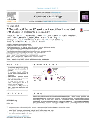
Fig. 1. A sequence alignment was used to construct a theoretical structural model of the catalytic domain of PfPAP. A) ClustalX alignment of PF3D7_1401300 (N-terminal
PEXEL region removed) against the prolyl aminopeptidase from S. marcescens (UniProtKB 032449). Catalytic triad residues (S249, D402, H430) are highlighted. B) Crystal structure of
prolyl aminopeptidase from S. marcescens (PDB code 1QTR). Catalytic residues are numbered and shown as Corey, Pauling, Koltun (CPK) surfaces. The protein backbone is shown as a
thin tube. C). Initial model structure of the putative prolyl aminopeptidase PF3D7_1401300. Catalytic residues are numbered and shown as CPK surfaces. The protein backbone is
shown as a thin tube.
F.L. da Silva et al. / Experimental Parasitology 169 (2016) 13e2116
5. addition of anti-rabbit FITC and anti-mouse Alexa Fluor 647. Sec-
ondary antibodies were washed from the slides and the nuclei
stained with 10 mg/ml of 40,6-diamidino-2-phenylindole (DAPI)
prior to mounting of slides. Images were taken on a Delta Vision
(DV) Elite Microscope with 100Â oil objective. Images were pro-
cessed using NIH ImageJ version 1.48c (http://imagej.nih.gov/ij/).
2.12. Atomic force microscopy
To investigate the mechanical properties of the infected and
uninfected RBCs, arrays of 20 Â 20 force curves on a 10 Â 10 mm
area were recorded on samples immersed in PBS employing an
MFP-3D (Asylum Research) atomic force microscope (AFM) in force
spectroscopy mode. The AFM was mounted on an anti-vibrational
table (Herzan) and operated within an acoustic isolation enclo-
sure (TMC, USA). The force curves were recorded using a SiNi
cantilever (Budget Sensors, Bulgaria) having a nominal spring
constant KN ¼ 0.06 N/m. Prior to use the cantilevers had been
calibrated against a glass slide, using the thermal vibration method
embedded in the AFM processing software. All experiments were
repeated 4 times in triplicate with the loading force kept constant
at 20 nN and the velocity at 1 mm/s. Force curve data were analysed
using IGOR software. The Young’s modulus, E, (±SD) was calculated
using the Hertz model.
2.13. Spleen mimic filtration
Parent 3D7 and 3D7-PAPKO parasites were synchronized to a 2 h
window (Lambros and Vanderberg, 1979). Spleen mimic filtration
was performed as previously described (Deplaine et al., 2011).
Briefly, parasite infected RBCs at 5% parasitemia were re-suspended
at 1% hematocrit in 1% AlbuMax II in PBS. The solution was flowed
over a 5 mm bead volume of calibrated metal microbeads ranging
in size from 5 to 25 mm at a rate of 60 ml/h. The percentage para-
sitemia pre and post filtration was assessed via Giemsa stained thin
blood films and used to calculate the percentage of parasites pre-
sent in the flow through (% flow). Three biological repeats were
performed.
2.14. Cytoadherence assays
Cytoadherence assays were performed using prefabricated
slides (ibidi GmbH). Slides were coated with 125 mg/ml of recom-
binant CD36 in PBS overnight prior to blocking with 1% BSA for
1 h at 37 C. The slides were washed with RPMI-HEPES (minus
NaHCO3). All assays were performed on a DV elite microscope with
environmental chamber set at 37 C.
Parasite infected RBCs at 3% parasitemia and 1% hematocrit in
RPMI-HEPES (minus NaHCO3) were flowed through the chamber
for 5 min at a pressure of 0.1 or 0.05 Pa prior to a further washing
(5 min) with RPMI-HEPES (minus NaHCO3). Washing and counting
was performed under the same conditions as binding. The number
of parasite infected RBCs bound in 20 fields were counted, and
expressed as parasites bound per mm2
(Crabb et al., 1997). Three
biological repeats were performed.
2.15. Electron microscopy
Parasite infected RBCs were embedded in 1% molten agarose in
0.1 M phosphate buffer. The agarose blocks were processed into
Epon resin using a Pelco 34700 Biowave Microwave Oven (Ted Pella
Inc., Redding, CA). Cells were post-fixed in aqueous potassium
ferricyanide-reduced osmium tetroxide and dehydrated in ethanol
prior to infiltration and embedment in Epon resin. Unstained ul-
trathin sections were observed and photographed using a JEOL 1011
transmission electron microscope (JEOL Ltd, Tokyo, Japan) equip-
ped with an Olympus Morada side-mounted digital camera
(Olympus, USA).
3. Results
3.1. In silico modelling of PfPAP
While no malaria parasite PAP structures currently exist in the
protein data bank (PDB), X-ray derived solid-state structures of
several other validated PAPs have been determined (http://www.
rcsb.org (Berman et al., 2000)). To provide additional evidence
that PfPAP is indeed a PAP its amino acid sequence was aligned with
a validated PAP derived from Serratia marcescens (Yoshimoto et al.,
Fig. 2. Purification of a functionally active recombinant PfPAP. A). Purification of
active rPfPAP. M, molecular size markers; S, soluble supernatant; W, washes; E, eluted
rPfPAP. Purified protein migrates at ~45 kDa. B). Immunoblot confirmed identity of
eluted purified rPfPAP with primary mouse anti-histidine antibody and secondary goat
anti-mouse horseradish peroxidase antibody. C). Enzyme assays with the fluorogenic
peptide substrate H-Pro-NHMec demonstrate that the rPfPAP exhibits typical
Michaelis-Menten enzymatic kinetics with a Km constant of 403.6 mM.
F.L. da Silva et al. / Experimental Parasitology 169 (2016) 13e21 17
6. 1999) (PDB code 1QTR; Fig. 1, panels A, B and C; 41% similarity with
PfPAP; with specific N-terminal PEXEL sequence removed). Co-
alignment of catalytically significant triad residues (Ser249,
Asp402, His430) and other highly conserved residues, character-
istic of PAPs, provided further evidence that PfPAP belongs to this
class of protein (Fig. 1A). The primary sequence similarity observed
in the alignment was also observed in the structural homology
model. Comparison of the PfPAP structural model with the X-ray
derived structure of S. marcescens PAP (PDB code 1QTR) revealed
that the spatial distributions of catalytic residues and other notable
residues (Fig. 1C) are highly conserved, further supporting the
proposed function of PF3D7_1401300.
3.2. Biochemical characterization of functionally active
recombinant PfPAP
PfPAP was expressed and purified as a recombinant protein
(rPfPAP) from bacterial cells. The enzyme resolved as a single pro-
tein of ~45 kDa (Fig. 2A) and immunoblotting confirmed expression
of the recombinant protein (Fig. 2B). Using the fluorogenic sub-
strate H-Pro-NHMec (7-amido-4-methylcoumarin), rPfPAP exhibi-
ted a Km of 403 mM and a Kcat/Km value 28.28 MÀ1
sÀ1
(Fig. 2C).
Experiments investigating the specificity of rPfPAP demonstrate a
low, but significant, level of activity when incubated with the flu-
orogenic substrate H-Ala-NHMec (2e5% compared to H-Pro-
NHMec) but no hydrolysis of H-Leu-NHMec or H-Glu-NHMec.
When the metal chelator o-phenanthroline (2 mM) was added,
only slight inhibition of rPfPAP activity was observed, confirming
the activity is specific and not due to contaminating bacterial
neutral aminopeptidases (not shown).
3.3. PfPAP is transcribed throughout the intraerythrocytic asexual
lifecycle and exported into the host RBC
Northern blot analysis indicated that Pf transcribes a single species
of mRNA with an apparent size of ~3 kbp throughout the intra-
erythrocytic life cycle (Fig. 3A). Quantitative analysis of transcription
suggested that peak expression occurs early in development (Fig. 3B).
Western blot analysis of parent 3D7 parasites with anti-PfPAP
antiserum revealed a single species with an apparent molecular
weight (MW) of ~40 kDa (Fig. 3C). Immunochemistry using an anti-
GFP antibody on a transgenic GFP-tagged PfPAP chimeric protein
also identified a single protein species (not shown). Immunofluo-
rescence analysis using anti-PfPAP antibody localized PfPAP to the
infected RBC cytoplasm which was confirmed in GFP-tagged PfPAP
transfected parasites (Fig. 3D).
3.4. Targeted gene disruption (TGD) of PfPAP changes the
viscoelastic properties of the infected RBC membrane
PCR analysis indicated that a single homologous recombination
of the 50 targeting sequence had occurred in the clone selected for
further study. Nonetheless this led to a truncation of the genomic
copy and Western blot analysis using antibodies to rPfPAP indicated
loss of PfPAP expression (Fig. 3C).
Genetic disruption of PF3D7_1401300 caused no obvious
changes in macroscopic phenotype, including life cycle length, or in
parasite viability in vitro. However, significant changes in the
viscoelastic properties of the infected RBC plasma membrane were
observed using AFM. The Young’s modulus value (E) was deter-
mined to be 760 ± 140 kPa for uninfected RBCs, and 1760 ± 710 kPa
for 3D7 early trophozoite infected RBCs. However, 3D7_PAP-KO
early trophozoite einfected RBCs generated a Young’s modulus
value of 800 ± 250 kPa .
A microbead filtration system that mimics the splenic micro-
circulation was employed to assess RBC deformability changes in
more detail (Fig. 4A). Tightly synchronized parasites were analysed
16, 18, 20 and 26 h post invasion. No significant difference between
the percentage of 3D7 and 3D7_PAP-KO parasites in the flow
through was seen at 16 h (64 ± 14% vs 58 ± 6%; relative to starting
parasitemia). However, 3D7_PAP-KO parasites were significantly
more filterable than 3D7 parasites at 18e26 h post-invasion (18 h:
64 ± 4% vs 41 ± 5%; 20 h: 58 ± 6% vs 26 ± 3%; and 26 h: 40 ± 5% vs
21 þ 3%; P ¼ 0.0001) (Fig. 4A).
The impact of PfPAP TGD on the expression and transport of
proteins thought to play a role in the trafficking of unique Pf
Fig. 3. PfPAP is transcribed throughout the intraerythrocytic asexual lifecycle and exported into the host RBC. A). Northern blot analysis of PF3D7_1401300 transcription, RNA
was probed with the full length coding region of the gene. R ¼ ring stage parasites. ET ¼ early trophozoite parasites, LT ¼ late trophozoite parasites, S ¼ schizont stage parasites.
Analysis reveals transcription in all stages. B). Quantitative analysis of PfPAP transcription relative to reference genes demonstrates highest transcription in early stages of the intra-
erythrocytic asexual life cycle C). Western blot of 3D7 and 3D7_PAP-KO parasites probed with anti PfPAP shows the loss of protein expression in the knockout line. REX1 protein
expression in both clones was used to demonstrate protein loading. C). Direct fluorescence of transgenic parasites expressing PF3D7_1401300 C-terminally tagged with GFP.
BF ¼ bight field, GFP ¼ GFP fluorescence, Nuclei ¼ nuclear staining with Hoechst, Merge ¼ merge of the previous images. Bar 5 mm.
F.L. da Silva et al. / Experimental Parasitology 169 (2016) 13e2118
7. cytoadherence proteins, such as PfEMP-1, to the infected RBC sur-
face was assessed. Data demonstrated that TGD of PfPAP does not
change the location of KAHRP, ring-exported protein-1 (REX1),
skeleton-binding protein-1 (SBP1) or PfEMP-3. Each of these pro-
teins was found at the RBC membrane or Maurer’s clefts of 3D7 and
3D7PAPKO parasites (Fig. 4B). Electron microscopy also confirmed
the presence of electron dense structures at the infected RBC
plasma membrane of both 3D7 (not shown) and 3D7PAPKO para-
sites (Fig. 4C), consistent with correct trafficking and delivery of
KAHRP.
The presence of PfEMP-1 at the surface of both 3D7 and
3D7PAPKO parasites was demonstrated by trypsin cleavage
(Fig. 5A). Cleavage products (75e100 kDa) were observed in
trypsin-treated (T) samples but were absent from samples with no
trypsin (P) or with trypsin plus inhibitor (i) (Fig. 5A). Consistent
with these data parent 3D7 and 3D7_PAP-KO infected RBCs
adhered to recombinant CD36 at similar levels under static condi-
tions (Fig. 5B). However, assessment of binding under physiological
flow conditions demonstrated that 3D7 infected RBCs bound at
500 ± 20 infected RBC/mm2
while the 3D7_PAP-KO infected RBCs
bound at a significantly lower rate (367 ± 16 infected RBC/mm2
;
P 0.0001). This significant decrease in binding was also seen at
the higher shear stress of 0.1 Pa. (Fig. 5C).
4. Discussion
In this work PF3D7_1401300-encoded PfPAP was verified as an
aminopeptidase with a preference for N-terminal proline sub-
strates and with a weaker specificity for substrates containing N-
terminal alanine. While the fine specificities of various S33 clan
Fig. 4. Targeted gene disruption of PfPAP changes the filterability of the infected RBCs but not the expression or localization of KAHRP, REX1, and PfEMP-3. A). Analysis of
filterability of 3D7_PAP-KO through micro beads designed to mimic the splenic microcirculation. Samples were measured at 16, 18, 20 and 26 h post invasion. Data are presented as
mean percentage of parasites present in the flow-through relative to the starting parasitemia (±SEM; from 3 triplicate experiments). B). Immunofluorescence using anti-KAHRP,
REX1, and PfEMP-3 antibodies on 3D7 and 3D7_PAP-KO show no difference in the location of these proteins. C). Electron microscopy of 3D7_PAP-KO showing electron dense
structures at the surface of the infected erythrocyte indicative of the presence of knobs. Bars are 5 mm.
F.L. da Silva et al. / Experimental Parasitology 169 (2016) 13e21 19
8. members can vary, a weak specificity for N-terminal alanine is not
uncommon in this group of enzymes. PAPs are not obligate proline
aminopeptidases, with members being capable of cleaving addi-
tional residues including alanine (Mahon et al., 2009). A sequence
comparison of PfPAP with the well described PAP protein from
Serratia marcescens (PDB code 1QTR) (Fig. 1, panels A, B and C) also
demonstrated the conserved location of catalytic triad residues in
PfPAP.
Particularly interesting features of PfPAP are that it is unique to
Pf and contains a protein export element (PEXEL) or vacuolar transit
sequence (VTS) (www.plasmo.db.org). The presence of an export
sequence suggests that unlike all other characterized Pf amino-
peptidases, this enzyme is transported into the RBC cytoplasm. One
of the important functions of exported proteins is to modify the
host RBC membrane to facilitate adhesion to blood vessel walls, an
event that underlies much of the pathophysiology of Pf infections.
For example a number of exported parasite proteins facilitate the
presentation of the adhesin, PfEMP-1, at the RBC surface. They
achieve this by reorganizing the host RBC membrane skeleton and
by forming raised structures, known as knobs. The surface pre-
sentation of PfEMP-1 at the knobby protrusions facilitates binding
to endothelial receptors, such as CD36 (Crabb et al., 1997). Intra-
erythrocytic maturation of Pf is also associated with RBC mem-
brane rigidification (Glenister et al., 2002). While KAHRP and
PfEMP-3 are responsible for about 50% of the observed rigidification
(Glenister et al., 2002), it is recognized that other structural pro-
teins and enzymes can contribute to the reorganization of the
membrane skeleton and rigidification (Glenister et al., 2002; Sanyal
et al., 2012).
To investigate the role of PfPAP in RBC re-modelling and
cytoadherence we examined the expression and location of PfPAP
within infected RBCs. We also performed a TGD of PfPAP. In these
studies the expression of PfEMP-1, REX1, KAHRP and PfEMP-3 was
examined as was host RBC rigidity, deformability and CD36-
mediated cytoadherence. Our data demonstrated that PfPAP is
exported into the host RBC cytoplasm (Fig. 3D) and that it is highly
expressed early in the parasite asexual intra-erythrocytic life cycle
(Fig. 3B), characteristics that support a role in RBC remodeling. They
also showed that while static CD36-mediated adhesion is not
changed by PfPAPKO, adherence is weaker under flow conditions
(Fig. 5). In addition the rigidity and filterability (Fig. 4) of host RBCs
infected with parasites no longer able to express PfPAP is reduced
when compared to wild-type parasites. Interestingly TGD of PfPAP
had no impact on the delivery of KAHRP or PfEMP-3 to the RBC
membrane skeleton (Fig. 4). It also had no impact on the delivery of
REX1 or PfEMP-1 to their known locations in the Maurer’s clefts and
RBC surface respectively (Figs. 4 and 5).
While the precise role of the PfPAP remains to be elucidated,
current data suggest that this protein plays a role in host RBC re-
modelling independent of PfEMP-1, PfEMP-3, KAHRP and REX1
and that this role results in reduced RBC rigidity and cytoadherence
under flow conditions. These data fit well with the non-essential
nature of PfPAP in vitro where parasites are not dependent on
cytoadherence and RBC deformability to survive. The ability of
parasites to cytoadhere and avoid splenic clearance in vivo is of no
advantage to parasites in vitro. Nevertheless, caution must be
exercised before drawing firm conclusions in this regard. The cur-
rent study did not investigate the expression of STEVOR (sub-
telomeric variant open reading frame) proteins in wild-type and
transgenic parasites and recent studies have demonstrated that
these proteins play a role in the deformability of host RBCs infected
with Pf gametocytes and asexual stages (Sanyal et al., 2012; Tiburcio
et al., 2012). In addition, while the PfEMP-1 variant expressed by
both wild-type and PfPAPKO parasites in this study demonstrated a
comparable preference for CD36 under static conditions (Fig. 5)
further studies examining the impact of these variant proteins on
RBC deformability and adherence under shear flow conditions were
not performed, however care was taken to ensure that CD36 spe-
cific PfEMP-1 binding variants were expressed by panning of par-
asites to CD36.
The current study verified PF3D7_1401300-encoded PfPAP as a
PAP and has provided the first insights into the functional role of
this exported protease. Although further work including the anal-
ysis of additional clones, an assessment of expressed stevor genes in
clones and an assessment of the impact of var gene expression on
this protein’s apparent role in RBC re-modelling is required to fully
elucidate the role of PfPAP in modifying the host RBC and to
determine its contribution to survival fitness in vivo, the current
data suggest that drugs designed to inhibit PfPAP may be useful in
preventing sequestration of the asexual stage in the micro-
capillaries and warrants further investigation as an anti-disease
drug target.
Fig. 5. Targeted gene disruption of PfPAP reduces CD36 cytoadhere under flow conditions A). Intact 3D7 and 3D7_PAP-KO infected RBCs were subjected to treatment with PBS
(P) or trypsin (T) or trypsin plus inhibitor (i), then extracted and subjected to SDS-PAGE and probed using an antibody recognizing the ATS domain of PfEMP-1. PfEMP-1 cleavage
products are indicated. B) 3D7 and 3D7_PAP-KO-infected RBCs adhere to recombinant CD36 at comparable levels under static conditions C). 3D7_PAP-KO parasites have a reduced
ability to cytoadhere to CD36 under flow conditions. Adherence under flow conditions was assessed at flow rates equivalent to shear stresses of 0.1 and 0.05 Pa. The mean numbers
(from 3 separate experiments) of parasites bound per mm2
are shown (±SEM).
F.L. da Silva et al. / Experimental Parasitology 169 (2016) 13e2120
9. Acknowledgements
This work was supported by the National Health Medical
Research Council of Australia (1098992, 1078065 and 0602541) and
the Australia Research Council (PD0666128 and DP110100624).
References
Aikawa, M., 1997. Studies on falciparum malaria with atomic-force and surface-
potential microscopes. Ann. Trop. Med. Parasitol. 91, 689e692.
Atkinson, C.T., Aikawa, M., 1990. Ultrastructure of malaria-infected erythrocytes.
Blood Cells 16, 351e368.
Augagneur, Y., Wesolowski, D., Tae, H.S., Altman, S., Ben Mamoun, C., 2012. Gene
selective mRNA cleavage inhibits the development of Plasmodium falciparum.
Proc. Natl. Acad. Sci. U. S. A. 109, 6235e6240.
Berman, H.M., Westbrook, J., Feng, Z., Gilliland, G., Bhat, T.N., Weissig, H.,
Shindyalov, I.N., Bourne, P.E., 2000. The protein data bank. Nucleic Acids Res. 28,
235e242.
Bolumar, T., Sanz, Y., Aristoy, M.C., Toldra, F., 2003. Purification and characterization
of a prolyl aminopeptidase from Debaryomyces hansenii. Appl. Environ. Micro-
biol. 69, 227e232.
Cooke, B.M., Mohandas, N., Coppel, R.L., 2004. Malaria and the red blood cell
membrane. Semin. Hematol. 41, 173e188.
Crabb, B.S., Cooke, B.M., Reeder, J.C., Waller, R.F., Caruana, S.R., Davern, K.M.,
Wickham, M.E., Brown, G.V., Coppel, R.L., Cowman, A.F., 1997. Targeted gene
disruption shows that knobs enable malaria-infected red cells to cytoadhere
under physiological shear stress. Cell 89, 287e296.
Cyrklaff, M., Sanchez, C.P., Kilian, N., Bisseye, C., Simpore, J., Frischknecht, F.,
Lanzer, M., 2011. Hemoglobins S and C interfere with actin remodeling in
Plasmodium falciparum-infected erythrocytes. Science 334, 1283e1286.
Deplaine, G., Safeukui, I., Jeddi, F., Lacoste, F., Brousse, V., Perrot, S., Biligui, S.,
Guillotte, M., Guitton, C., Dokmak, S., Aussilhou, B., Sauvanet, A., Cazals
Hatem, D., Paye, F., Thellier, M., Mazier, D., Milon, G., Mohandas, N., Mercereau-
Puijalon, O., David, P.H., Buffet, P.A., 2011. The sensing of poorly deformable red
blood cells by the human spleen can be mimicked in vitro. Blood 117, e88e95.
Dixon, M.W., Hawthorne, P.L., Spielmann, T., Anderson, K.L., Trenholme, K.R.,
Gardiner, D.L., 2008. Targeting of the ring exported protein 1 to the Maurer’s
clefts is mediated by a two-phase process. Traffic 9, 1316e1326.
Felipe, M.S., Andrade, R.V., Arraes, F.B., Nicola, A.M., Maranhao, A.Q., Torres, F.A.,
Silva-Pereira, I., Pocas-Fonseca, M.J., Campos, E.G., Moraes, L.M., Andrade, P.A.,
Tavares, A.H., Silva, S.S., Kyaw, C.M., Souza, D.P., Pereira, M., Jesuino, R.S.,
Andrade, E.V., Parente, J.A., Oliveira, G.S., Barbosa, M.S., Martins, N.F.,
Fachin, A.L., Cardoso, R.S., Passos, G.A., Almeida, N.F., Walter, M.E., Soares, C.M.,
Carvalho, M.J., Brigido, M.M., PbGenome, N., 2005. Transcriptional profiles of
the human pathogenic fungus Paracoccidioides brasiliensis in mycelium and
yeast cells. J. Biol. Chem. 280, 24706e24714.
Glenister, F.K., Coppel, R.L., Cowman, A.F., Mohandas, N., Cooke, B.M., 2002.
Contribution of parasite proteins to altered mechanical properties of malaria-
infected red blood cells. Blood 99, 1060e1063.
Khilji, M.A., Akrawi, A.F., Bailey, G.S., 1979. Purification and partial characterisation
of a bovine kidney aminotripeptidase (capable of cleaving prolyl-glycylglycine).
Mol. Cell Biochem. 23, 45e52.
Kyes, S., Pinches, R., Newbold, C., 2000. A simple RNA analysis method shows var
and rif multigene family expression patterns in Plasmodium falciparum. Mol.
Biochem. Parasitol. 105, 311e315.
Kyes, S.A., Rowe, J.A., Kriek, N., Newbold, C.I., 1999. Rifins: a second family of clonally
variant proteins expressed on the surface of red cells infected with Plasmodium
falciparum. Proc. Natl. Acad. Sci. U. S. A. 96, 9333e9338.
Lambros, C., Vanderberg, J.P., 1979. Synchronization of Plasmodium falciparum
erythrocytic stages in culture. J. Parasitol. 65, 418e420.
Larkin, M.A., Blackshields, G., Brown, N.P., Chenna, R., McGettigan, P.A.,
McWilliam, H., Valentin, F., Wallace, I.M., Wilm, A., Lopez, R., Thompson, J.D.,
Gibson, T.J., Higgins, D.G., 2007. Clustal W and clustal X version 2.0.
Bioinformatics 23, 2947e2948.
Mahon, C.S., O’Donoghue, A.J., Goetz, D.H., Murray, P.G., Craik, C.S., Tuohy, M.G.,
2009. Characterization of a multimeric, eukaryotic prolyl aminopeptidase: an
inducible and highly specific intracellular peptidase from the non-pathogenic
fungus Talaromyces emersonii. Microbiology 155, 3673e3682.
Maier, A.G., Cooke, B.M., Cowman, A.F., Tilley, L., 2009. Malaria parasite proteins that
remodel the host erythrocyte. Nat. Rev. Microbiol. 7, 341e354.
Marti, M., Good, R.T., Rug, M., Knuepfer, E., Cowman, A.F., 2004. Targeting malaria
virulence and remodeling proteins to the host erythrocyte. Science 306,
1930e1933.
Miller, L.H., Baruch, D.I., Marsh, K., Doumbo, O.K., 2002. The pathogenic basis of
malaria. Nature 415, 673e679.
Nash, G.B., O’Brien, E., Gordon-Smith, E.C., Dormandy, J.A., 1989. Abnormalities in
the mechanical properties of red blood cells caused by Plasmodium falciparum.
Blood 74, 855e861.
Phillips, J.C., Braun, R., Wang, W., Gumbart, J., Tajkhorshid, E., Villa, E., Chipot, C.,
Skeel, R.D., Kale, L., Schulten, K., 2005. Scalable molecular dynamics with
NAMD. J. Comput. Chem. 26, 1781e1802.
Roseler, A., Prieto, J.H., Iozef, R., Hecker, B., Schirmer, R.H., Kulzer, S., Przyborski, J.,
Rahlfs, S., Becker, K., 2012. Insight into the selenoproteome of the malaria
parasite Plasmodium falciparum. Antioxid. Redox Signal 17, 534e543.
Sanyal, S., Egee, S., Bouyer, G., Perrot, S., Safeukui, I., Bischoff, E., Buffet, P.,
Deitsch, K.W., Mercereau-Puijalon, O., David, P.H., Templeton, T.J., Lavazec, C.,
2012. Plasmodium falciparum STEVOR proteins impact erythrocyte mechanical
properties. Blood 119, e1e8.
Schoenen, F.J., Weiner, W.S., Baillargeon, P., Brown, C.L., Chase, P., Ferguson, J., Fer-
nandez-Vega, V., Ghosh, P., Hodder, P., Krise, J.P., Matharu, D.S.,
Neuenswander, B., Porubsky, P., Rogers, S., Skinner-Adams, T., Sosa, M., Spicer, T.,
To, J., Tower, N.A., Trenholme, K.R., Wang, J., Whipple, D., Aube, J., Rosen, H.,
White, E.L., Dalton, J.P., Gardiner, D.L., 2010. Inhibitors of the Plasmodium fal-
ciparum M18 Aspartyl Aminopeptidase. Probe Reports from the NIH Molecular
Libraries Program, Bethesda (MD).
Shi, H., Liu, Z., Li, A., Yin, J., Chong, A.G., Tan, K.S., Zhang, Y., Lim, C.T., 2013. Life cycle-
dependent cytoskeletal modifications in Plasmodium falciparum infected
erythrocytes. PLoS One 8, e61170.
Spielmann, T., Gardiner, D.L., Beck, H.P., Trenholme, K.R., Kemp, D.J., 2006. Organi-
zation of ETRAMPs and EXP-1 at the parasite-host cell interface of malaria
parasites. Mol. Microbiol. 59, 779e794.
Stack, C.M., Lowther, J., Cunningham, E., Donnelly, S., Gardiner, D.L., Trenholme, K.R.,
Skinner-Adams, T.S., Teuscher, F., Grembecka, J., Mucha, A., Kafarski, P., Lua, L.,
Bell, A., Dalton, J.P., 2007. Characterization of the Plasmodium falciparum M17
leucyl aminopeptidase. A protease involved in amino acid regulation with po-
tential for antimalarial drug development. J. Biol. Chem. 282, 2069e2080.
Tiburcio, M., Niang, M., Deplaine, G., Perrot, S., Bischoff, E., Ndour, P.A., Silvestrini, F.,
Khattab, A., Milon, G., David, P.H., Hardeman, M., Vernick, K.D., Sauerwein, R.W.,
Preiser, P.R., Mercereau-Puijalon, O., Buffet, P., Alano, P., Lavazec, C., 2012.
A switch in infected erythrocyte deformability at the maturation and blood
circulation of Plasmodium falciparum transmission stages. Blood 119, e172e180.
Trager, W., Jensen, J.B., 1976. Human malaria parasites in continuous culture. Sci-
ence 193, 673e675.
Watermeyer, J.M., Hale, V.L., Hackett, F., Clare, D.K., Cutts, E.E., Vakonakis, I.,
Fleck, R.A., Blackman, M.J., Saibil, H.R., 2016. A spiral scaffold underlies
cytoadherent knobs in Plasmodium falciparum-infected erythrocytes. Blood 127,
343e351.
Waters, S.P., Dalling, M.J., 1983. Purification and characterization of an imino-
peptidase from the primary leaf of wheat (Triticum aestivum L.). Plant Physiol.
73, 1048e1054.
World Health Organization, 2015. World Malaria Report. World Health Organiza-
tion, Geneva.
Wu, Y., Sifri, C.D., Lei, H.H., Su, X.Z., Wellems, T.E., 1995. Transfection of Plasmodium
falciparum within human red blood cells. Proc. Natl. Acad. Sci. U. S. A. 92,
973e977.
Yoshimoto, T., Kabashima, T., Uchikawa, K., Inoue, T., Tanaka, N., Nakamura, K.T.,
Tsuru, M., Ito, K., 1999. Crystal structure of prolyl aminopeptidase from Serratia
marcescens. J. Biochem. 126, 559e565.
F.L. da Silva et al. / Experimental Parasitology 169 (2016) 13e21 21