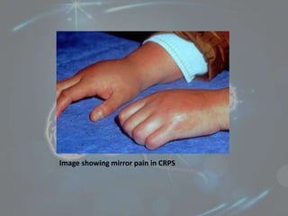
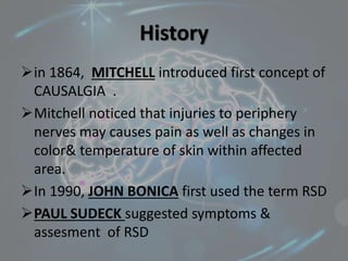
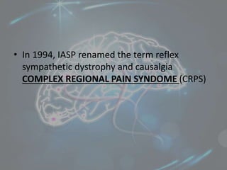
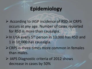
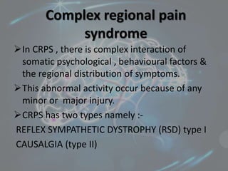
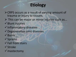
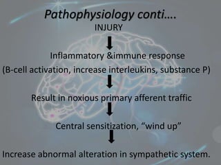
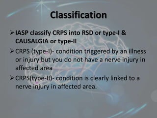
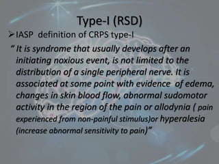
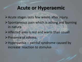
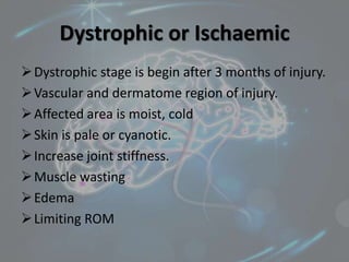
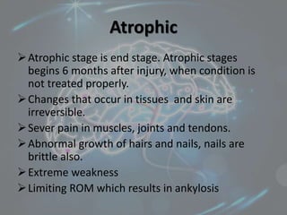
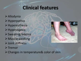
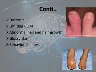
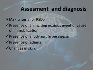
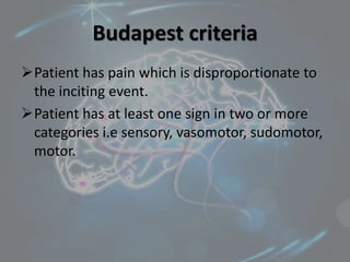
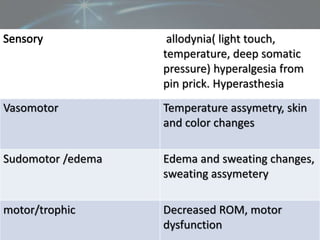
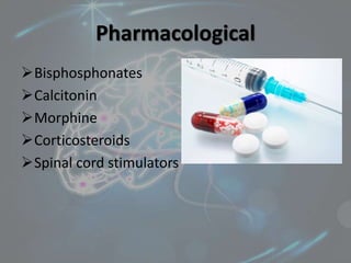
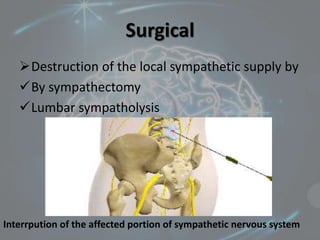
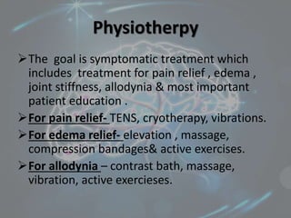
Reflex Sympathetic Dystrophy
and
Causalgia
By- kajal sansoya
Introduction
Reflex sympathetic dystrophy (RSD) is a
chronic condition that causes persistent
burning pain , swelling and abnormal nerve
activity affected upper or lower limb followed
by any major or minor injury .
RSD is also known as complex regional pain
syndrome (CRPS)
Commonly occurs to proximal or contralateral
limb or also occur as mirror pain.
Image showing mirror pain in CRPS
Terminology
Algodystrophy
Sudeck’s atrophy
Complex regional pain syndrome
Shoulder-hand-syndrome(SHS)
Sympathetically-maintained pain(SMP)
Causalgia
Post traumatic dystrophy
History
in 1864, MITCHELL introduced first concept of
CAUSALGIA .
Mitchell noticed that injuries to periphery
nerves may causes pain as well as changes in
color& temperature of skin within affected
area.
In 1990, JOHN BONICA first used the term RSD
PAUL SUDECK suggested symptoms &
assesment of RSD
• In 1994, IASP renamed the term reflex
sympathetic dystrophy and causalgia
COMPLEX REGIONAL PAIN SYNDOME (CRPS)
Epidemiology
 According to IASP incidence of RSD or CRPS
occurs at any age. Number of cases reported
for RSD is more than causalgia.
In USA every 5th person in 10,000 has RSD and
1 in 10,000 has causalgia.
CRPS is three times more common in females
than males.
 IAPS Diagnostic criteria of 2012 shows
decrease in cases by 50%
Complex regional pain
syndrome
In CRPS , there is complex interaction of
somatic psychological , behavioural factors &
the regional distribution of symptoms.
This abnormal activity occur because of any
minor or major injury.
CRPS has two types namely :-
REFLEX SYMPATHETIC DYSTROPHY (RSD) type I
CAUSALGIA (type II)
Etiology
CRPS occurs as a result of varying amount of
trauma or injury to tissues.
 This can be major or minor injuries such as...
Blunt injuries
Inflammatory diseases
Degenerative joint diseases
Burns
Sprains
Fall from stairs
Stroke
mastectomy
Pathophysiology conti….
INJURY
Inflammatory &immune response
(B-cell activation, increase interleukins, substance P)
Result in noxious primary afferent traffic
Central sensitization, “wind up”
Increase abnormal alteration in sympathetic system.
Classification
IASP classify CRPS into RSD or type-I &
CAUSALGIA or type-II
CRPS (type-I)- condition triggered by an illness
or injury but you do not have a nerve injury in
affected area
CRPS(type-II)- condition is clearly linked to a
nerve injury in affected area.
Type-I (RSD)
IASP definition of CRPS type-I
“ It is syndrome that usually develops after an
initiating noxious event, is not limited to the
distribution of a single peripheral nerve. It is
associated at some point with evidence of edema,
changes in skin blood flow, abnormal sudomotor
activity in the region of the pain or allodynia ( pain
experienced from non-painful stimulus)or hyperalesia
(increase abnormal sensitivity to pain)”
Points to remember…RSD
 Develops after an Noxious event
 Not limited to single peripheral nerve and no
nerve injury at affected area.
 Presence of allodynia & hyperalesia, changes
in skin color& temperature.
“ reflex nature is unclear, however the role of
sympathetic system is also not well
understood..so term CRPS used”
RSD often poorly
misunderstood by conditions
such as..
Pain and altered sensations
Motor disturbance and soft tissues changes
Vasomotor, autonomic changes
Psychological distrubances
Type-II CAUSALGIA
Causalgia derives from Greek word causos
which describes the burning pain sometimes
associated with neuropathy pain
Occur due to any major or minor trauma or
lesion followed by nerve injury
Causalgia
Syndrome develops after a nerve injury.
Spontaneous pain or allodynia &
hyperalesia
Evidence of pain, edema, skin changes
 Abnormal sudomotor(sweat glands)
activity in the region of the pain since the
inciting event.
Stages of progression
There are three stages of progression of
condition namely…
First stages/acute/hyperaemic
Second/dystrophic/ischaemic
Third/atrophic
Acute or Hyperaemic
Acute stages lasts few weeks after injury.
Spontaneous pain which is aching and burning
in nature.
Affected area is red and warm than usuall
Presence of edema.
Hyperpathia – painful syndrome caused by
increase reaction to stimulus
Dystrophic or Ischaemic
Dystrophic stage is begin after 3 months of injury.
Vascular and dermatome region of injury.
Affected area is moist, cold
Skin is pale or cyanotic.
Increase joint stiffness.
Muscle wasting
Edema
Limiting ROM
Atrophic
Atrophic stage is end stage. Atrophic stages
begins 6 months after injury, when condition is
not treated properly.
Changes that occur in tissues and skin are
irreversible.
Sever pain in muscles, joints and tendons.
Abnormal growth of hairs and nails, nails are
brittle also.
Extreme weakness
Limiting ROM which results in ankylosis
Clinical features
Allodynia
Hyperpathia
Hyperesthesia
Hyperalgesia
Sweating/edema
Muscle weaking
Joint stiffness
Tremor
Changes in temperature& color of skin
Conti..
Dystonia
Limiting ROM
Abnormal nail and hair growth
Glossy skin
Behavioral illness
Assesment and diagnosis
IASP criteria for RSD:-
Presence of an inciting noxious event or cause
of immobilization
Presence of allodynia , hyperalgesia
Presence of edema
Changes in skin.
Budapest criteria
Patient has pain which is disproportionate to
the inciting event.
Patient has at least one sign in two or more
categories i.e sensory, vasomotor, sudomotor,
motor.
Sensory
allodynia( light touch,
temperature, deep somatic
pressure) hyperalgesia from
pin prick. Hyperasthesia
Vasomotor Temperature assymetry, skin
and color changes
Sudomotor /edema Edema and sweating changes,
sweating assymetery
motor/trophic Decreased ROM, motor
dysfunction
Investigation
X-rays – demonstrate patchy demineralization
in affected area.
MRI
Shows soft tissues swelling , joint effusion,
periarticular marrow.
Arrow indicates joint contracture &effusion
Investigation conti..
Bone scans shows changes that occur in early
stages.
EMG &NCV are used to check the conductive
properties of nerve.
Thermograph – used to check the skin
temperature within affected area .
Stage 1 – increased temperature
Stage 2- normalized or warm temperature
Stage 3- decreased temperature.
Thermography shows temperature difference in affected areas
Management
Prevention is best treatment in such cases.
However, medical and surgical interventions
reported to be benefit in RSD and CAUSALGIA
Treatment includes – pharmacological
intervention , surgical and physiotherapy
management.
Pharmacological
Bisphosphonates
Calcitonin
Morphine
Corticosteroids
Spinal cord stimulators
Surgical
Destruction of the local sympathetic supply by
By sympathectomy
Lumbar sympatholysis
Interrpution of the affected portion of sympathetic nervous system
Physiotherpy
The goal is symptomatic treatment which
includes treatment for pain relief , edema ,
joint stiffness, allodynia & most important
patient education .
For pain relief- TENS, cryotherapy, vibrations.
For edema relief- elevation , massage,
compression bandages& active exercises.
For allodynia – contrast bath, massage,
vibration, active exercieses.
For joint stiffness- active and passive
exercises, ultrasound, gentle stretching, cold,
heat fermentation, biofeedback activities,
splinting.
Thank you
Happy reading 

Reflex sympathetic dystrophy and causalgia

  • 1.
  • 2.
    Introduction Reflex sympathetic dystrophy(RSD) is a chronic condition that causes persistent burning pain , swelling and abnormal nerve activity affected upper or lower limb followed by any major or minor injury . RSD is also known as complex regional pain syndrome (CRPS) Commonly occurs to proximal or contralateral limb or also occur as mirror pain.
  • 3.
  • 4.
    Terminology Algodystrophy Sudeck’s atrophy Complex regionalpain syndrome Shoulder-hand-syndrome(SHS) Sympathetically-maintained pain(SMP) Causalgia Post traumatic dystrophy
  • 5.
    History in 1864, MITCHELLintroduced first concept of CAUSALGIA . Mitchell noticed that injuries to periphery nerves may causes pain as well as changes in color& temperature of skin within affected area. In 1990, JOHN BONICA first used the term RSD PAUL SUDECK suggested symptoms & assesment of RSD
  • 6.
    • In 1994,IASP renamed the term reflex sympathetic dystrophy and causalgia COMPLEX REGIONAL PAIN SYNDOME (CRPS)
  • 7.
    Epidemiology  According toIASP incidence of RSD or CRPS occurs at any age. Number of cases reported for RSD is more than causalgia. In USA every 5th person in 10,000 has RSD and 1 in 10,000 has causalgia. CRPS is three times more common in females than males.  IAPS Diagnostic criteria of 2012 shows decrease in cases by 50%
  • 8.
    Complex regional pain syndrome InCRPS , there is complex interaction of somatic psychological , behavioural factors & the regional distribution of symptoms. This abnormal activity occur because of any minor or major injury. CRPS has two types namely :- REFLEX SYMPATHETIC DYSTROPHY (RSD) type I CAUSALGIA (type II)
  • 9.
    Etiology CRPS occurs asa result of varying amount of trauma or injury to tissues.  This can be major or minor injuries such as... Blunt injuries Inflammatory diseases Degenerative joint diseases Burns Sprains Fall from stairs Stroke mastectomy
  • 10.
    Pathophysiology conti…. INJURY Inflammatory &immuneresponse (B-cell activation, increase interleukins, substance P) Result in noxious primary afferent traffic Central sensitization, “wind up” Increase abnormal alteration in sympathetic system.
  • 11.
    Classification IASP classify CRPSinto RSD or type-I & CAUSALGIA or type-II CRPS (type-I)- condition triggered by an illness or injury but you do not have a nerve injury in affected area CRPS(type-II)- condition is clearly linked to a nerve injury in affected area.
  • 12.
    Type-I (RSD) IASP definitionof CRPS type-I “ It is syndrome that usually develops after an initiating noxious event, is not limited to the distribution of a single peripheral nerve. It is associated at some point with evidence of edema, changes in skin blood flow, abnormal sudomotor activity in the region of the pain or allodynia ( pain experienced from non-painful stimulus)or hyperalesia (increase abnormal sensitivity to pain)”
  • 13.
    Points to remember…RSD Develops after an Noxious event  Not limited to single peripheral nerve and no nerve injury at affected area.  Presence of allodynia & hyperalesia, changes in skin color& temperature. “ reflex nature is unclear, however the role of sympathetic system is also not well understood..so term CRPS used”
  • 14.
    RSD often poorly misunderstoodby conditions such as.. Pain and altered sensations Motor disturbance and soft tissues changes Vasomotor, autonomic changes Psychological distrubances
  • 15.
    Type-II CAUSALGIA Causalgia derivesfrom Greek word causos which describes the burning pain sometimes associated with neuropathy pain Occur due to any major or minor trauma or lesion followed by nerve injury
  • 16.
    Causalgia Syndrome develops aftera nerve injury. Spontaneous pain or allodynia & hyperalesia Evidence of pain, edema, skin changes  Abnormal sudomotor(sweat glands) activity in the region of the pain since the inciting event.
  • 17.
    Stages of progression Thereare three stages of progression of condition namely… First stages/acute/hyperaemic Second/dystrophic/ischaemic Third/atrophic
  • 18.
    Acute or Hyperaemic Acutestages lasts few weeks after injury. Spontaneous pain which is aching and burning in nature. Affected area is red and warm than usuall Presence of edema. Hyperpathia – painful syndrome caused by increase reaction to stimulus
  • 19.
    Dystrophic or Ischaemic Dystrophicstage is begin after 3 months of injury. Vascular and dermatome region of injury. Affected area is moist, cold Skin is pale or cyanotic. Increase joint stiffness. Muscle wasting Edema Limiting ROM
  • 20.
    Atrophic Atrophic stage isend stage. Atrophic stages begins 6 months after injury, when condition is not treated properly. Changes that occur in tissues and skin are irreversible. Sever pain in muscles, joints and tendons. Abnormal growth of hairs and nails, nails are brittle also. Extreme weakness Limiting ROM which results in ankylosis
  • 21.
  • 22.
    Conti.. Dystonia Limiting ROM Abnormal nailand hair growth Glossy skin Behavioral illness
  • 23.
    Assesment and diagnosis IASPcriteria for RSD:- Presence of an inciting noxious event or cause of immobilization Presence of allodynia , hyperalgesia Presence of edema Changes in skin.
  • 24.
    Budapest criteria Patient haspain which is disproportionate to the inciting event. Patient has at least one sign in two or more categories i.e sensory, vasomotor, sudomotor, motor.
  • 25.
    Sensory allodynia( light touch, temperature,deep somatic pressure) hyperalgesia from pin prick. Hyperasthesia Vasomotor Temperature assymetry, skin and color changes Sudomotor /edema Edema and sweating changes, sweating assymetery motor/trophic Decreased ROM, motor dysfunction
  • 26.
    Investigation X-rays – demonstratepatchy demineralization in affected area.
  • 27.
    MRI Shows soft tissuesswelling , joint effusion, periarticular marrow. Arrow indicates joint contracture &effusion
  • 28.
    Investigation conti.. Bone scansshows changes that occur in early stages. EMG &NCV are used to check the conductive properties of nerve. Thermograph – used to check the skin temperature within affected area . Stage 1 – increased temperature Stage 2- normalized or warm temperature Stage 3- decreased temperature.
  • 29.
    Thermography shows temperaturedifference in affected areas
  • 30.
    Management Prevention is besttreatment in such cases. However, medical and surgical interventions reported to be benefit in RSD and CAUSALGIA Treatment includes – pharmacological intervention , surgical and physiotherapy management.
  • 31.
  • 32.
    Surgical Destruction of thelocal sympathetic supply by By sympathectomy Lumbar sympatholysis Interrpution of the affected portion of sympathetic nervous system
  • 33.
    Physiotherpy The goal issymptomatic treatment which includes treatment for pain relief , edema , joint stiffness, allodynia & most important patient education . For pain relief- TENS, cryotherapy, vibrations. For edema relief- elevation , massage, compression bandages& active exercises. For allodynia – contrast bath, massage, vibration, active exercieses.
  • 35.
    For joint stiffness-active and passive exercises, ultrasound, gentle stretching, cold, heat fermentation, biofeedback activities, splinting.
  • 36.