Downloaded 141 times


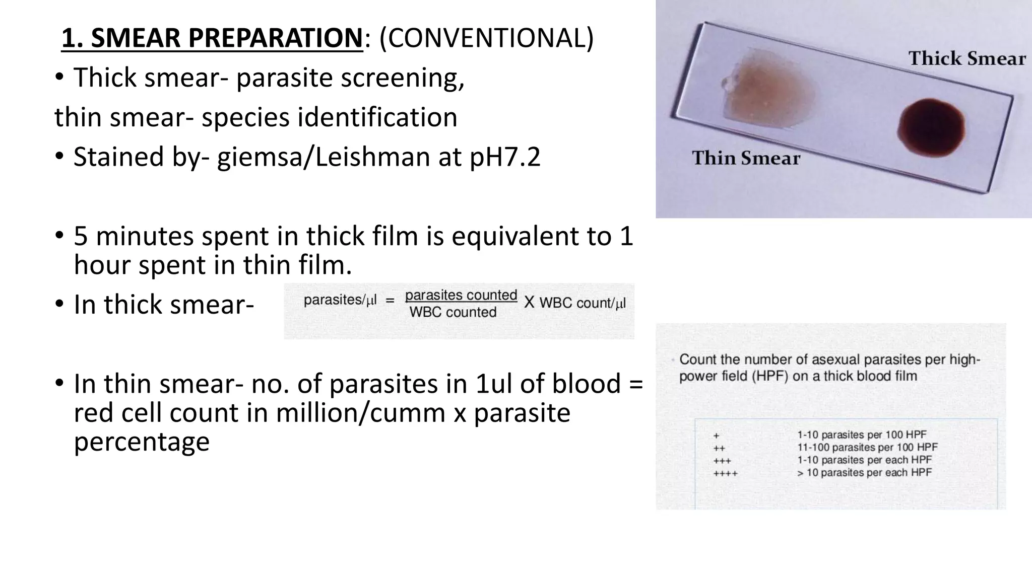


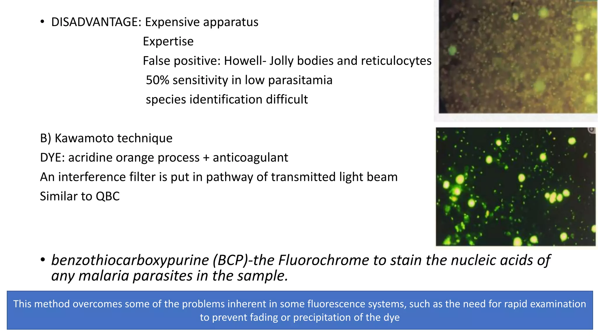


1. Several techniques are used to diagnose hemoparasite infections including microscopy, rapid diagnostic tests, serological tests, and molecular methods. Microscopy remains the standard but has limitations like low sensitivity and requiring experienced technicians. 2. Rapid diagnostic tests detect parasites' antigens and are sensitive when parasitemia is high, but can remain positive for weeks after treatment. Molecular methods like PCR are most sensitive and specific but are complex and time-consuming. 3. Flow cytometry is a promising technique for malaria diagnosis as it can distinguish infected red blood cells from white blood cells using DNA-binding dyes and evaluate drug susceptibility rapidly based on parasite growth.


