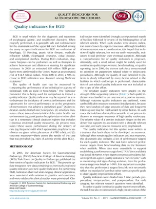
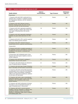
The document outlines quality indicators for GI endoscopic procedures established by a task force. It defines quality indicators and describes the methodology used to develop and update the indicators. The indicators are divided into pre-procedure, intra-procedure, and post-procedure categories. Key areas addressed include appropriate indication, informed consent, risk assessment, management of medications, and timeliness. Performance targets are established when data is available. The ultimate goal is to use these indicators to drive continuous quality improvement efforts and ensure high quality endoscopy care for patients.
![3. Faigel DO, Pike IM, Baron TH, et al. Quality indicators for gastrointes-
tinal endoscopic procedures: an introduction. Am J Gastroenterol
2006;101:866-72.
4. Guyatt G, Sinclair J, Cook D, et al. Moving from evidence to action.
Grading recommendationsda qualitative approach. In: Users’ guides
to the medical literature. Chicago: AMA Press; 2002, p. 599-608.
5. ASGE Standards of Practice Committee; Early DS, Ben-Menachem T,
Decker GA, et al. Appropriate use of GI endoscopy. Gastrointest Endosc
2012;75:1127-31.
6. ASGE Standards of Practice Committee. Appropriate use of gastroin-
testinal endoscopy. American Society for Gastrointestinal Endoscopy.
Gastrointest Endosc 2000;52:831-7.
7. Froehlich F, Repond C, Mullhaupt B, et al. Is the diagnostic yield of up-
per GI endoscopy improved by the use of explicit panel-based appro-
priateness criteria? Gastrointest Endosc 2000;52:333-41.
8. de Bosset V, Froehlich F, Rey JP, et al. Do explicit appropriateness
criteria enhance the diagnostic yield of colonoscopy? Endoscopy
2002;34:360-8.
9. Bersani G, Rossi A, Ricci G, et al. Do ASGE guidelines for the appropriate
use of colonoscopy enhance the probability of finding relevant pathol-
ogies in an open access service? Dig Liver Dis 2005;37:609-14.
10. Morini S, Hassan C, Meucci G, et al. Diagnostic yield of open access co-
lonoscopy according to appropriateness. Gastrointest Endosc 2001;54:
175-9.
11. Mahajan RJ, Marshall JB. Prevalence of open-access gastrointestinal
endoscopy in the United States. Gastrointest Endosc 1997;46:21-6.
12. Mahajan RJ, Barthel JS, Marshall JB. Appropriateness of referrals for
open-access endoscopy. How do physicians in different medical spe-
cialties do? Arch Intern Med 1996;156:2065-9.
13. Keren D, Rainis T, Stermer E, et al. A nine-year audit of open-access up-
per gastrointestinal endoscopic procedures: results and experience of
a single centre. Can J Gastroenterol 2011;25:83-8.
14. Standards of Practice Committee; Zuckerman MJ, Shen B, Harrison ME
3rd, et al. Informed consent for GI endoscopy. Gastrointest Endosc
2007;66:213-8.
15. Cotton PB. Analysis of 59 ERCP lawsuitsdmainly about indications.
Gastrointest Endosc 2006;63:378; 82, quiz 464.
16. ASGE Standards of Practice Committee. Quality improvement of
gastrointestinal endoscopy: guidelines for clinical application. From
the ASGE. American Society for Gastrointestinal Endoscopy. Gastroint-
est Endosc 1999;49:842-4.
17. Standards of Practice Committee; Lichtenstein DR, Jagannath S, Baron
TH, et al. Sedation and anesthesia in GI endoscopy. Gastrointest En-
dosc 2008;68:205-16.
18. American Society of Anesthesiologists. Standards, Quality indicators,
Statements and Other Documents. Available at: https://www.asahq.
org/For-Members/Standards-Guidelines-and-Statements.aspx.
19. American Society of Anesthesiologists Committee. Practice guidelines
for preoperative fasting and the use of pharmacologic agents to
reduce the risk of pulmonary aspiration: application to healthy patients
undergoing elective procedures: an updated report by the American
Society of Anesthesiologists Committee on Standards and Practice Pa-
rameters. Anesthesiology 2011;114:495-511.
20. Huffman M, Unger RZ, Thatikonda C, et al. Split-dose bowel prepara-
tion for colonoscopy and residual gastric fluid volume: an observa-
tional study. Gastrointest Endosc 2010;72:516-22.
21. Enestvedt BK, Eisen GM, Holub J, et al. Is the American Society of An-
esthesiologists classification useful in risk stratification for endoscopic
procedures? Gastrointest Endosc 2013;77:464-71.
22. Charles RJ, Chak A, Cooper GS, et al. Use of open access in GI endos-
copy at an academic medical center. Gastrointest Endosc 1999;50:
480-5.
23. ASGE Standards of Practice Committee; Banerjee S, Shen B, Baron TH,
et al. Antibiotic prophylaxis for GI endoscopy. Gastrointest Endosc
2008;67:791-8.
24. Wilson W, Taubert KA, Gewitz M, et al. Prevention of infective endocar-
ditis: guidelines from the American Heart Association: a guideline from
the American Heart Association Rheumatic Fever, Endocarditis and Ka-
wasaki Disease Committee, Council on Cardiovascular Disease in the
Young, and the Council on Clinical Cardiology, Council on Cardiovascu-
lar Surgery and Anesthesia, and the Quality of Care and Outcomes
Research Interdisciplinary Working Group. J Am Dent Assoc
2007;138:739; 45, 747-60.
25. Antibiotic Prophylaxis for Patients after Total Joint Replacement Infor-
mation Statement from the American Academy of Orthopedic Sur-
geons. Antibiotic Prophylaxis for Bacteremia in Patients with Joint
Replacements [no authors listed]. Statement 1033. February 2009,
revised June, 2010, retired December 17, 2012.
26. American Academy of Orthopaedic Surgeons/American Dental Associ-
ation. Prevention of orthopaedic implant infection in patients under-
going dental procedures. Evidence-based guideline and evidence
report [no authors listed]. Available at: http://www.aaos.org/research/
guidelines/PUDP/PUDP_guideline.pdf. Accessed August 26, 2014.
27. Piraino B, Bailie GR, Bernardini J, et al. Peritoneal dialysis-related
infections recommendations: 2005 update. Perit Dial Int 2005;25:107-31.
28. American Society of Anesthesiologists. Continuum of depth of sedation:
definition of general anesthesia and levels of sedation/analgesia.
Available at: http://www.asahq.org/For-Members/Standards-Guidelines-
and-Statements.aspx. Accessed August 26, 2014.
29. ASGE Standards of Practice Committee; Anderson MA, Ben-Menachem
T, Gan SI, et al. Management of antithrombotic agents for endoscopic
procedures. Gastrointest Endosc 2009;70:1060-70.
30. Lorenzo-Zuniga V, Moreno de Vega V, Domenech E, et al. Endoscopist
experience as a risk factor for colonoscopic complications. Colorectal
Dis 2010;12:e273-7.
31. Eisen GM, Baron TH, Dominitz JA, et al. Methods of granting hospital
privileges to perform gastrointestinal endoscopy. Gastrointest Endosc
2002;55:780-3.
32. Wexner SD, Litwin D, Cohen J, et al. Principles of privileging and
credentialing for endoscopy and colonoscopy. Gastrointest Endosc
2002;55:145-8.
33. Eisen GM, Dominitz JA, Faigel DO, et al. Quality indicators for creden-
tialing and granting privileges for endoscopic ultrasound. Gastrointest
Endosc 2001;54:811-4.
34. Faigel DO, Baron TH, Adler DG, et al. ASGE guideline: guidelines for
credentialing and granting privileges for capsule endoscopy. Gastro-
intest Endosc 2005;61:503-5.
35. ASGE Standards of Practice Committee. ASGE Quality indicators for
clinical application. Methods of privileging for new technology in
gastrointestinal endoscopy. American Society for Gastrointestinal
Endoscopy. Gastrointest Endosc 1999;50:899-900.
36. Standards of practice committee; Dominitz JA, Ikenberry SO, Anderson
MA, et al. Renewal of and proctoring for endoscopic privileges. Gastro-
intest Endosc 2008;67:10-6.
37. Imperiale TF, Wagner DR, Lin CY, et al. Risk of advanced proximal neo-
plasms in asymptomatic adults according to the distal colorectal find-
ings. N Engl J Med 2000;343:169-74.
38. Lieberman DA, Weiss DG, Bond JH, et al. Use of colonoscopy to screen
asymptomatic adults for colorectal cancer. Veterans Affairs Coopera-
tive Study Group 380. N Engl J Med 2000;343:162-8.
39. Schoenfeld P, Cash B, Flood A, et al. Colonoscopic screening of average-
risk women for colorectal neoplasia. N Engl J Med 2005;352:2061-8.
40. Lieberman D, Nadel M, Smith RA, et al. Standardized colonoscopy
reporting and data system: report of the Quality Assurance Task Group
of the National Colorectal Cancer Roundtable. Gastrointest Endosc
2007;65:757-66.
41. Hewett DG, Rex DK. Improving colonoscopy quality through health-
care payment reform. Am J Gastroenterol 2010;105:1925-33.
42. American Society of Anesthesiologists Task Force on Sedation and
Analgesia by Non-Anesthesiologists. Practice guidelines for sedation
and analgesia by non-anesthesiologists. Anesthesiology2002;96:1004-17.
43. Waring JP, Baron TH, Hirota WK, et al. Quality indicators for conscious
sedation and monitoring during gastrointestinal endoscopy. Gastroint-
est Endosc 2003;58:317-22.
www.giejournal.org Volume 81, No. 1 : 2015 GASTROINTESTINAL ENDOSCOPY 15
Quality indicators for all GI endoscopic procedures](https://image.slidesharecdn.com/qualityinendoscopyset-211107013256/85/Quality-in-endoscopy-set-15-320.jpg)
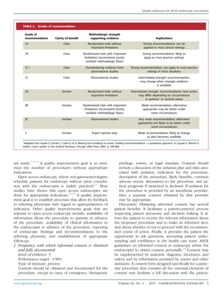
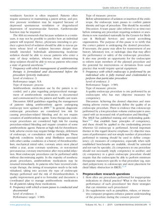
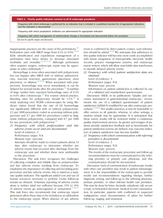
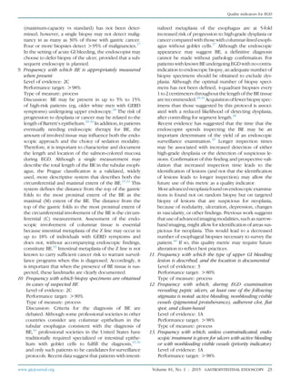
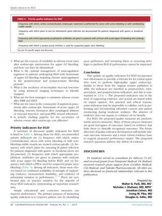
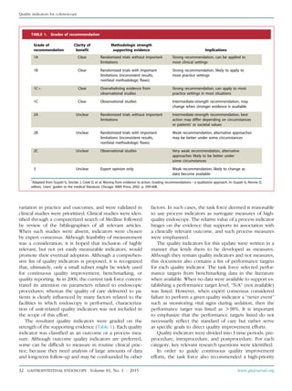
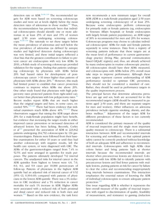
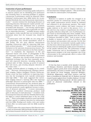
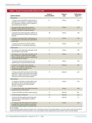
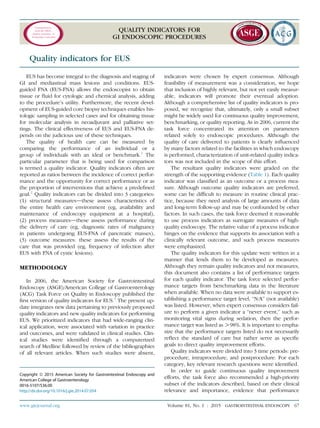
![indicators described, based on their clinical relevance and
importance, on evidence that performance of the indicator
varies significantly in clinical practice, and feasibility of mea-
surement (a function of the number of procedures needed
to obtain an accurate measurement with narrow confidence
intervals [CI] and the ease of measurement). A useful
approach for individual endoscopists is to first measure their
performances withregardtothesepriorityindicators.Quality
improvement efforts would then move to different quality in-
dicators ifendoscopists areperformingabove recommended
thresholds, or the employer and/or teaching center could
institute corrective measures and remeasure performance
of low-level performers.
Recognizing that certain quality indicators are common
to all GI endoscopic procedures, such items are presented
in detail in a separate document, similar to the process
in 2006.5
The preprocedure, intraprocedure, and postpro-
cedure indicators common to all endoscopy are listed in
Table 2. Those common factors will be discussed only
in this document insofar as the discussion needs to be
modified specifically to relate to EGD.
Preprocedure quality indicators
The preprocedure period includes all contact between
members of the endoscopy team and the patient before
the administration of sedation or insertion of the endo-
scope. Common issues for all endoscopic procedures dur-
ing this period include: appropriate indication, informed
consent, risk assessment, formulation of a sedation plan,
management of prophylactic antibiotics and antithrom-
botic drugs, and timeliness of the procedure.5
Preproce-
dure quality indicators specific to EGD include the
following:
1. Frequency with which EGD is performed for an indi-
cation that is included in a published standard list
of appropriate indications, and the indication is
documented
Level of evidence: 1Cþ
Performance target: O80%
Type of measure: process
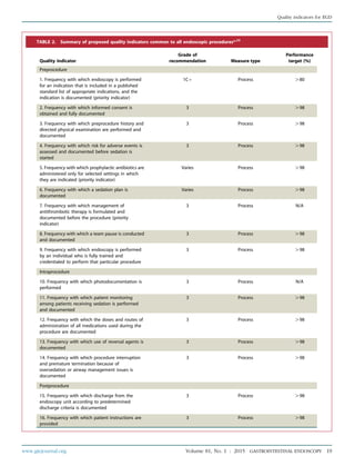
Discussion: The accepted indications for EGD are re-
viewed in detail in a recently updated document by
the ASGE Standards of Practice Committee (Table 3).6
The indications for EGD have expanded to include
endoscopic therapy for Barrett’s esophagus (BE), intra-
operative evaluation of reconstructed anatomic recon-
structions typical of modern foregut surgery, and
management of operative adverse events. Performing
EGD for an accepted indication is associated with a statis-
tically higher rate of clinically relevant findings.7,8
In one
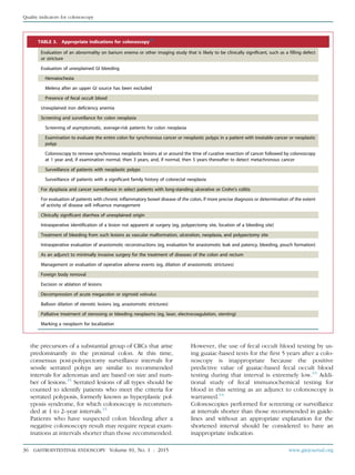
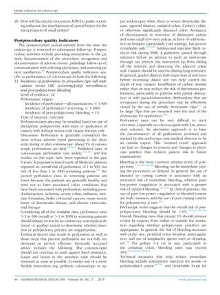
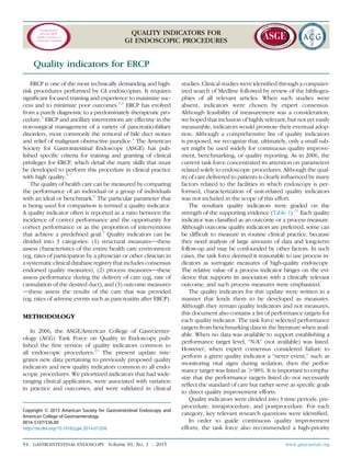
TABLE 1. Grades of recommendation*
Grade of
recommendation
Clarity of
benefit
Methodologic strength
supporting evidence Implications
1A Clear Randomized trials without
important limitations
Strong recommendation; can be
applied to most clinical settings
1B Clear Randomized trials with important
limitations (inconsistent results,
nonfatal methodologic flaws)
Strong recommendation, likely to
apply to most practice settings
1Cþ Clear Overwhelming evidence from
observational studies
Strong recommendation; can apply to
most practice settings in most situations
1C Clear Observational studies Intermediate-strength recommendation;
may change when stronger evidence
is available
2A Unclear Randomized trials without
important limitations
Intermediate-strength recommendation;
best action may differ depending on
circumstances or patients’ or societal values
2B Unclear Randomized trials with important
limitations (inconsistent results,
nonfatal methodologic flaws)
Weak recommendation; alternative
approaches may be better under some
circumstances
2C Unclear Observational studies Very weak recommendation; alternative
approaches likely to be better under
some circumstances
3 Unclear Expert opinion only Weak recommendation, likely to
change as data become available
*Adapted from Guyatt G, Sinclair J, Cook D, et al. Moving from evidence to action. Grading recommendationsda qualitative approach. In: Guyatt G, Rennie D,
editors. Users’ guides to the medical literature. Chicago: AMA Press; 2002. p. 599-608.
Quality indicators for EGD
18 GASTROINTESTINAL ENDOSCOPY Volume 81, No. 1 : 2015 www.giejournal.org](https://image.slidesharecdn.com/qualityinendoscopyset-211107013256/85/Quality-in-endoscopy-set-18-320.jpg)
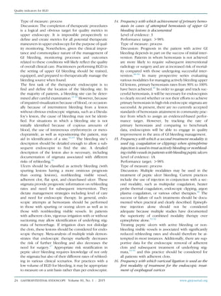
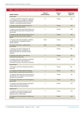
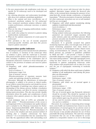
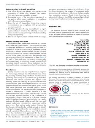
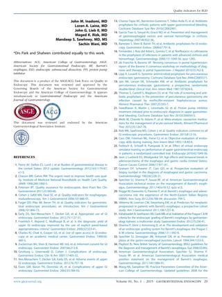
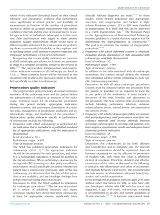
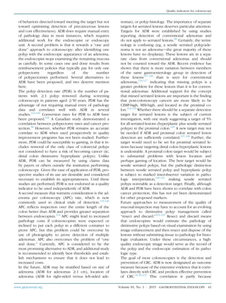
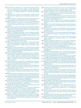
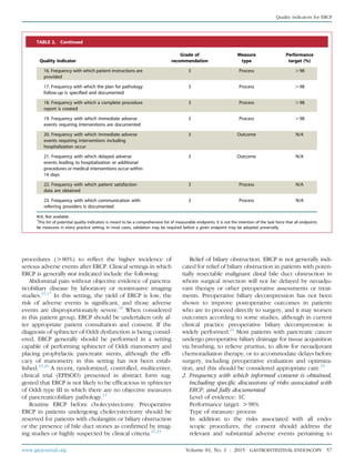
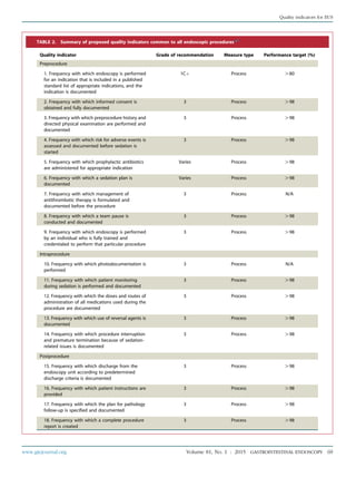
![21. Frequency with which patients with evidence of
recurrent bleeding from peptic ulcer disease after
endoscopic treatment undergo repeat upper
endoscopy
Level of evidence: 1B
Performance target: O98%
Type of measure: process
Discussion: Despite adequate endoscopic therapy for a
bleeding peptic ulcer, rebleeding can occur in up to one
third of patients. Repeat endoscopy for recurrent
bleeding is effective and should be done unless contra-
indicated.71,72
This should be documented and commu-
nicated with the primary providers. Routine second-look
endoscopy in the absence of rebleeding is not
recommended.26,72,73
22. Frequency that patients are contacted to document
the occurrence of adverse events after EGD
Level of evidence: 3
Performance target: N/A
Type of measure: process
Discussion: As more therapeutic EGD procedures occur
(EMR, endoscopic submucosal dissection [ESD]), endo-
scopists should develop a mechanism to capture and
track not only immediate but also delayed endoscopic
adverse events (from 14 days to 1 month). Such a prac-
tice would promote patient safetyda principle
supported by the ASGE, ACG, American Gastroentero-
logical Association, and the Institute of Medicine.11,74,75
Tracked adverse events should include cardiopulmo-
nary events, infections, perforation, bleeding, and
abdominal pain requiring medical attention or interven-
tion. In the future, individual adverse events could be
developed into separate quality indicators once further
data are obtained for benchmarking. For EGD, these
might include specific adverse event rates such as skin
infections after PEG tube placement, aspiration pneu-
monia after EGD with hemostasis, and stricture forma-
tion after esophageal mucosal resection or ablation.
Postprocedure research questions
1. What is the long-term outcome from following surveil-
lance recommendations for BE, and how will targeted
biopsy techniques that use new technology affect the
yield and efficacy of surveillance?
2. Are there variations in rebleeding rates from peptic ul-
cer disease after endoscopic therapy, and can this be
used to identify high performers of quality upper
endoscopy?
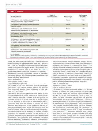
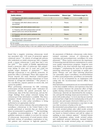
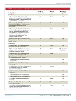
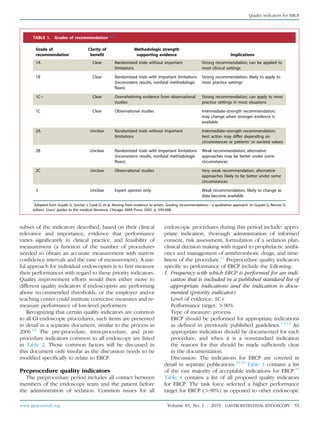
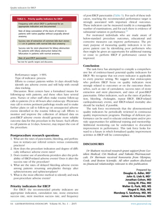
TABLE 4. Continued
Quality indicator
Grade of
recommendation Type of measure
Performance
target (%)
16. Frequency with which variceal ligation is used as
the first modality of treatment for the endoscopic
treatment of esophageal varices
1A Process O98
17. Frequency with which at least 4 intestinal
biopsies are done from patients in whom celiac
disease is suspected
1C Process O90
Postprocedure
18. Frequency with which PPI therapy is
recommended for patients who underwent dilation
for peptic esophageal strictures
1A Process O98
19. Frequency with which patients diagnosed with
gastric or duodenal ulcers are instructed to take PPI
medication or an H2 antagonist
1A Process O98
20. Frequency with which plans to test for H pylori
infection are documented for patients diagnosed
with gastric or duodenal ulcers (priority indicator)
1A Process O98
21. Frequency with which patients with evidence of
rebleeding from peptic ulcer disease after
endoscopic treatment undergo repeat upper
endoscopy
1B Process O98
22. Frequency with which patients are contacted to
document the occurrence of adverse events after
EGD
3 Process N/A
PPI, Proton pump inhibitor.
*
This list of potential quality indicators was meant to be a comprehensive listing of measurable endpoints. It is not the intention of the task force that all
endpoints be measured in every practice setting. In most cases, validation may be required before a given endpoint may be universally adopted.
www.giejournal.org Volume 81, No. 1 : 2015 GASTROINTESTINAL ENDOSCOPY 27
Quality indicators for EGD](https://image.slidesharecdn.com/qualityinendoscopyset-211107013256/85/Quality-in-endoscopy-set-27-320.jpg)
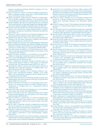
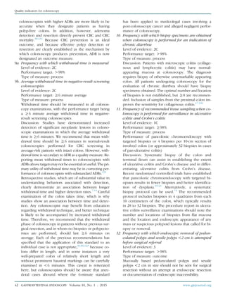
![Abbreviations: ACG, American College of Gastroenterology; ADR,
adenoma detection rate; APC, adenoma per colonoscopy; ASGE,
American Society for Gastrointestinal Endoscopy; CRC, colorectal
cancer; PDR, polyp detection rate.
This document is a product of the ASGE/ACG Task Force on Quality in
Endoscopy. This document was reviewed and approved by the
Governing Boards of the American Society for Gastrointestinal
Endoscopy and the American College of Gastroenterology. It appears
simultaneously in Gastrointestinal Endoscopy and the American
Journal of Gastroenterology.
This document was reviewed and endorsed by the American
Gastroenterological Association Institute.
REFERENCES
1. Centers for Disease Control and Prevention. Vital signs: colorectal
cancer screening test usedUnited States, 2012. MMWR Morb Mortal
Wkly Rep 2013;62:881-8.
2. Peery AF, Dellon ES, Lund J, et al. Burden of gastrointestinal disease in
the United States: 2012 update. Gastroenterology 2012;143:1179-87.
3. McLachlan SA, Clements A, Austoker J. Patients’ experiences and re-
ported barriers to colonoscopy in the screening contextda system-
atic review of the literature. Patient Educ Couns 2012;86:137-46.
4. Harewood GC, Sharma VK, de Garmo P. Impact of colonoscopy prep-
aration quality on detection of suspected colonic neoplasia. Gastro-
intest Endosc 2003;58:76-9.
5. Froehlich F, Wietlisbach V, Gonvers JJ, et al. Impact of colonic
cleansing on quality and diagnostic yield of colonoscopy: the Euro-
pean Panel of Appropriateness of Gastrointestinal Endoscopy Euro-
pean multicenter study. Gastrointest Endosc 2005;61:378-84.
6. Rex DK, Imperiale TF, Latinovich DR, et al. Impact of bowel prepara-
tion on efficiency and cost of colonoscopy. Am J Gastroenterol
2002;97:1696-700.
7. Rex DK. Colonoscopic withdrawal technique is associated with ade-
noma miss rates. Gastrointest Endosc 2000;51:33-6.
8. Lee RH, Tang RS, Muthusamy VR, et al. Quality of colonoscopy with-
drawal technique and variability in adenoma detection rates (with
videos). Gastrointest Endosc 2011;74:128-34.
9. Barclay R, Vicari JJ, Johanson JF, et al. Variation in adenoma detection
rates and colonoscopic withdrawal times during screening colonos-
copy [abstract]. Gastrointest Endosc 2005;61:AB107.
10. Sanchez W, Harewood GC, Petersen BT. Evaluation of polyp detection
in relation to procedure time of screening or surveillance colonos-
copy. Am J Gastroenterol 2004;99:1941-5.
11. Fatima H, Rex DK, Rothstein R, et al. Cecal insertion and withdrawal
times with wide-angle versus standard colonoscopes: a randomized
controlled trial. Clin Gastroenterol Hepatol 2008;6:109-14.
12. Simmons DT, Harewood GC, Baron TH, et al. Impact of endoscopist
withdrawal speed on polyp yield: implications for optimal colonos-
copy withdrawal time. Aliment Pharmacol Ther 2006;24:965-71.
13. Lim G, Viney SK, Chapman BA, et al. A prospective study of
endoscopist-blinded colonoscopy withdrawal times and polyp detec-
tion rates in a tertiary hospital. N Z Med J 2012;125:52-9.
14. Lin OS, Kozarek RA, Arai A, et al. The effect of periodic monitoring and
feedback on screening colonoscopy withdrawal times, polyp detec-
tion rates, and patient satisfaction scores. Gastrointest Endosc
2010;71:1253-9.
15. Lieberman DA, Rex DK, Winawer SJ, et al. Guidelines for colonoscopy
surveillance after screening and polypectomy: a consensus update by
the US Multi-Society Task Force on Colorectal Cancer. Gastroenter-
ology 2012;143:844-57.
16. Kaminski MF, Regula J, Kraszewska E, et al. Quality indicators for co-
lonoscopy and the risk of interval cancer. N Engl J Med 2010;362:
1795-803.
17. Rubin CE, Haggitt RC, Burmer GC, et al. DNA aneuploidy in colonic bi-
opsies predicts future development of dysplasia in ulcerative colitis.
Gastroenterology 1992;103:1611-20.
18. Jess T, Simonsen J, Jorgensen KT, et al. Decreasing risk of colorectal
cancer in patients with inflammatory bowel disease over 30 years.
Gastroenterology 2012;143:375-81.
19. Kiesslich R, Fritsch J, Holtmann M, et al. Methylene blue-aided chro-
moendoscopy for the detection of intraepithelial neoplasia and colon
cancer in ulcerative colitis. Gastroenterology 2003;124:880-8.
20. Rutter MD, Saunders BP, Schofield G, et al. Pancolonic indigo carmine
dye spraying for the detection of dysplasia in ulcerative colitis. Gut
2004;53:256-60.
21. Wu L, Li P, Wu J, et al. The diagnostic accuracy of chromoendoscopy
for dysplasia in ulcerative colitis: meta-analysis of six randomized
controlled trials. Colorectal Dis 2012;14:416-20.
22. Chukmaitov A, Bradley CJ, Dahman B, et al. Association of polypec-
tomy techniques, endoscopist volume, and facility type with colonos-
copy complications. Gastrointest Endosc 2013;77:436-46.
23. Baxter NN, Goldwasser MA, Paszat LF, et al. Association of colonos-
copy and death from colorectal cancer. Ann Intern Med 2009;150:
1-8.
24. Brenner H, Chang-Claude J, Seiler CM, et al. Does a negative screening
colonoscopy ever need to be repeated? Gut 2006;55:1145-50.
25. Lakoff J, Paszat LF, Saskin R, et al. Risk of developing proximal versus
distal colorectal cancer after a negative colonoscopy: a population-
based study. Clin Gastroenterol Hepatol 2008;6:1117-21.
26. Singh H, Nugent Z, Mahmud SM, et al. Predictors of colorectal cancer
after negative colonoscopy: a population-based study. Am J Gastro-
enterol 2010;105:663-73.
27. Singh H, Nugent Z, Demers AA, et al. The reduction in colorectal can-
cer mortality after colonoscopy varies by site of the cancer. Gastroen-
terology 2010;139:1128-37.
28. Brenner H, Chang-Claude J, Seiler CM, et al. Protection from colorectal
cancer after colonoscopy: a population-based, case-control study.
Ann Intern Med 2011;154:22-30.
29. Rex DK, Rahmani EY, Haseman JH, et al. Relative sensitivity of colo-
noscopy and barium enema for detection of colorectal cancer in clin-
ical practice. Gastroenterology 1997;112:17-23.
30. Baxter N, Sutradhar R, Forbes DD, et al. Analysis of administrative data
finds endoscopist quality measures asociated with post-colonoscopy
colorectal cancer. Gastroenterology 2011;140:65-72.
31. Rabeneck L, Paszat LF, Saskin R. Endoscopist specialty is associated
with incident colorectal cancer after a negative colonoscopy. Clin
Gastroenterol Hepatol 2010;8:275-9.
32. Baxter NN, Warren JL, Barrett MJ, et al. Association between colonos-
copy and colorectal cancer mortality in a US cohort according to
site of cancer and colonoscopist specialty. J Clin Oncol 2012;30:
2664-9.
33. Ko CW, Dominitz JA, Green P, et al. Specialty differences in polyp
detection, removal, and biopsy during colonoscopy. Am J Med
2010;123:528-35.
34. Pox CP, Altenhofen L, Brenner H, et al. Efficacy of a nationwide
screening colonoscopy program for colorectal cancer. Gastroenter-
ology 2012;142:1460-7.
35. Petersen BT. Quality assurance for endoscopists. Best Pract Res Clin
Gastroenterol 2011;25:349-60.
36. Rex DK, Petrini JL, Baron TH, et al. Quality indicators for colonoscopy.
Gastrointest Endosc 2006;63:S16-28.
www.giejournal.org Volume 81, No. 1 : 2015 GASTROINTESTINAL ENDOSCOPY 49
Quality indicators for colonoscopy](https://image.slidesharecdn.com/qualityinendoscopyset-211107013256/85/Quality-in-endoscopy-set-49-320.jpg)
![37. Faigel DO, Pike IM, Baron TH, et al. Quality indicators for gastrointes-
tinal endoscopic procedures: an introduction. Gastrointest Endosc
2006;63:S3-9.
38. Rizk MK, Sawhney MS, Cohen J, et al. Quality indicators common to all
GI endoscopic procedures. Gastrointest Endosc 2015;81:3-16.
39. Early DS, Ben-Menachem T, Decker GA, et al. Appropriate use of GI
endoscopy. Gastrointest Endosc 2012;75:1127-31.
40. Balaguer F, Llach J, Castells A, et al. The European panel on the appro-
priateness of gastrointestinal endoscopy guidelines colonoscopy in
an open-access endoscopy unit: a prospective study. Aliment Phar-
macol Ther 2005;21:609-13.
41. Vader JP, Pache I, Froehlich F, et al. Overuse and underuse of colonos-
copy in a European primary care setting. Gastrointest Endosc 2000;52:
593-9.
42. de Bosset V, Froehlich F, Rey JP, et al. Do explicit appropriateness
criteria enhance the diagnostic yield of colonoscopy? Endoscopy
2002;34:360-8.
43. Terraz O, Wietlisbach V, Jeannot JG, et al. The EPAGE internet guide-
line as a decision support tool for determining the appropriateness
of colonoscopy. Digestion 2005;71:72-7.
44. Morini S, Hassan C, Meucci G, et al. Diagnostic yield of open access
colonoscopy according to appropriateness. Gastrointest Endosc
2001;54:175-9.
45. Bersani G, Rossi A, Ricci G, et al. Do ASGE guidelines for the appro-
priate use of colonoscopy enhance the probability of finding rele-
vant pathologies in an open access service? Dig Liver Dis 2005;37:
609-14.
46. Baron TH, Kimery BD, Sorbi D, et al. Strategies to address increased
demand for colonoscopy: guidelines in an open endoscopy practice.
Clin Gastroenterol Hepatol 2004;2:178-82.
47. Rex DK, Johnson DA, Anderson JC, et al. American College of Gastro-
enterology guidelines for colorectal cancer screening 2008. Am J Gas-
troenterol 2009;104:739-50.
48. Levin B, Lieberman DA, McFarland B, et al. Screening and surveillance
for the early detection of colorectal cancer and adenomatous polyps,
2008: a joint guideline from the American Cancer Society, the US
Multi-Society Task Force on Colorectal Cancer, and the American Col-
lege of Radiology. Gastroenterology 2008;134:1570-95.
49. Brenner H, Chang-Claude J, Seiler CM, et al. Long-term risk of colo-
rectal cancer after negative colonoscopy. J Clin Oncol 2011;29:
3761-7.
50. Imperiale TF, Glowinski EA, Lin-Cooper C, et al. Five-year risk of colo-
rectal neoplasia after negative screening colonoscopy. N Engl J Med
2008;359:1218-24.
51. Rex DK, Cummings OW, Helper DJ, et al. 5-year incidence of ade-
nomas after negative colonoscopy in asymptomatic average-risk per-
sons [see comment]. Gastroenterology 1996;111:1178-81.
52. Selby JV, Friedman GD, Quesenberry CP Jr, et al. A case-control study
of screening sigmoidoscopy and mortality from colorectal cancer.
N Engl J Med 1992;326:653-7.
53. Newcomb PA, Storer BE, Morimoto LM, et al. Long-term efficacy of
sigmoidoscopy in the reduction of colorectal cancer incidence.
J Natl Cancer Inst 2003;95:622-5.
54. Goodwin JS, Singh A, Reddy N, et al. Overuse of screening colono-
scopy in the Medicare population. Arch Intern Med 2011;171:1335-43.
55. Mysliwiec PA, Brown ML, Klabunde CN, et al. Are physicians doing too
much colonoscopy? A national survey of colorectal surveillance after
polypectomy. Ann Intern Med 2004;141:264-71.
56. Saini SD, Nayak RS, Kuhn L, et al. Why don’t gastroenterologists follow
colon polyp surveillance guidelines? Results of a national survey.
J Clin Gastroenterol 2009;43:554-8.
57. Burke C, Issa M, Church J. A nationwide survey of post-polypectomy
surveillance colonoscopy: too many too soon! Gastroenterology
2005;128:A566.
58. Boolchand V, Singh J, Olds G, et al. Colonoscopy surveillance after
polypectomy: a national survey study of primary care physicians.
Am J Gastroenterol 2005;100:S384-5.
59. Kim ER, Sinn DH, Kim JY, et al. Factors associated with adherence to
the recommended postpolypectomy surveillance interval. Surg En-
dosc 2012;26:1690-5.
60. Shah TU, Voils CI, McNeil R, et al. Understanding gastroenterologist
adherence to polyp surveillance guidelines. Am J Gastroenterol
2012;107:1283-7.
61. Schoen RE, Pinsky PF, Weissfeld JL, et al. Utilization of surveillance
colonoscopy in community practice. Gastroenterology 2010;138:
73-81.
62. Khashab M, Eid E, Rusche M, et al. Incidence and predictors of “late”
recurrences after endoscopic piecemeal resection of large sessile
adenomas. Gastrointest Endosc 2009;70:344-9.
63. Finkelstein S, Bini EJ. Annual fecal occult blood testing can be safely
suspended for up to 5 years after a negative colonoscopy in asymp-
tomatic average-risk patients [abstract]. Gastrointest Endosc 2005;61:
AB250.
64. Bampton PA, Sandford JJ, Cole SR, et al. Interval faecal occult blood
testing in a colonoscopy based screening programme detects addi-
tional pathology. Gut 2005;54:803-6.
65. Katzka I, Brody RS, Morris E, et al. Assessment of colorectal cancer risk
in patients with ulcerative colitis: experience from a private practice.
Gastroenterology 1983;85:22-9.
66. Friedman S, Rubin PH, Bodian C, et al. Screening and surveillance
colonoscopy in chronic Crohn’s colitis. Gastroenterology 2001;120:
820-6.
67. Connell WR, Talbot IC, Harpaz N, et al. Clinicopathological character-
istics of colorectal carcinoma complicating ulcerative colitis. Gut
1994;35:1419-23.
68. Karlen P, Kornfeld D, Brostrom O, et al. Is colonoscopic surveillance
reducing colorectal cancer mortality in ulcerative colitis? A popula-
tion based case control study. Gut 1998;42:711-4.
69. Bernstein CN, Weinstein WM, Levine DS, et al. Physicians’ perceptions
of dysplasia and approaches to surveillance colonoscopy in ulcerative
colitis. Am J Gastroenterol 1995;90:2106-14.
70. Eaden JA, Ward BA, Mayberry JF. How gastroenterologists screen for
colonic cancer in ulcerative colitis: an analysis of performance. Gas-
trointest Endosc 2000;51:123-8.
71. Kornbluth A, Sachar DB. Ulcerative colitis practice guidelines in adults:
American College Of Gastroenterology, Practice Parameters Commit-
tee. Am J Gastroenterol 2010;105:501-23.
72. Provenzale D, Onken J. Surveillance issues in inflammatory bowel dis-
ease: ulcerative colitis. J Clin Gastroenterol 2001;32:99-105.
73. Herrinton LJ, Liu L, Levin TR, et al. Incidence and mortality of colo-
rectal adenocarcinoma in persons with inflammatory bowel disease
from 1998 to 2010. Gastroenterology 2012;143:382-9.
74. Rutter MD, Saunders BP, Wilkinson KH, et al. Cancer surveillance in
longstanding ulcerative colitis: endoscopic appearances help predict
cancer risk. Gut 2004;53:1813-6.
75. Winawer S, Fletcher R, Rex D, et al. Colorectal cancer screening and
surveillance: clinical guidelines and rationaledupdate based on
new evidence. Gastroenterology 2003;124:544-60.
76. Lieberman D, Nadel M, Smith RA, et al. Standardized colonoscopy re-
porting and data system: report of the Quality Assurance Task Group
of the National Colorectal Cancer Roundtable. Gastrointest Endosc
2007;65:757-66.
77. Wexner SD, Beck DE, Baron TH, et al. A consensus document on
bowel preparation before colonoscopy: prepared by a Task Force
from the American Society of Colon and Rectal Surgeons (ASCRS),
the American Society for Gastrointestinal Endoscopy (ASGE), and
the Society of American Gastrointestinal and Endoscopic Surgeons
(SAGES). Surg Endosc 2006;20:1161.
78. Larsen M, Hills N, Terdiman J. The impact of the quality of colon prep-
aration on follow-up colonoscopy recommendations. Am J Gastroen-
terol 2011;106:2058-62.
79. Manno M, Pigo F, Manta R, et al. Bowel preparation with polyethylene
glycol electrolyte solution: optimizing the splitting regimen. Dig Liver
Dis 2012;44:576-9.
50 GASTROINTESTINAL ENDOSCOPY Volume 81, No. 1 : 2015 www.giejournal.org
Quality indicators for colonoscopy](https://image.slidesharecdn.com/qualityinendoscopyset-211107013256/85/Quality-in-endoscopy-set-50-320.jpg)
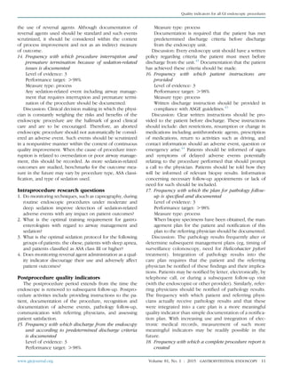
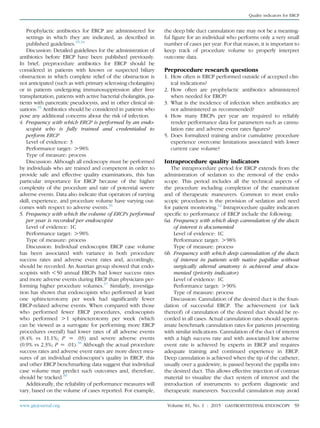
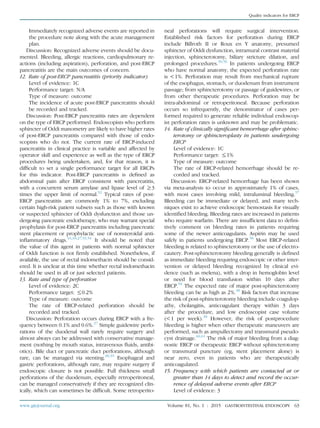
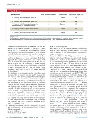
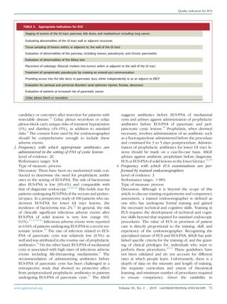
![each specific ERCP procedure. Informed consent for
ERCP should focus on at least 6 possible adverse out-
comes: (1) pancreatitis, (2) hemorrhage, (3) infection,
(4) cardiopulmonary events, (5) allergic reaction, and
(6) perforation. It is also advisable that patients be
informed of the possibility that the procedure may not
be successful and that additional procedures may be
warranted. The patient should be informed that adverse
events could be severe in nature.
Discussion: Some ERCP adverse events are unique from
those that occur with standard luminal endoscopy. A review
of the adverse events specific to ERCP has been published
previously.26
The expected rate of post-ERCP pancreatitis
is generally between 1% and 7% for most average-risk pa-
tients.27-30
There are several situations in which this rate
may be significantly higher, most notably in patients with
known or suspected sphincter of Oddi dysfunction. Adverse
events in these patients can approach 20% to 30%, with
severe pancreatitis also being more likely.31
Numerous factors, both patient-related and
procedure-related, may influence the risk for post-
ERCP pancreatitis and need to be taken into account
when endoscopists are planning for the procedure
and obtaining informed consent. Cholangitis occurs
in !1% of patients after ERCP, and cholecystitis com-
plicates 0.2% to 0.5% of ERCPs. Hemorrhage is most
commonly an adverse event of endoscopic sphincterot-
omy and has been reported to occur in 0.8% to 2% of
cases. Perforations may be guidewire-induced, sphinc-
terotomy-induced, or endoscope-induced. The overall
incidence of perforation during ERCP has been re-
ported to be 0.1% to 0.6%.32
3. Frequency with which appropriate antibiotics for
ERCP are administered for settings in which they are
indicated
Level of evidence: 2B
Performance target: O98%
Type of measure: process
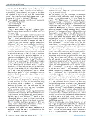
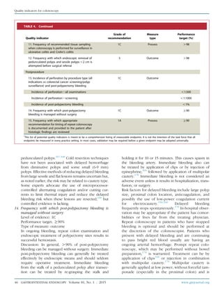
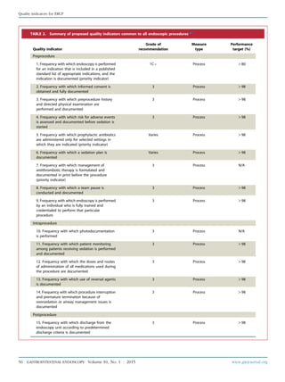
TABLE 3. Appropriate indications for ERCP15
The jaundiced patient suspected of having biliary obstruction (appropriate therapeutic maneuvers should be performed during the
procedure)
The patient without jaundice whose clinical and biochemical or imaging data suggest pancreatic duct or biliary tract disease
Evaluation of signs or symptoms suggesting pancreatic malignancy when results of direct imaging (eg, EUS, US, computed tomography
[CT], magnetic resonance imaging [MRI]) are equivocal or normal
Evaluation of pancreatitis of unknown etiology
Preoperative evaluation of the patient with chronic pancreatitis and/or pseudocyst
Evaluation of the sphincter of Oddi by manometry
Empirical biliary sphincterotomy without sphincter of Oddi manometry is not recommended in patients with suspected type III sphincter
of Oddi dysfunction
Endoscopic sphincterotomy:
Choledocholithiasis.
Papillary stenosis or sphincter of Oddi dysfunction
To facilitate placement of biliary stents or dilation of biliary strictures
Sump syndrome
Choledochocele involving the major papilla
Ampullary carcinoma in patients who are not candidates for surgery
Facilitate access to the pancreatic duct
Stent placement across benign or malignant strictures, fistulae, postoperative bile leak, or in high-risk patients with large unremovable
common duct stones
Dilation of ductal strictures
Balloon dilation of the papilla
Nasobiliary drain placement
Pancreatic pseudocyst drainage in appropriate cases
Tissue sampling from pancreatic or bile ducts
Ampullectomy of adenomatous neoplasms of the major papilla
Therapy of disorders of the biliary and pancreatic ducts
Faciliation of cholangioscopy and/or pancreatoscopy
58 GASTROINTESTINAL ENDOSCOPY Volume 81, No. 1 : 2015 www.giejournal.org
Quality indicators for ERCP](https://image.slidesharecdn.com/qualityinendoscopyset-211107013256/85/Quality-in-endoscopy-set-58-320.jpg)
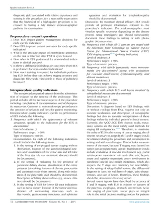
![varies significantly in clinical practice, and feasibility of
measurement (a function of the number of procedures
needed to obtain an accurate measurement with narrow
confidence intervals [CI] and the ease of measurement).
A useful approach for individual endoscopists is to first
measure their performance with regard to these priority in-
dicators. Quality improvement efforts would then move
to different quality indicators if endoscopists are perform-
ing above recommended thresholds, or the employer
and/or teaching center could institute corrective measures
and remeasure performance of low-level performers.
Recognizing that certain quality indicators are common
to all GI endoscopic procedures, such items are presented
in detail in a separate document, similar to the process
in 2006.4
The preprocedure, intraprocedure, and postpro-
cedure indicators common to all endoscopy are listed in
Table 2. Those common factors will be discussed in
this document only insofar as the discussion needs to be
modified specifically related to EUS.
Preprocedure quality indicators
The preprocedure period includes all contact between
members of the endoscopy team with the patient
before the administration of sedation. Common issues
for all endoscopic procedures during this period include:
appropriate indication, informed consent, risk assessment,
formulation of a sedation plan, clinical decision making
with regard to prophylactic antibiotics and management
of antithrombotic drugs, and timeliness of the procedure.5
Preprocedure quality indicators specific to performance of
EUS include the following:
1. Frequency with which EUS is performed for an in-
dication that is included in a published standard list
of appropriate indications, and the indication is
documented
Level of evidence: 1C
Performance target: O80%
Type of measure: process
The ASGE has published appropriate indications for
EUS (Table 3).6
An appropriate indication should be
documented for each procedure, and, when it is not a
standard indication listed in the current ASGE Appro-
priate Use of GI Endoscopy guideline, it should be justi-
fied in the documentation.
Discussion: Acceptable indications for EUS have been
published recently.6,7
Although there are many in-
stances in which EUS can be performed, the value of
the procedure in the care of any particular patient de-
pends on its impact on management, improvement in
outcomes, and the superiority of EUS over other avail-
able imaging or surgical procedures. This implies a
certain degree of clinical judgment in choosing when
and if to perform EUS in relation to other procedures,
making rigid indications impractical. Expert opinion
TABLE 1. Grades of recommendation*
Grade of
recommendation
Clarity of
benefit
Methodologic strength
supporting evidence Implications
1A Clear Randomized trials without important
limitations
Strong recommendation; can be applied to
most clinical settings
1B Clear Randomized trials with important
limitations (inconsistent results,
nonfatal methodologic flaws)
Strong recommendation; likely to apply to
most practice settings
1Cþ Clear Overwhelming evidence from
observational studies
Strong recommendation; can apply to most
practice settings in most situations
1C Clear Observational studies Intermediate-strength recommendation; may
change when stronger evidence is available
2A Unclear Randomized trials without
important limitations
Intermediate-strength recommendation; best
action may differ depending on circumstances
or patients’ or societal values
2B Unclear Randomized trials with important
limitations (inconsistent results,
nonfatal methodologic flaws)
Weak recommendation; alternative approaches
may be better under some circumstances
2C Unclear Observational studies Very weak recommendation; alternative approaches
likely to be better under some circumstances
3 Unclear Expert opinion only Weak recommendation; likely to change as data
become available
*
Adapted from Guyatt G, Sinclair J, Cook D, et al. Moving from evidence to action. Grading recommendationsda qualitative approach. In: Guyatt G, Rennie D,
editors. Users’ guides to the medical literature. Chicago: AMA Press; 2002. p. 599-608.
Quality indicators for EUS
68 GASTROINTESTINAL ENDOSCOPY Volume 81, No. 1 : 2015 www.giejournal.org](https://image.slidesharecdn.com/qualityinendoscopyset-211107013256/85/Quality-in-endoscopy-set-68-320.jpg)
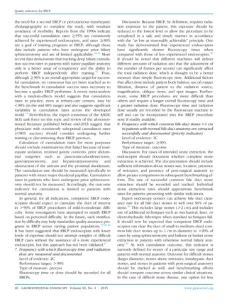
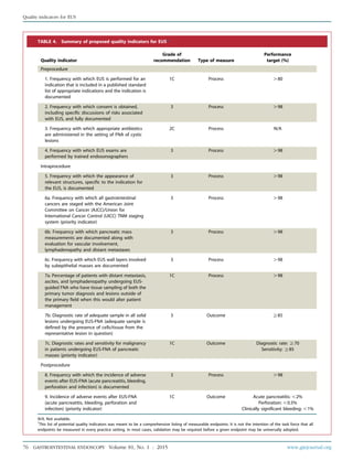
![with pancreatic cancer. In pancreatic cancer, results
from contemporary studies have reported accuracy of
T staging ranging from 62% to 67%, 45-48
with earlier
studies reporting higher accuracy rates (85%-94%).49-51
In the absence of distant metastasis, the presence
and degree of contact between the tumor and the peri-
pancreatic vessels is of paramount importance in
determining surgical resectability. In a meta-analysis,
the sensitivity and specificity of EUS in diagnosing
vascular invasion was 73% (95% CI, 68.8-76.9) and
90% (95% CI, 87.9-92.2).52
Results from available data
with regard to accuracy of EUS in predicting vascular
invasion are variable, with a wide range suggesting
the operator dependency and variability. The task
force acknowledges this and hence does not make ac-
curacy of vascular invasion as a quality indicator but
recommends documentation of vascular invasion as a
quality indicator. Similarly, variable rates of accuracy
for N staging have been reported in pancreatic cancer
(range 40%-85%).45,47,48,50,51,53,54
In esophageal cancer,
sensitivity and specificity of EUS for T staging has
ranged from 81% to 92% and 94% to 99%, respec-
tively.55
Although the role of EUS has been questioned
in the setting of Barrett’s-esophagus–related neoplasia
(high-grade dysplasia and intramucosal cancer),56,57
EUS has moderate accuracy rates in differentiating
mucosal (T1a) versus submucosal (T1b) esophageal can-
cer, although this is largely being supplanted by EMR
and/or endoscopic submucosal dissection and direct pa-
thology staging.58
Sensitivity and specificity of EUS for N
staging was 80% (95% CI, 75-84) and 70% (95% CI, 65-
75) in a meta-analysis.59
In gastric cancer, a recent
meta-analysis reported high accuracy rates in differenti-
ating T1-2 from T3-4 disease (sensitivity 86% [95% CI,
81-90] and specificity 91% [95% CI, 89-93]. EUS for
lymph node status was less reliable sensitivity 69%
[95% CI, 63-74] and specificity 84% [95% CI, 81-88]).60
The sensitivity and specificity for T staging in rectal can-
cer was 88% and 98% for T1, 81% and 96% for T2, 96%
and 91% for T3, and 95% and 98% for T4 cancer, respec-
tively.61
However, recent studies have questioned these
high accuracy rates and have suggested that magnetic
resonance imaging may have similar accuracy rates in
the T and N staging of rectal cancer.62,63
7a. Percentage of patients with distant metastasis, ascites,
and lymphadenopathy undergoing EUS-guided FNA
who have tissue sampling of both the primary tumor
and lesions outside of the primary field when this
would alter patient management
Level of evidence: 1C
Performance target: O98%
Type of measure: process
7b. Diagnostic rate of adequate sample in all solid le-
sions undergoing EUS-FNA (adequate sample is
defined by the presence of cells and/or tissue from
the representative lesion in question)
Level of evidence: 3
Performance target: R85%
Type of measure: outcome
7c. Diagnostic rates and sensitivity for malignancy in
patients undergoing EUS-FNA of pancreatic masses
(priority indicator)
Level of evidence: 1C
Performance target: Diagnostic rate of malignancy
in patients undergoing EUS-FNA of all pancreatic
masses, R70% and sensitivity of malignancy among
patients with pancreatic cancer, R85%
Type of measure: outcome
Discussion: The additional clinical information ob-
tained from FNA can increase the diagnostic accuracy
of EUS significantly by confirming a pathologic diag-
nosis, by obtaining more accurate nodal staging in ma-
lignancy, and by yielding fluid for various analyses,
including chemical analyses, tumor markers, and bac-
terial and/or fungal stains or culture. FNA is not feasible
or appropriate in all conditions. Sampling a lymph
node by traversing the primary tumor with the FNA
needle should be avoided, because this may result in
a false-positive lymph node cytology result and can
potentially seed a previously benign lymph node with
malignant cells from the primary tumor. The need
for pretreatment FNA of pancreas tumors is variable.
The primary value of FNA is to confirm malignancy,
particularly when chemoradiotherapy is considered
prior to or in lieu of surgery or to exclude lesions
such as metastases to the pancreas, mass-forming
pancreatitis, non-adenocarcinoma histology, and lym-
phoma. However, when FNA is appropriate, the endo-
sonographer should make every effort to obtain
adequate cytologic material to confirm a diagnosis.
Accuracy of EUS-FNA has been evaluated in several
studies in patients with cancers of the pancreas, esoph-
agus, stomach, bile duct, and rectum. Data from these
studies provide a benchmark for quality performance
measurement in EUS. A multicenter, retrospective study
that included 1075 patients who underwent EUS-FNA
of solid pancreatic masses at 21 centers (81% academic)
with 41 endosonographers reported an overall diag-
nostic rate of malignancy of 71% (95% CI, 69-74).64
Sensitivity and specificity that uses the criterion
standard of either surgical pathology or long-term
follow-up are ideal benchmarks for pancreatic EUS-
FNA performance. A recent meta-analysis that included
studies that met this criterion reported a pooled sensi-
tivity of 85% (95% CI, 84-86) and specificity of 98%
(95% CI, 97-99), with higher accuracy of EUS-FNA
reported in prospective, multicenter studies.65
In the setting of esophageal cancer in the thoracic
esophagus, malignant celiac axis lymph nodes no
longer confer M1a status and, per the new staging sys-
tem, a regional lymph node has been redefined to
include any paraesophageal node extending from
www.giejournal.org Volume 81, No. 1 : 2015 GASTROINTESTINAL ENDOSCOPY 73
Quality indicators for EUS](https://image.slidesharecdn.com/qualityinendoscopyset-211107013256/85/Quality-in-endoscopy-set-73-320.jpg)
![cervical nodes to celiac nodes.66
EUS-FNA for lymph
node staging in esophageal cancer is an accurate stag-
ing modality with sensitivity of 83% (95% CI, 70-93),
specificity of 93% (95% CI, 77%-99%), and accuracy
of 87% (95% CI, 77-94) as reported in a prospective
study that included 76 consecutive patients with path-
ologic evaluation of resected lymph nodes.67
Retro-
spective studies that focused primarily on celiac
lymph nodes reported sensitivity of 88% to 100%, spec-
ificity of 100%, and accuracy rates ranging from 87% to
100% for detection of lymph node metastases.68-71
Several studies have reported the use of EUS-FNA for
the diagnosis of cholangiocarcinoma in the setting of
indeterminate extrahepatic strictures. Reported sensi-
tivity ranges from 29% to 89%72-77
with a higher sensi-
tivity reported for distal compared with proximal
strictures (81% vs 59%; P Z .04) in a single study.77
The conventional criteria for malignant lymph nodes
at EUS (size O1 cm, round, hypoechoic, and homoge-
nous) have a poor predictive value in malignant lymph-
adenopathy associated with cholangiocarcinoma.78
Hence, given the potential for avoiding unnecessary
neoadjuvant therapy and staging laparotomy, a low
threshold for sampling lymphadenopathy in this situa-
tion should be maintained. EUS-FNA should be per-
formed only when results are likely to alter decision
making (primary surgical resection or definitive or
neoadjuvant chemoradiation). EUS-FNA also should
be performed in patients with suspected distant metas-
tases, given the potential to significantly change pa-
tient management.
The involvement of an on-site cytopathologist during
EUS-FNA may help limit the number of FNA passes
taken and increase the overall diagnostic accuracy of
the procedure, although data are inconclusive.9,79-85
The impact of on-site cytopathology evaluation in
terms of diagnostic yield, number of passes, repeat
procedures, and procedure time has not been studied
in a randomized, controlled trial. However, it is recog-
nized that not all endosonographers will have access to
this degree of service. Therefore, for situations in
which an on-site cytopathologist or cytotechnologist
is not available, 5 to 7 FNA passes for pancreas masses
and 2 to 4 passes for lymph nodes or suspected liver
metastases are advised.86-88
Other methods to increase
cytologic adequacy and accuracy have not been defin-
itively shown to be superior. EUS-FNA can be per-
formed by using 25-gauge, 22-gauge, or 19-gauge
needles. Randomized, controlled trials comparing 25-
gauge and 22-gauge needles demonstrated no differ-
ence in diagnostic accuracy between the two
groups.89-91
A recent meta-analysis of 8 studies
involving 1292 patients undergoing EUS-FNA (25-
gauge, 565 patients and 22-gauge, 799 patients)
showed that a 25-gauge needle was more sensitive
than a 22-gauge needle for diagnosing pancreatic ma-
lignancy (pooled sensitivity, 25-gauge: 0.93 [95% CI,
0.91-0.96] vs 22-gauge: 0.85 [95% CI, 0.82-0.88]).92
A
randomized, controlled trial comparing 19-gauge and
22-gauge needle systems in patients undergoing EUS-
FNA of pancreatic masses demonstrated a higher diag-
nostic accuracy rate and the presence of superior
cellular material by using the 19-gauge needle. Howev-
er, a significantly lower technical success rate was re-
ported by using the 19-gauge needle system.93
Large
needle gauges (19-gauge) provide a larger specimen
but are limited to transgastric biopsy in most cases
and for EUS-guided interventions such as pseudocyst
drainage. Few randomized, controlled trials have
demonstrated no advantage in the routine use of a sty-
let during EUS-FNA.94-96
In recent years, the technique
of performing EUS-FNA passes without the use of a sty-
let has gained popularity but has not been adopted by
all endosonographers. Use of traditional true-cut bi-
opsy has not been shown to be superior to FNA and
is associated with a high failure rate in transduodenal
puncture.9
Recent availability of small-gauge core bi-
opsy needles (25-gauge and 22-gauge) and flexible
19-gauge needles offers an opportunity for research.
Intraprocedure research questions
1. What are the thresholds for accurate T and N staging of
GI malignancies?
2. How do community practices compare with academic
centers with regard to EUS staging and EUS-FNA
accuracy?
3. Under what circumstances does FNA change patient
management?
4. What is the optimal technique for performing EUS-FNA,
and what are the variables that impact obtaining
adequate specimens?
5. How does on-site cytopathology evaluation during EUS-
FNA impact diagnostic yield, number of passes, repeat
procedures, and procedure time?
6. What are the optimal methods for tissue processing of
FNA specimens?
Postprocedure quality indicators
The postprocedure period extends from the time the
endoscope is removed to subsequent follow-up. Postpro-
cedure activities include providing instructions to the pa-
tient, documentation of the procedure, recognition and
documentation of adverse events, pathology follow-up,
communication with referring physicians, and assessing
patient satisfaction.5
Postprocedure quality indicators
specific to performance of EUS include the following:
8. Frequency with which the incidence of adverse events
after EUS-FNA (acute pancreatitis, bleeding, perfora-
tion, and infection) is documented
Level of evidence: 3
Performance target: O98%
Type of measure: process
74 GASTROINTESTINAL ENDOSCOPY Volume 81, No. 1 : 2015 www.giejournal.org
Quality indicators for EUS](https://image.slidesharecdn.com/qualityinendoscopyset-211107013256/85/Quality-in-endoscopy-set-74-320.jpg)
![9. Incidence of adverse events after EUS-FNA (acute
pancreatitis, bleeding, perforation, and infection) (pri-
ority indicator)
Level of evidence: 1C
Performance target: acute pancreatitis !2%, perfora-
tion !0.5%, clinically significant bleeding !1%
Type of measure: outcome
Discussion
A. Overall and specific adverse event rates. The overall
safety of EUS-FNA is well-established, with a low overall
adverse event rate. The main adverse events include acute
pancreatitis, bleeding, and infection. Two other adverse
events that merit mention include tumor seeding and
false-positive EUS-FNA cytology results.
Variable rates of morbidity related to EUS-FNA have been
reported, ranging from 0% to 2.5%.13-15,19-21
A recent system-
atic review that included 10,941 patients reported an overall
EUS-FNA specific morbidity rate of 0.98% (107/10,941) and
mortality rate of 0.02% (2/10,941).10
Patients undergoing
EUS-FNA of the pancreas for evaluation of pancreatic masses,
cystic lesions, or lesions of the pancreatic duct are at risk of
developing pancreatitis, likely as a result of direct tissue
injury as the needle traverses pancreatic tissue. The inci-
dence of pancreatitis in this setting, including data from pro-
spective series, has ranged between 0% and 2%.19,21,23-26
The
rate of pancreatitis was 0.44% (36/8246) in a systematic re-
view, mild-moderate severity in most patients.10
Acute clini-
cally significant bleeding related to EUS-FNA is a rare adverse
event, and incidence has ranged from 0 to 0.5%.10,13-15,19-21
Mild intraluminal bleeding has been reported in up to 4%
of cases,97
extraluminal bleeding in 1.3% to 2.6% of
cases,26,98
and intracystic bleeding in up to 6% of cases dur-
ing EUS-FNA of pancreatic cysts.99
The risk of clinically signif-
icant infectious adverse events after EUS-FNA of solid lesions
is very low (range 0%-0.6%).13-15,19-22
Infectious adverse
events were reported in 5 of 10,941 (0.04%) patients in a
recent systematic review.10
The rate of infection related to
EUS-FNA of pancreatic cysts is relatively low (0.5%) and is
attributed to the routine use of prophylactic antibiotics.10
On the other hand, EUS-FNA of mediastinal cysts is associ-
ated with high rates of infectious adverse events including
life-threatening mediastinitis.8
B. Tumor seeding after EUS-FNA. Needle track seeding
or implantation metastasis has been reported after EUS-
FNA and deserves special mention. This adverse event
has been described as case reports.27-31
However, the
true incidence of this adverse event is difficult to assess
because of the high mortality of patients ineligible for
potentially curable therapy. In addition, tumor seeding
may occur at sites that are outside the field of primary
resection. In a prospective study of 140 patients undergo-
ing EUS, which included patients with cancer and benign
lesions, the luminal fluid aspirated through the accessory
channel before and after FNA was submitted for cytologic
analysis. Cytology examination of the luminal fluid showed
positive results for malignancy in 48% of patients and 10%
in patients with extraluminal cancer. Post-FNA luminal fluid
cytology was unexpectedly positive in 3 of 26 pancreatic
cancer patients. This suggests that EUS-FNA may withdraw
malignant cells from the tumor into the GI lumen and
potentially cause seeding from the target organ.32
Another
retrospective study demonstrated a higher rate of perito-
neal carcinomatosis related to pancreatic cancer in patients
undergoing percutaneously guided FNA compared with
EUS-FNA (16.3% vs 2.2%; P! .025).100
The concern for tu-
mor seeding is of greatest relevance in patients with sus-
pected cholangiocarcinoma and EUS-FNA of the primary
tumor and is considered as a contraindication to liver trans-
plantation for cholangiocarcinoma. A recent study evalu-
ated the incidence of tumor seeding in 191 patients with
locally unresectable hilar cholangiocarcinoma undergoing
liver transplant evaluation. There were 16 patients who un-
derwent transperitoneal FNA (16 percutaneous, 3 EUS)d6
were positive for malignancy, 9 negative, and 1 had equiv-
ocal results. During operative staging, peritoneal metas-
tasis was seen in 5 of 6 (83%) patients with positive FNA
versus 0 of 9 (0%) with negative FNA. Peritoneal metastasis
was significantly higher in patients with positive preopera-
tive FNA compared with those not undergoing transperito-
neal sampling (5/6 [83%] vs 14/175 [8%]; P Z .009).33
C. False-positive EUS-FNA cytology results. The inci-
dence of false-positive EUS-FNA cytology results
ranges from 1.1% to 5.3%.101-103
In a study that matched
377 EUS-FNA cytology results of positive or suspicious
with surgical specimens in patients who had not received
any neoadjuvant chemoradiation, a false-positive rate of
5.3% (increased to 7.2% if false-suspicious included)
was reported. The false positive rate was higher in non-
pancreatic FNA compared with pancreatic FNA (15% vs
2.2%; P Z .0001). Discordant results were then blindly as-
sessed by 3 cytopathologists, and reasons for false-positive
results included epithelial cell contamination and patho-
logy misinterpretation.101
Another retrospective study that
involved 367 patients with solid pancreatic lesions in
whom EUS-FNA cytology results were positive or suspicious
for malignancy resulting in surgical resection, the false pos-
itive rate was 1.1% (3.8% if false-suspicious included).
These false-positive results were attributed to pathology
misinterpretation in the setting of chronic pancreatitis.102
D. Risk factors for adverse events related to EUS-FNA.
Given the rarity of EUS-FNA–related adverse events,
studies assessing predictors for adverse events are
hampered by the lack of power to evaluate risk factors.
Prospective studies report a higher cumulative FNA-
related morbidity rate compared with retrospective studies
(59/3426 [1.72%] vs 48/7515 [0.64%]). These findings hold
true for FNA-related adverse events of pancreatic lesions
(mass and cystic lesion).10
EUS-FNA of cystic lesions in
the pancreas is associated with a higher rate of adverse
events compared with EUS-FNA of solid lesions, although
it is still quite low.10,13
The number of passes is not associ-
ated with the risk of adverse events.9
Similarly, needle
www.giejournal.org Volume 81, No. 1 : 2015 GASTROINTESTINAL ENDOSCOPY 75
Quality indicators for EUS](https://image.slidesharecdn.com/qualityinendoscopyset-211107013256/85/Quality-in-endoscopy-set-75-320.jpg)
![79. Klapman JB, Logrono R, Dye CE, et al. Clinical impact of on-site cyto-
pathology interpretation on endoscopic ultrasound-guided fine nee-
dle aspiration. Am J Gastroenterol 2003;98:1289-94.
80. Iglesias-Garcia J, Dominguez-Munoz JE, Abdulkader I, et al. Influence
of on-site cytopathology evaluation on the diagnostic accuracy of
endoscopic ultrasound-guided fine needle aspiration (EUS-FNA) of
solid pancreatic masses. Am J Gastroenterol 2011;106:1705-10.
81. Cleveland P, Gill KR, Coe SG, et al. An evaluation of risk factors for
inadequate cytology in EUS-guided FNA of pancreatic tumors and
lymph nodes. Gastrointest Endosc 2010;71:1194-9.
82. Cherian PT, Mohan P, Douiri A, et al. Role of endoscopic ultrasound-
guided fine-needle aspiration in the diagnosis of solid pancreatic
and peripancreatic lesions: Is onsite cytopathology necessary? HPB
(Oxford) 2010;12:389-95.
83. Moller K, Papanikolaou IS, Toermer T, et al. EUS-guided FNA of solid
pancreatic masses: high yield of 2 passes with combined histologic-
cytologic analysis. Gastrointest Endosc 2009;70:60-9.
84. Turner BG, Cizginer S, Agarwal D, et al. Diagnosis of pancreatic
neoplasia with EUS and FNA: a report of accuracy. Gastrointest
Endosc 2010;71:91-8.
85. Schmidt RL, Witt BL, Matynia AP, et al. Rapid on-site evaluation
increases endoscopic ultrasound-guided fine-needle aspiration
adequacy for pancreatic lesions. Dig Dis Sci 2012;58:872-82.
86. Erickson RA, Sayage-Rabie L, Beissner RS. Factors predicting the
number of EUS-guided fine-needle passes for diagnosis of pancreatic
malignancies. Gastrointest Endosc 2000;51:184-90.
87. LeBlanc JK, Ciaccia D, Al-Assi MT, et al. Optimal number of EUS-
guided fine needle passes needed to obtain a correct diagnosis.
Gastrointest Endosc 2004;59:475-81.
88. Wallace MB, Kennedy T, Durkalski V, et al. Randomized controlled
trial of EUS-guided fine needle aspiration techniques for the detection
of malignant lymphadenopathy. Gastrointest Endosc 2001;54:441-7.
89. Camellini L, Carlinfante G, Azzolini F, et al. A randomized clinical
trial comparing 22G and 25G needles in endoscopic ultrasound-guided
fine-needle aspiration of solid lesions. Endoscopy 2011;43:709-15.
90. Fabbri C, Polifemo AM, Luigiano C, et al. Endoscopic ultrasound-
guided fine needle aspiration with 22- and 25-gauge needles in solid
pancreatic masses: a prospective comparative study with randomisa-
tion of needle sequence. Dig Liver Dis 2011;43:647-52.
91. Siddiqui UD, Rossi F, Rosenthal LS, et al. EUS-guided FNA of solid
pancreatic masses: a prospective, randomized trial comparing 22-
gauge and 25-gauge needles. Gastrointest Endosc 2009;70:1093-7.
92. Madhoun MF, Wani SB, Rastogi A, et al. The diagnostic accuracy of
22-gauge and 25-gauge needles in endoscopic ultrasound-guided
fine needle aspiration of solid pancreatic lesions: a meta-analysis.
Endoscopy 2013;45:86-92.
93. Song TJ, Kim JH, Lee SS, et al. The prospective randomized, controlled
trial of endoscopic ultrasound-guided fine-needle aspiration using
22G and 19G aspiration needles for solid pancreatic or peripancreatic
masses. Am J Gastroenterol 2010;105:1739-45.
94. Wani S, Early D, Kunkel J, et al. Diagnostic yield of malignancy during
EUS-guided FNA of solid lesions with and without a stylet: a prospec-
tive, single blind, randomized, controlled trial. Gastrointest Endosc
2012;76:328-35.
95. Rastogi A, Wani S, Gupta N, et al. A prospective, single-blind, random-
ized, controlled trial of EUS-guided FNA with and without a stylet.
Gastrointest Endosc 2011;74:58-64.
96. Sahai AV, Paquin SC, Gariepy G. A prospective comparison of endo-
scopic ultrasound-guided fine needle aspiration results obtained in
the same lesion, with and without the needle stylet. Endoscopy
2010;42:900-3.
97. Voss M, Hammel P, Molas G, et al. Value of endoscopic ultrasound
guided fine needle aspiration biopsy in the diagnosis of solid pancre-
atic masses. Gut 2000;46:244-9.
98. Affi A, Vazquez-Sequeiros E, Norton ID, et al. Acute extraluminal hem-
orrhage associated with EUS-guided fine needle aspiration: frequency
and clinical significance. Gastrointest Endosc 2001;53:221-5.
99. Varadarajulu S, Eloubeidi MA. Frequency and significance of acute in-
tracystic hemorrhage during EUS-FNA of cystic lesions of the
pancreas. Gastrointest Endosc 2004;60:631-5.
100. Micames C, Jowell PS, White R, et al. Lower frequency of peritoneal
carcinomatosis in patients with pancreatic cancer diagnosed by EUS-
guided FNA vs. percutaneous FNA. Gastrointest Endosc 2003;58:690-5.
101. Gleeson FC, Kipp BR, Caudill JL, et al. False positive endoscopic ultra-
sound fine needle aspiration cytology: incidence and risk factors. Gut
2010;59:586-93.
102. Siddiqui AA, Kowalski TE, Shahid H, et al. False-positive EUS-guided
FNA cytology for solid pancreatic lesions. Gastrointest Endosc
2011;74:535-40.
103. Schwartz DA, Unni KK, Levy MJ, et al. The rate of false-positive results
with EUS-guided fine-needle aspiration. Gastrointest Endosc 2002;56:
868-72.
104. Gerke H, Rizk MK, Vanderheyden AD, et al. Randomized study
comparing endoscopic ultrasound-guided Trucut biopsy and fine
needle aspiration with high suction. Cytopathology 2010;21:44-51.
105. Kipp BR, Pereira TC, Souza PC, et al. Comparison of EUS-guided FNA
and Trucut biopsy for diagnosing and staging abdominal and medi-
astinal neoplasms. Diagn Cytopathol 2009;37:549-56.
106. Wittmann J, Kocjan G, Sgouros SN, et al. Endoscopic ultrasound-guided
tissue sampling by combined fine needle aspiration and trucut needle
biopsy: a prospective study. Cytopathology 2006;17:27-33.
107. Bang JY, Hebert-Magee S, Trevino J, et al. Randomized trial
comparing the 22-gauge aspiration and 22-gauge biopsy needles
for EUS-guided sampling of solid pancreatic mass lesions. Gastroint-
est Endosc 2012;76:321-7.
108. Ngamruengphong S, Li F, Zhou Y, et al. EUS and survival in patients
with pancreatic cancer: a population-based study. Gastrointest En-
dosc 2010;72:78-83, 83 e1-2.
109. Das A, Chak A, Sivak MV Jr, et al. Endoscopic ultrasonography and
prognosis of esophageal cancer. Clin Gastroenterol Hepatol 2006;4:
695-700.
110. Annema JT, Versteegh MI, Veselic M, et al. Endoscopic ultrasound-
guided fine-needle aspiration in the diagnosis and staging of lung
cancer and its impact on surgical staging. J Clin Oncol 2005;23:
8357-61.
111. Wani S, Das A, Rastogi A, et al. Endoscopic ultrasonography in esoph-
ageal cancer leads to improved survival rates: results from a popula-
tion-based study. Cancer 2014 Sep 18. doi: 10.1002/cncr.29043. [Epub
ahead of print] PMID:25236485.
112. Faigel DO, Pike IM, Baron TH, et al. Quality indicators for gastrointes-
tinal endoscopic procedures: an introduction. Am J Gastroenterol
2006;101:866-72.
113. Hewett DG, Rex DK. Improving colonoscopy quality through health-
care payment reform. Am J Gastroenterol 2010;105:1925-33.
80 GASTROINTESTINAL ENDOSCOPY Volume 81, No. 1 : 2015 www.giejournal.org
Quality indicators for EUS](https://image.slidesharecdn.com/qualityinendoscopyset-211107013256/85/Quality-in-endoscopy-set-80-320.jpg)


