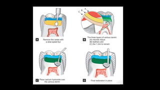
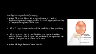
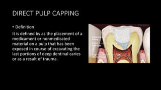
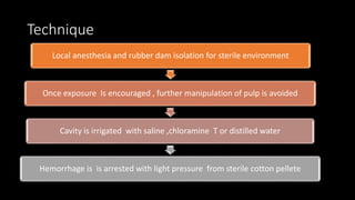
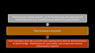
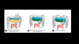
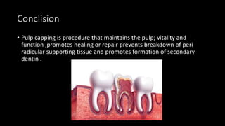
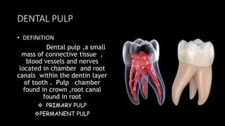
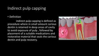
DENTAL PULP AND
PULP CAPPING
PEDIATRIC DENTISTRY
DENTAL PULP
• DEFINITION
Dental pulp ,a small
mass of connective tissue ,
blood vessels and nerves
located in chamber and root
canals within the dentin layer
of tooth . Pulp chamber
found in crown ,root canal
found in root
 PRIMARY PULP
PERMANENT PULP
DIFFERENCE BETWEEN PRIMARY AND
PERMANENT PULP
PRIMARY PULP PERMANENT PULP
Greater thickness of dentin over pulpal
wall of occlusal fossa.
Less covering dentin
Large pulp chambers Small pulp chambers
Pulp horns are high ,especially the mesial
pulp horn in case primary first molar
Pulp horns are low
Accessary canals seen in furcation areas Accessary canals seen in apical region
Continuation ……
PRIMARY PULP PERMANENT PULP
No regressive changes seen Regressive changes seen as pulp stones
and calcifications
Root canals are ribbon like Root canals are more torturous and
curved
High degree of vascularity and cellularity Comparatively less degree of vascularity
and cellularity
High potential for repair Comparatively less potential for repair
Indirect pulp capping
• Definition
indirect pulp capping is defined as
procedure where in small amount carious
dentin is retained in deep areas of cavity
to avoid exposure of pulp , followed by
placement of a suitable medications and
restorative material that seals the carious
dentin and pulp recovery.
• Objective of indirect pulp capping
• Arresting the carious process
• Promoting dentin sclerosis
• Stimulating formation of tertiary dentin
• Remineralization of carious dentin
Indications
• • Deep carious lesion, which are close to, but
• not involving the pulp in vital primary or
• young permanent teeth
• • No mobility
• • When pulp inflammation is seen as nominal
• and there is a definite layer of affected
• dentin after removal of infected dentin
Contraindications
• Sharp, penetrating pulpalgia indicating acute pulpal
inflammation
• Prolonged spontaneous pain particularly at night
• Mobility of the tooth
• Discoloration of the tooth
• Negative reaction of electric pulp testing
Single
appointment
procedure Two appointment
procedure
Single appoitnment procedure
Use local anesthesia and rubber dam isolation
Establish cavity with high speed handpiece
Remove all caries using caries detector dye i.e infected
dentin has to be removed
Stop excarvation as soon as the firm resistance if sound
dentist felt
If there is probabliy of exposure while remove further
caries then a conventional approach is choosen by
placing a hard set calcium hydroxide and temporization
the too
Cavity flushed with saline and dried with cotton p;oints
Site is covered with calcium hydroxide [ Ca OH 2]
Reminder cavity is filled with reinforced zinc oxide
eug;enol [ZOE] cement
Final restoration done followed by placement of
crown
Two appointment procedure
after first appointment [6 to8 weeks later ]
Between the appointment history must be negative and
temporary restoration should be intact
If reparative dentin bridge is formed , a permanent
restoration restoration followed by full coverage
restoration is chosen
If there is some amount of caries , remaining on re
entry carefully removed ,now somewhat sclerotic may
reveal a sound base of dentin without pulp exposure
Previous remaining carious dentin will have become
dried out, flaky and easily removed
The area around the potential exposed will be appear
whitish and may be soft ; which is pre dentin do not
disturb this area .
The cavity is prepared is washed out and dried gently
Cover the entire floor with calcium hydroxide
Base is build up with reinforced ZOE cement or GIC
Final restoration is then placed
Histological Changes after Pulp Capping
• After 24 hours: Necrotic zone adjacent to calcium
hydroxide paste is separated from healthy pulp tissue by
a deep staining basophilic layer.
• After 7 days: Increase in cellular and fibroblasticactivity.
• After 14 days: Partly calcified fibrous tissue lined by
odontoblastic cells is seen below the calcium protienate
zone; disappearance of necrotic zone.
• After 28 days: Zone of new dentin
• Sequelae/Outcome of IPC
• Three distinct types of new dentin formation take place1
1. Cellular fibrillar dentin—first 2 months
2. Globular dentin—3 months
3. Tubular dentin (uniform mineralized dentin)
1/5th of reparative dentin formation begins in less
than 30 days
After 3 months, 0.1 mm is formed
DIRECT PULP CAPPING
• Definition
It is defined by as the placement of a
medicament or nonmedicated
material on a pulp that has been
exposed in course of excavating the
last portions of deep dentinal caries
or as a result of trauma.
• Objective
• To create new dentin in the area of the exposure and
• subsequent healing of the pulp
• Indications
• Small mechanical exposure surrounded by sound dentin
in asymptomatic vital primary teeth or young permanent teeth.
• Exposure should have bright red hemorrhage that is
• easily controlled by dry cotton pellet with minimal pressure.
• True pin point exposure
• Contraindications
Severe toothache at night
Spontaneous pain
Tooth mobility
Radiographic appearance of pulp, periradicular degeneration.
Excess of hemorrhage at the time of exposure
Serous exudate from the exposure
External/internal root resorption
Swelling and fistula
Technique
Local anesthesia and rubber dam isolation for sterile environment
Once exposure Is encouraged , further manipulation of pulp is avoided
Cavity is irrigated with saline ,chloramine T or distilled water
Hemorrhage is is arrested with light pressure from sterile cotton pellete
Place the pulp capping material , on the exposed pulp with application of
minimal pressure so as to avoid forcing material into pulp chamber
Place temporary restoration
final restoration done aft successful pulp capping which done by demineralization
of dentin bridge . Maintenance of pulp vitality ,lack of pain and minimal
inflammatory response
Medicatioms and materials used in
pulp capping
Calcium hydroxide
Carticosteroids and antibiotics
Collagen fibers
4 META adhesives
Isobutyl cyanoacrylate
Denatured albumin
Mineral trioxide aggiregate
Lasers
Bone morphogenic proteins
Conclision
• Pulp capping is procedure that maintains the pulp; vitality and
function ,promotes healing or repair prevents breakdown of peri
radicular supporting tissue and promotes formation of secondary
dentin .
DENTAL PULP AND PULP CAPPING.pptx

DENTAL PULP AND PULP CAPPING.pptx

  • 1.
    DENTAL PULP AND PULPCAPPING PEDIATRIC DENTISTRY
  • 2.
    DENTAL PULP • DEFINITION Dentalpulp ,a small mass of connective tissue , blood vessels and nerves located in chamber and root canals within the dentin layer of tooth . Pulp chamber found in crown ,root canal found in root  PRIMARY PULP PERMANENT PULP
  • 3.
    DIFFERENCE BETWEEN PRIMARYAND PERMANENT PULP PRIMARY PULP PERMANENT PULP Greater thickness of dentin over pulpal wall of occlusal fossa. Less covering dentin Large pulp chambers Small pulp chambers Pulp horns are high ,especially the mesial pulp horn in case primary first molar Pulp horns are low Accessary canals seen in furcation areas Accessary canals seen in apical region
  • 4.
    Continuation …… PRIMARY PULPPERMANENT PULP No regressive changes seen Regressive changes seen as pulp stones and calcifications Root canals are ribbon like Root canals are more torturous and curved High degree of vascularity and cellularity Comparatively less degree of vascularity and cellularity High potential for repair Comparatively less potential for repair
  • 7.
    Indirect pulp capping •Definition indirect pulp capping is defined as procedure where in small amount carious dentin is retained in deep areas of cavity to avoid exposure of pulp , followed by placement of a suitable medications and restorative material that seals the carious dentin and pulp recovery.
  • 8.
    • Objective ofindirect pulp capping • Arresting the carious process • Promoting dentin sclerosis • Stimulating formation of tertiary dentin • Remineralization of carious dentin
  • 9.
    Indications • • Deepcarious lesion, which are close to, but • not involving the pulp in vital primary or • young permanent teeth • • No mobility • • When pulp inflammation is seen as nominal • and there is a definite layer of affected • dentin after removal of infected dentin
  • 10.
    Contraindications • Sharp, penetratingpulpalgia indicating acute pulpal inflammation • Prolonged spontaneous pain particularly at night • Mobility of the tooth • Discoloration of the tooth • Negative reaction of electric pulp testing
  • 11.
  • 12.
    Single appoitnment procedure Uselocal anesthesia and rubber dam isolation Establish cavity with high speed handpiece Remove all caries using caries detector dye i.e infected dentin has to be removed Stop excarvation as soon as the firm resistance if sound dentist felt If there is probabliy of exposure while remove further caries then a conventional approach is choosen by placing a hard set calcium hydroxide and temporization the too
  • 13.
    Cavity flushed withsaline and dried with cotton p;oints Site is covered with calcium hydroxide [ Ca OH 2] Reminder cavity is filled with reinforced zinc oxide eug;enol [ZOE] cement Final restoration done followed by placement of crown
  • 14.
    Two appointment procedure afterfirst appointment [6 to8 weeks later ] Between the appointment history must be negative and temporary restoration should be intact If reparative dentin bridge is formed , a permanent restoration restoration followed by full coverage restoration is chosen If there is some amount of caries , remaining on re entry carefully removed ,now somewhat sclerotic may reveal a sound base of dentin without pulp exposure Previous remaining carious dentin will have become dried out, flaky and easily removed
  • 15.
    The area aroundthe potential exposed will be appear whitish and may be soft ; which is pre dentin do not disturb this area . The cavity is prepared is washed out and dried gently Cover the entire floor with calcium hydroxide Base is build up with reinforced ZOE cement or GIC Final restoration is then placed
  • 17.
    Histological Changes afterPulp Capping • After 24 hours: Necrotic zone adjacent to calcium hydroxide paste is separated from healthy pulp tissue by a deep staining basophilic layer. • After 7 days: Increase in cellular and fibroblasticactivity. • After 14 days: Partly calcified fibrous tissue lined by odontoblastic cells is seen below the calcium protienate zone; disappearance of necrotic zone. • After 28 days: Zone of new dentin
  • 18.
    • Sequelae/Outcome ofIPC • Three distinct types of new dentin formation take place1 1. Cellular fibrillar dentin—first 2 months 2. Globular dentin—3 months 3. Tubular dentin (uniform mineralized dentin) 1/5th of reparative dentin formation begins in less than 30 days After 3 months, 0.1 mm is formed
  • 19.
    DIRECT PULP CAPPING •Definition It is defined by as the placement of a medicament or nonmedicated material on a pulp that has been exposed in course of excavating the last portions of deep dentinal caries or as a result of trauma.
  • 20.
    • Objective • Tocreate new dentin in the area of the exposure and • subsequent healing of the pulp • Indications • Small mechanical exposure surrounded by sound dentin in asymptomatic vital primary teeth or young permanent teeth. • Exposure should have bright red hemorrhage that is • easily controlled by dry cotton pellet with minimal pressure. • True pin point exposure
  • 21.
    • Contraindications Severe toothacheat night Spontaneous pain Tooth mobility Radiographic appearance of pulp, periradicular degeneration. Excess of hemorrhage at the time of exposure Serous exudate from the exposure External/internal root resorption Swelling and fistula
  • 22.
    Technique Local anesthesia andrubber dam isolation for sterile environment Once exposure Is encouraged , further manipulation of pulp is avoided Cavity is irrigated with saline ,chloramine T or distilled water Hemorrhage is is arrested with light pressure from sterile cotton pellete
  • 23.
    Place the pulpcapping material , on the exposed pulp with application of minimal pressure so as to avoid forcing material into pulp chamber Place temporary restoration final restoration done aft successful pulp capping which done by demineralization of dentin bridge . Maintenance of pulp vitality ,lack of pain and minimal inflammatory response
  • 25.
    Medicatioms and materialsused in pulp capping Calcium hydroxide Carticosteroids and antibiotics Collagen fibers 4 META adhesives Isobutyl cyanoacrylate Denatured albumin Mineral trioxide aggiregate Lasers Bone morphogenic proteins
  • 26.
    Conclision • Pulp cappingis procedure that maintains the pulp; vitality and function ,promotes healing or repair prevents breakdown of peri radicular supporting tissue and promotes formation of secondary dentin .